+ Serum Protein Electrophoresis with Immunofixation
|
|
|
- Meagan Leonard
- 6 years ago
- Views:
Transcription
1 + Serum Protein Electrophoresis with Immunofixation Dr.Ajay Phadke Centre Head SRL Diagnostics-Dr.Avinash Phadke s Lab
2 + What is electrophoresis? Electrophoresis is a method of separating proteins based on their physical properties. Proteins can be separated using a buffered solid medium (agarose electrophoresis ) or using only the liquid phase (capillary electrophoresis) The net charge (positive or negative) and the size and shape of the protein commonly are used in differentiating various serum proteins. A negatively charged particle usually travels to the positively charged electrode(gel EP) In Capillary EP? Negatively charged particle travels to the negatively charged electrode(cathode) WHY?
3 + Capillary Electrophoresis in Minicap/Capillarys Anode + DETECTION OF PROTEINS EOF Protein migration INJECTION OF SERUM Electro migration Positive charges of the buffer solution Negative charges of capillarywall The Electro-Osmotic Flow (EOF) is a stronger force than the Electrical Field. As a result, all proteins are carriedtowards the cathodic end of the capillary.
4 + The complementary positively charged ions in the surrounding buffer are free to move under the electromotive force, and they carry with them molecules of the solvent water. This buffer flow is termed electro-osmosis or endosmosis, which also carries the proteins with it to some extent by mechanical flow, not by charge. The actual distance traveled by a particular protein migrating in an electrical field is determined by the electromotive force (a feature of the protein itself and the ph) and the electro- osmotic force (a function primarily of the support medium). When the electro-osmotic force is greater than the electrophoretic force acting on weakly anionic proteins (e.g., γ-globulins), those proteins move from the application point toward the cathode, even though their charge is slightly negative.

5 Electrophoretic System in + Minicap/Capillarys Capillarys Electrophoresis Principle Thermic bridge Temperature Controlled by Peltier device Detector Migration Deuterium lamp Capillary in thermoconductive resin Cathode - High Voltage Anode +
6 + When do doctors ask for SPE? Unexplained anemia / weakness / fatigue / ESR Unexplained renal insufficiency Heavy proteinuria in patient >40yrs Bence Jones proteinuria Hypercalcaemia Hypergammaglobulinemia Immunoglobulin deficiency Peripheral neuropathy (5% will have MGUS) Recurrent infections Unexplained bone pain / pathologic fracture / lytic lesion-
7 + 1.Elderly patient with suspicion for MM i.e bone pain, lytic lesion 2. fever for >1 month 3. ESR increase, persistent anemia, fatigue 4. CRP high 5.Heavy proteinuria in adults 6.persistent increase in calcium 7.Peripheral neuropathy since a percentage have MGUS

8 Capillarys 2 Minicap

9 Carousel 28 positions

10 + Reagent Compartment Easy access to consumables: reagent cups, waste & reagents containers (2 buffer vials on board)
11 + Fraction identification α -1 acidglycoprotein α -1 antitrypsin TBG, Transcortin Haptoglobin α -2 macroglobulin Ceruloplasmin Gammaglobulins C3 complement B Lipoprotein Hemopexin Transferrin

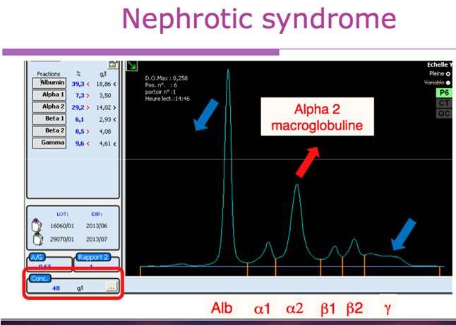
12 Normal Gaussian aspect in gamma & No increase or additional deformation/peak in gamma, beta 1, beta 2 and alpha 2 Alb α1 α2 β1 β2 γ
13 Bis albuminemia Causes: genetic, lipoproteins, biliary pigments, antibiotics, products of con

14
15 + In diabetes mellitus, patients with Type 2-2 haptoglobin have shown higher risk for vascular complications, perhaps owing to different ability to clear hemoglobin, thus leading to altered iron handling and heightened oxidative load Serum haptoglobin rises in response to stress, infection, acute inflam- mation, or tissue necrosis,
16 + Fibrinogen levels become elevated with the other acute phase reactants, occasionally to over 1.0 g/l. In such instances, the erythrocyte sedimentation rate (ESR) is also markedly elevated owing to fibrinogen content directly. Fibrinogen levels also rise with pregnancy and use of contraceptive medications. Low levels generally indicate extensive activation of coagulation with consumption of fibrinogen.
17 Congenital deficiency of A1AT A1AT = 0.17g/L (normal range: g/l) Alb α1 α2 β1 β2 γ
18 + Because of the rapid dynamics of its synthesis and clearance, prealbumin is considered to be a better early indicator of change in nutritional status Presence of a distinct band of prealbumin is used only as a landmark to confirm that the specimen was likely CSF. A protein band frequently appears in the pre-albumin position of of serum from patients who have had heparin therapy. In the circulation, heparin activates and releases lipoprotein lipase activity, which attacks triglycerides in lipoprotein fractions.
19
20 + Albumin Albumin concentrations are vital to the understanding and interpretation of calcium and magnesium levels because these ions are bound to albumin, and so decreases in albumin are directly responsible for depression of their concentrations
21 C3 (and also C4) concentration is a convenient marker for assessing disease activity in rheumatic disorders such as lupus erythematosus and rheumatoid arthritis. C4 is not appreciated on serum protein electrophoresis because its concentration is normally only about one-fifth that of C3. Both C3 and C4 are now easily quantitated by nephelometry for monitoring rheumatic disease activity
22

23

24

25
26 Haptoglobin phenotypes in alpha 2 zone Phenotype I - I Phenotype I - II Phenotype II - II
27 Monoclonal peak in Gamma Alb α1 α2 β1 β2 γ
28 Monoclonal peak in beta Alb α1 α2 β1 β2 γ

29 Oligoclonal pattern Alb α1 α2 β1 β2 γ
30 + Immunofixation & immunotyping Principle: Apply the patients sample on gel. Separate the sample. Add antibody. If positive = on washing this sample remains because of large size of complex. Immunotyping : similar principle. Automated, not labour intensive. BASIC DIFFERENCE: way how sample is processed. WE MIX sample with antibody before processing.complex is made EVEN BEFORE SEPERATION TAKES PLACE. Then injected into capillary. Monoclonal complex will MIGRATE SLOWLY and will NOT form a peak. THEREFORE, in IF you are looking for the band to be PRESENT. While in IT you are looking for it to be ABSENT! IFE is Very labour intensive
31 + IEP: serum applied to aggel in wells. EP. Antisera added. 24 hr incubation. ARCS formed IFE:Sample on solid matrix IT: NO GEL. Migration in buffered medium. Mono-specific antisera. REDUCTION technique. Antisera binds to Immunoglobulin. Heavy, large molecule created. Pulled OUT of viewing area. If PEAK DETECTEd, just click on immunotyping after selecting dilution. Hypgogamma : Ig<0.8g/L(1/10) Std :Ig g/l(1/20) Hypergamma: Ig >2.0g/L(1/40)
32 Monoclonal peak or polyclonal increase in gamma? Pointedpeak Rounded top Narrow basement Monoclonal peak IT Complete substraction of the peakwith one antiserumagainst a heavychain and a light chain Large basement Polyclonalincrease IT Complete substractionwith the antiserumagainst a heavychain and partial substractionwith the antiseraagainst kappa and lambda
33 + Normal sample Zoom


34 ELP G A ~ 80% IgG ~ 15% IgA M K L ~ 5% IgM 2/3 Kappa 1/3 Lambda

35 Abnormal peak in gamma The peak disappears in Ig G The peak disappears in Kappa Conclusion: Detection of monoclonal Ig G Kappa
36

37 IgG lambda

38
39 Zoom


40 IgG kappa
41 Fusion between beta 1& 2

42 IgA lambda

43

44 IgG lambda + free light chains lambda
45 + When interpreting IT, always consider: «If removingsomething, whatisremaining?» In eachwindow, removing one specific class of IgG highlights what is happening with the residual immunoglobulins that remain after substraction

46 When removing the IgM, the whole peak disappears When removing the Kappa a slight peak in the residual Lambda remains When removing the Lambda Ig M a peak Kappa in the + residual Ig M Kappa Lambda remains There are 2 monoclonal peaks, one Kappa and one Lambda. Both disappear when removing the IgM
47
48 Zoom

49 IgM kappa
50 + IgM Kappa+ IgM Lambda + oligoclonal profile in IgG IgM L IgM K


51 + IgG IgM Before BME treatment
52 + Treatment with Beta Mercaptoethanol (BME) to depolymerize a monoclonal Ig : * Prepare a 1% BME reducing solution : 1) 90µL H2O + 10µL BME 2) 10µL 1/10 diluted BME + 90µL Fluidil (ref 4587) * 100µL of 1% BME reducing solution + 300µL serum * Incubate 10 mn at room temperature Analyze immediately the treated sample in Capillarys or Minicap without any delay To separate co-migrating Igs of different types (Ig M vs Ig G)

53 + IgG kappa + IgM Lambda IgG K IgM L After BME treatment
54 + One or Two Ig G kappa?

55 + Smoothing 0 Change of the smoothing option 2 IgG kappa Smoothing 2
56 Polyclonal increase or monoclonal Ig in gamma? Zoom
57 Polyclonal increase or monoclonal Ig in gamma?
58 +

59 + Polyclonal increase of Ig G
60 + Oligoclonal profile Oligoclonal: Presence of multiple faint peaks or distorsions This profile is observed in: Autoimmune diseases (Rheumatoid arthritis, Gougerot Sjögren syndrome, Lupus erythematosus) Infectious (viral, bacterial, parasitic) diseases Autoimmune reactions (Patients with transplants or patients under immuno- suppressive treatments) Oligoclonal profile is linked with a dysregulation of immune system


61 + Oligoclonal profile on Hydragel IF
62 Zoom
63 Oligoclonal profile in Ig G (with major Ig G lambda to recontrol)

64
65 + Hints and tips for IT interpretation Examine carefully all IT curves without a zoom to verify the correct overlapping on albumin and the zone of interest between ELP and antisera curves Verify that the correct sample dilution has been used Compare the residual heavy and light chains after substraction and their position to verify additional presence of other monoclonal Ig If there is no correspondence between heavy and light chains, complete the test with an immunofixation to check for free light chains and/or IgD, IgE
66 + Once a monoclonal gammopathy is identified by serum protein electrophoresis, multiple myeloma must be differentiated from other causes of this type of gammopathy. (Waldenström smacroglobulinemia, smoldering multiple myeloma, monoclonal gammopathy of undetermined significance, plasma cell leukemia, heavy chain disease)
67 + sizeof the M-protein spike. Although this spike is usually greater than 3 g per dl in patients with multiple myeloma, up to one fifth of patients with this tumor may have an M-protein spike of less than 1 g per dl. 10 Hypogammaglobulinemia on serum protein electrophoresis occurs in about 10 percent of patients with multiple myeloma who do not have a serum M-protein spike. 11 Most of these patients have a large amount of Bence Jones protein (monoclonal free kappa or lambda chain) in their urine. 11 Thus, the size of the M-protein spike is not helpful in excluding multiple myeloma. If multiple myeloma still is considered clinically in a patient who does not have an M-protein spike on serum protein electrophoresis, urine protein electrophoresis should be performed.
68 + With the addition of sodium sulfate, sodium sulfite, ammonium sulfate, or methanol, the globulins tend to precipitate, leaving albumin in solution. By measuring total protein in the original serum and protein in the precipitate or the supernatant, values for albumin and globulin can be derived.
69 + 1.Elderly patient with suspicion for MM i.e bone pain, lytic lesion 2. fever for >1 month 3. ESR increase, persistent anemia, fatigue 4. CRP high 5.Heavy proteinuria in adults 6.persistent increase in calcium 7.Peripheral neuropathy since a percentage have MGUS

70 +
71 + When does one advise urine EP The following conditions (to list a few) warrant urine protein electrophoresis: 1) monoclonal protein in serum is >1.5 g/dl, 2) monoclonal free light chains are detected in serum, 3) hypogammaglobulinemia is present in serum; 4) serum electrophoresis shows nephrotic pattern. In the context of screening, the serum FLC assay in combination with serum protein electrophoresis (PEL) and immunofixation yields high sensitivity, and negates the need for 24-h urine studies for diagnoses other than light chain amyloidosis (AL)....once diagnosis of a plasma cell disorder is made, 24-h urine studies are required for all patients. For AL screening, however, the urine IFE should still be done in addition to the serum tests including the serum FLC. The FLC assay cannot replace the 24-h urine protein electrophoresis for monitoring myeloma patients with measurable urinary M proteins.
72
73 What history is important? What would you report?
74
75 Increase in alpha1, alpha 2 Advise renal profile,upe and IT
76 65 year old patient. Weakness, What do you see on the graph. What will you advise?
77 50 year old female What is your impression? What would you advise?
78 30 Year old Female. What is your opinion? What history will you take
79 Hb: 8.0 RDW: 20.3 Retic N Ferritin : 3.0 Normal range( ) B 12 : 254
80 What is important in this graph?
81

82 +
diag.of gammop Dr. A Sarrafnejad PhD Dep. Immunology School of public health TUMS
 laboratory diag.of gammop Dr. A Sarrafnejad PhD Dep. Immunology School of public health TUMS Sarrafnejad@tums.ac.ir Arne Wilhelm Kaurin Tiselius Nobel Prize for Chemistry (1948) Electrophoresis ٤ After
Typical bands found on serum gel electrophoresis:
 Gel Electrophoresis LD Recognise EPG patterns typical of other body fluids including urine and CSF Identify patterns of changes including - Paraproteins - Hypogamma - Acute phase - Circulating immune complexes
What do you do, with an M protein?
 What do you do, with an M protein? Cancer Day for Primary Care Emily Rimmer MD FRCPC January 31, 2014 emily.rimmer@cancercare.mb.ca Disclosure of Potential for Conflict of Interest Name of presenter: Emily
Southern Derbyshire Shared Care Pathology Guidelines. Diagnosis and Management of Myeloma
 Southern Derbyshire Shared Care Pathology Guidelines Diagnosis and Management of Myeloma When to screen for Myeloma and related disorders Not recommended to screen the normal population Clinical Symptoms,
Electrophoresis. Assays... INTERLAB ASSAYS. Instrument. Software Easy data management thanks to innovative Elfolab software. General Characteristic
 Software Easy data management thanks to innovative Elfolab software. Instrument Complete walk-away automation. Initial 52 results available within 50 minutes. Impressive 208 Serum Proteins samples per
Elevated Immunoglobulins and Paraproteins
 Elevated Immunoglobulins and Paraproteins NWL Pathology GP Study Afternoon Thursday 19 th October 2017 Dr Aristeidis Chaidos Consultant Haematologist and Honorary Senior Clinical Lecturer Hammersmith Hospital,
SERUM PROTEIN ELECTROPHORESIS
 SERUM PROTEIN ELECTROPHORESIS ABD-ALLA BSC - OMDURMAN AHLIA HIGH DOPLOMA DGREE - ELZAEM EL-AZHARY FORMER HEAD OF HEMATOLOGY & BLOOD BANK MINISTRY OF HEALTH LABORATORY ADMINISTRATION KHARTOUM STATE MARKETING
Multiple myeloma (MM) & related disorders
 & related disorders") Multiple myeloma (MM) & related disorders Plasma cell neoplasms Six major variants: (1) Multiple myeloma (2) Solitary plasmacytoma (3) Lymphoplasmacytic lymphoma They secrete a single complete or partial
Multiple myeloma (MM) & related disorders Plasma cell neoplasms Six major variants: (1) Multiple myeloma (2) Solitary plasmacytoma (3) Lymphoplasmacytic lymphoma They secrete a single complete or partial
WG-ICQA Harmonisation of Interpretive Commenting EQA
 WG-ICQA Harmonisation of Interpretive Commenting EQA WG-ICQA Subgroup: Harmonisation of reporting of protein electrophoresis and serum FLC, and quantification of small monoclonal proteins Improved patient
بسم اهلل الرمحن الرحيم
 بسم اهلل الرمحن الرحيم Definition Multiple myeloma is a neoplastic plasma cell dyscrasia (PCD) that generally produced a monoclonal immunoglobulin protein, characterized by a clinical pentad: (a) anemia;
Laboratory Persistence and Clinical Progression of Small Monoclonal Abnormalities
 Immunopathology / Persistence of IFE MGUS Laboratory Persistence and Clinical Progression of Small Monoclonal Abnormalities David L. Murray, MD, PhD, 1 Justin L. Seningen, MD, 1 Angela Dispenzieri, MD,
EDUCATIONAL COMMENTARY LABORATORY EVALUATION OF IMMUNOPROLIFERATIVE DISORDERS
 Commentary provided by: Louann W. Lawrence, Dr.PH, MLS (ASCP) Professor Emeritus, Retired Louisiana State University Health Sciences Center New Orleans, LA EDUCATIONAL COMMENTARY LABORATORY EVALUATION
SAS-MX IFE-1 Kit Instructions for Use
 SAS-MX IFE-1 Kit Instructions for Use Queensway South, Team Valley Trading Estate, 100300 Gateshead, Tyne and Wear, NE11 0SD, United Kingdom Tel: +44 (0)191 482 8440 Fax: +44 (0)191 482 8442 Email: info@helena-biosciences.com
Urine Protein Electrophoresis and Immunoelectrophoresis Using Unconcentrated or Minimally Concentrated Urine Samples
 Immunopathology / Electrophoresis of Unconcentrated Urine Samples Urine Protein Electrophoresis and Immunoelectrophoresis Using Unconcentrated or Minimally Concentrated Urine Samples Anja C. Roden, MD,
Serum Free Light Chain (FLC) Measurement Can Aid Capillary Zone Electrophoresis in Detecting Subtle FLC-Producing M Proteins
 Measurement Can Aid Capillary Zone Electrophoresis in Detecting Subtle FLC-Producing M Proteins") Immunopathology / DETECTING SUBTLE FLC-PRODUCING M PROTEINS Serum Free Light Chain (FLC) Measurement Can Aid Capillary Zone Electrophoresis in Detecting Subtle FLC-Producing M Proteins Nasir A, Bakshi,
Immunopathology / DETECTING SUBTLE FLC-PRODUCING M PROTEINS Serum Free Light Chain (FLC) Measurement Can Aid Capillary Zone Electrophoresis in Detecting Subtle FLC-Producing M Proteins Nasir A, Bakshi,
Serum heavy-light chain analysis (Hevylite): clinical applications for multiple myeloma
: clinical applications for multiple myeloma") Serum heavy-light chain analysis (Hevylite): clinical applications for multiple myeloma Kelly Endean PhD Scientific Affairs Manager, The Binding Site Focus of this talk An introduction to heavy-light chain
Serum heavy-light chain analysis (Hevylite): clinical applications for multiple myeloma Kelly Endean PhD Scientific Affairs Manager, The Binding Site Focus of this talk An introduction to heavy-light chain
Analytically coherent
 ZO M OCTOBER 2017 N 7 sebia FLC Free light chain testing coherent with electrophoretic methods Analytically coherent SPE: 3.1 g/l FLC Nephelometry: 32.2 g/l FLC ELISA: 3.1 g/l Principle The sebia FLC Kappa
DIAGNOSIS OF MYELOMA BASED ON THE 2014 INTERNATIONAL MYELOMA WORKING GROUP
 INDONESIAN JOURNAL OF CLINICAL PATHOLOGY AND MEDICAL LABORATORY Majalah Patologi Klinik Indonesia dan Laboratorium Medik Page 196 2018 March; 24(2): 196-200 p-issn 0854-4263 e-issn 2477-4685 Available
Guidelines for the diagnosis of Multiple Myeloma Ass.lec.: Dr. Karam T. Agha M.Sc.Pathology
 Guidelines for the diagnosis of Multiple Myeloma 2014 By:British Committee for Standards in Haematology (BCSH) Ass.lec.: Dr. Karam T. Agha M.Sc.Pathology Diagnosis, prognostic factors and disease monitoring
June Laboratory Evaluation of Plasma Cell Neoplasms Stan McCormick, MD
 June 2011 Laboratory Evaluation of Plasma Cell Neoplasms Stan McCormick, MD In this issue we review the laboratory work up of patients suspected of having a plasma cell neoplasm. As the range in clinical
Report of Survey conducted by IFCC WG Harmonisation of Interpretive Commenting EQA (WG-ICQA) subgroup:
 subgroup:") Report of Survey conducted by IFCC WG Harmonisation of Interpretive Commenting EQA (WG-ICQA) subgroup: Results of an international survey of the reporting of protein electrophoresis and serum free light
Report of Survey conducted by IFCC WG Harmonisation of Interpretive Commenting EQA (WG-ICQA) subgroup: Results of an international survey of the reporting of protein electrophoresis and serum free light
Nephelometry and turbidimetry are liquid based immunoassays based on the measurement of scattered or absorbed light.
 1 Nephelometry and turbidimetry are liquid based immunoassays based on the measurement of scattered or absorbed light. Light scattering is the physical phenomenon resulting from the interaction of light
Diagnosis and Follow-up of Multiple Myeloma and Related Disorders: The role of the laboratory
 Diagnosis and Follow-up of Multiple Myeloma and Related Disorders: The role of the laboratory Anne L Sherwood, PhD Director of Scientific Affairs The Binding Site, Inc. Learning Objectives Compare traditional
chronic leukemia lymphoma myeloma differentiated 14 September 1999 Transformed Pre- Ig Surface Surface Secreted B- ALL Macroglobulinemia Myeloma
 Disease Usual phenotype acute leukemia precursor chronic leukemia lymphoma myeloma differentiated Pre- B-cell B-cell Transformed B-cell Plasma cell Ig Surface Surface Secreted Major malignant counterpart
Assessment of Serum Free Light Chain Assays for Plasma Cell Disorder Screening in a Veterans Affairs Population
 Available online at www.annclinlabsci.org Annals of Clinical & Laboratory Science, vol 36, no. 2, 2006 157 Assessment of Serum Free Light Chain Assays for Plasma Cell Disorder Screening in a Veterans Affairs
Relatively Restricted Migration of Polyclonal IgG4 May Mimic a Monoclonal Gammopathy in IgG4-Related Disease
 Relatively Restricted Migration of Polyclonal 4 May Mimic a Monoclonal Gammopathy in 4-Related Disease Joannes F. M. Jacobs, MD, PhD, 1,2 Renate G. van der Molen, PhD, 1 and David F. Keren, MD, PhD 3 From
Laboratories and the New IMWG Myeloma Guidelines
 Laboratories and the New IMWG Myeloma Guidelines David F. Keren, M.D. Professor of Pathology Division Director, Clinical Pathology The University of Michigan dkeren@med.umich.edu Speaker Disclosure In
2. Relay characteristics of proteins and protein electrophoresis / fractionation.
 UNIT: Proteins 15prot_elec.wpd Task Electrophoresis Objectives Upon completion of this exercise, the student will be able to: 1. Review electrophoresis information as presented in class. 2. Relay characteristics
ABC of laboratory techniques for diagnosis and follow-up of monoclonal gammopathies. An Hendrickx MSc. Scientific Advisor
 ABC of laboratory techniques for diagnosis and follow-up of monoclonal gammopathies An Hendrickx MSc. Scientific Advisor In this talk introduction biology of free light chains laboratorium investigation
The Incidence and Significance of Pseudoparaproteins in a Community Hospital
 Annals o f Clinical & Laboratory Science, vol. 30, no. 3, 2000 289 The Incidence and Significance of Pseudoparaproteins in a Community Hospital Stephen Lewis Strobel Department of Pathology, St. Vincent
Electron Beam Sterilization of the Agarose Gel Used for Electrophoresis
 Electron Beam Sterilization of the Agarose Gel Used for Electrophoresis D. Ighigeanu 1, D. Martin 1, E. Manaila 1, D.E. Stan 2, I. V. Baciu 3, G. Craciun 1, C. Oproiu 1, N. Iacob 1 1 National Institute
SIGNIFICANCE OF GAMMA GLOBULINS IN MULTIPLE MYELOMA THROUGH SERUM ELECTROPHORETIC PATTERN
 WORLD JOURNAL OF PHARMACY AND PHARMACEUTICAL SCIENCES Kumar et al. SJIF Impact Factor 6.647 Volume 6, Issue 7, 1211-1218 Research Article ISSN 2278 4357 SIGNIFICANCE OF GAMMA GLOBULINS IN MULTIPLE MYELOMA
CHAPTER 3 ANTIBODY STRUCTURE I
 CHAPTER 3 ANTIBODY STRUCTURE I See APPENDIX: (3) OUCHTERLONY ANALYSIS; (6), EQUILIBRIUM DIALYSIS; (7) CROSS-REACTIVITY Electrophoretic separation of serum proteins identifies the GAMMA-GLOBULIN fraction
Diagnosis and Follow-up of Multiple Myeloma and Related Disorders: The role of the laboratory
 Diagnosis and Follow-up of Multiple Myeloma and Related Disorders: The role of the laboratory Anne L Sherwood, PhD Director of Scientific Affairs The Binding Site, Inc. Learning Objectives Compare traditional
Convenient and Effective Method for Removing Fibrinogen from Serum Specimens before Protein Electrophoresis
 Clinical Chemistry 49:6 868 872 (2003) Proteomics and Protein Markers Convenient and Effective Method for Removing Fibrinogen from Serum Specimens before Protein Electrophoresis Ling L. Qiu, 1 Stanley
Applicazioni della misura delle Free Light Chain nella pratica clinica. Maria Teresa Petrucci
 Applicazioni della misura delle Free Light Chain nella pratica clinica Maria Teresa Petrucci Serum free light chain immunoassay Heavy chain Kappa Light chain Hidden surface Lambda Exposed surface Serum
Understanding MGUS and Smoldering Multiple Myeloma
 Multiple Myeloma Cancer of the Bone Marrow Understanding MGUS and Smoldering Multiple Myeloma u-mgus+smm_en_2017_i1 12650 Riverside Drive, Suite 206 North Hollywood, CA 91607 USA Telephone: 800-452-CURE
IMMUNOCHEMICAL TECHNIQUES
 24 IMMUNOCHEMICAL TECHNIQUES 24.1 INTRODUCTION All vertebrates have advanced immune system. The more complex the organism the more advanced the immune system. The immune system of mammals has evolved over
Jerry A. Katzmann, 1* Raynell J. Clark, 1 Roshini S. Abraham, 1 Sandra Bryant, 1 James F. Lymp, 1 Arthur R. Bradwell, 2 and Robert A. Kyle 1.
 Clinical Chemistry 48:9 1437 1444 (2002) Enzymes and Protein Markers Serum Reference Intervals and Diagnostic Ranges for Free and Free Immunoglobulin Light Chains: Relative Sensitivity for Detection of
Suppression of Polyclonal Immunoglobulin Production by M-proteins Shows Isotype Specificity
 274 Annals of Clinical & Laboratory Science, vol. 31, no. 3, 2001 Suppression of Polyclonal Immunoglobulin Production by M-proteins Shows Isotype Specificity Liang Wang and David C. Young Department of
This Infosheet explains what Monoclonal Gammopathy of Undetermined Significance (MGUS) is and how it is diagnosed and managed.
 is and how it is diagnosed and managed.") MGUS This Infosheet explains what Monoclonal Gammopathy of Undetermined Significance (MGUS) is and how it is diagnosed and managed. What is MGUS? Monoclonal gammopathy of undetermined significance, or
MGUS This Infosheet explains what Monoclonal Gammopathy of Undetermined Significance (MGUS) is and how it is diagnosed and managed. What is MGUS? Monoclonal gammopathy of undetermined significance, or
The monoclonal gammopathies constitute a group of
 Sequence of Testing for Monoclonal Gammopathies The first test for recognition of monoclonal gammopathies should be serum protein electrophoresis with highresolution agarose gel. Serum protein electrophoresis
Attribution: University of Michigan Medical School, Department of Microbiology and Immunology
 Attribution: University of Michigan Medical School, Department of Microbiology and Immunology License: Unless otherwise noted, this material is made available under the terms of the Creative Commons Attribution
Controls & Calibrators Immunology Control & Coagulation
 Controls & Calibrators Immunology Control & Coagulation Immunology Control & Coagulation Human Serum based controls designed to monitor the precision of proteins and other analyte test methods, including
Capillary Zone Electrophoresis in the Evaluation of Serum Protein Abnormalities
 Capillary Zone Electrophoresis in the Evaluation of Serum Protein Abnormalities David F. Keren, MD Key Words: Capillary zone electrophoresis; M-protein; Serum protein electrophoresis; monoclonal gammopathy;
BN II The power of choice
 BN II The power of choice BN II The intelligent system for The BN II, with more than 1500 instruments sold so far, is one of the latest additions to the successful range of BN systems. This third generation
Index. Quinzanini, M. Departments of Rheumatology, Immunology & Allergology, Azienda Ospedaliera Spedali Civili, Brescia, Italy
 Poster from the Congress EUROMEDLAB Barcelona, 1-5 June 2003 15 th IFCC-FESCC European Congress of Clinical Chemistry and Laboratory Medicine 22 nd National Congress of the Spanish Society of Clinical
Immunoglobulin / free light chain ratio
 Immunoglobulin / free light chain ratio 1 1 2 Kiyotaka FUJITA Katsunori TAKAHASHI Ikunosuke SAKURABAYASHI FLC IgG, IgA, IgM, IgD, IgE 5 1 1 H 1 L H,,,, L 2 monoclonal immunoglobulinemia : M L L H 40 H
So we can separate antigens into their components and allow them to react with their antibodies
 Ag-ab reactions As for single immunodiffusion, double immunodiffusion can be also combined with electrophoresis to speed up the reaction, and in this case the test is called immunoelectrophoresis. So electrophoresis
LAM PRINCIPLE SPECIMEN. REF (150 tests) ANNUAL REVIEW Reviewed by: Date. Date INTENDED USE
 ANNUAL REVIEW Reviewed by: Date. Date INTENDED USE") IMMAGE Immunochemistry Systems Chemistry Information Sheet Copyright 2010 Beckman Coulter, Inc. Lambda Light Chain REF 446470 (150 tests) For In Vitro Diagnostic Use ANNUAL REVIEW Reviewed by: Date Reviewed
IMMAGE Immunochemistry Systems Chemistry Information Sheet Copyright 2010 Beckman Coulter, Inc. Lambda Light Chain REF 446470 (150 tests) For In Vitro Diagnostic Use ANNUAL REVIEW Reviewed by: Date Reviewed
Serum protein electrophoresis and immunofixation by a semiautomated electrophoresis system
 Clinical Chemistry 44:5 944 949 (1998) Enzymes and Protein Markers Serum protein electrophoresis and immunofixation by a semiautomated electrophoresis system Xavier Bossuyt, * Ann Bogaerts, Gilberte Schiettekatte,
Laboratory Procedure Handout. IMMUNOGLOBULIN M IgM
 KING ABDUL AZIZ UNIVERSITY FACULTY OF APPLEID MEDICAL SCIENCES DEPARTMENT OF MEDICAL LABORATORY TECHNOLOGY Laboratory Procedure Handout IMMUNOGLOBULIN M IgM Immunodiffusion single diffusion precipitation
University Journal of Pre and Para Clinical Sciences
 ISSN 2455 2879 Volume 3 Issue 1 2017 A case report of false positive Bence Jones proteinuria. SIVA SRINIVASAN Department of Biochemistry, MADRAS MEDICAL COLLEGE AND GOVERNMENT GENERAL HOSPITAL Abstract
Performance of the Sebia CAPILLARYS 2 for Detection and Immunotyping of Serum Monoclonal Paraproteins
 Immunopathology / SEBIA CAPILLARYS 2 PARAPROTEIN DETECTION Performance of the Sebia CAPILLARYS 2 for Detection and Immunotyping of Serum Monoclonal Paraproteins Zhaohai Yang, MD, PhD, Keith Harrison, MD,
SPIFE Ultra ImmunoFix Procedure
 Cat. No. 3445, 3445T, 3446, 3446T, 3447, 3447T INTENDED USE SPIFE Ultra ImmunoFix is intended for the qualitative identification of monoclonal gammopathies in serum, cerebrospinal fluid (CSF) or urine
Test Name Results Units Bio. Ref. Interval. Packed Cell Volume (PCV) %
 %") Lab No Mr DUMMY-----Z289 LLT12448 Age 15 Years Gender 16/4/2018 22600M 16/4/2018 23529M 27/6/2018 73049M Ref By Test Results Units Bio Ref Interval ANEMIA ANEL 1 COMLETE BLOOD COUNT (CBC) (Electrical Impedance
Lab No Mr DUMMY-----Z289 LLT12448 Age 15 Years Gender 16/4/2018 22600M 16/4/2018 23529M 27/6/2018 73049M Ref By Test Results Units Bio Ref Interval ANEMIA ANEL 1 COMLETE BLOOD COUNT (CBC) (Electrical Impedance
Evaluation of Sebia s Capillarys 2 flex-piercing system in. level monoclonal protein in serum and urine of patients
 Peer reviewed ORIGINAL ARTICLE Evaluation of Sebia s Capillarys 2 flex-piercing system in comparison with Hydrasys gel system, in identifying low level monoclonal protein in serum and urine of patients
Restrooms Cell phones on silent/vibrate. Refreshments. Patient resource materials
 Restrooms Cell phones on silent/vibrate Refreshments Patient resource materials This presentation should not replace discussions with your healthcare provider, but seeks to provide information and resources
Q1 What is your occupation/role? (select all that apply) Answered: 310 Skipped: 4 0% 10% 20% 30% 40% 50% 60% 70% 80% 90% 100%
 Answered: 310 Skipped: 4 0% 10% 20% 30% 40% 50% 60% 70% 80% 90% 100%") Q1 What is your occupation/role? (select all that apply) Answered: 310 Skipped: 4 ANSWER CHOICES Pathologist Total Respondents: 310 Pathologist Physician (non-radiolo... Medical Director Technologist/Te
Q1 What is your occupation/role? (select all that apply) Answered: 310 Skipped: 4 ANSWER CHOICES Pathologist Total Respondents: 310 Pathologist Physician (non-radiolo... Medical Director Technologist/Te
SWOG ONCOLOGY RESEARCH PROFESSIONAL (ORP) MANUAL GENERAL FORMS AND GUIDELINES MYELOMA FORMS CHAPTER 16D REVISED: SEPTEMBER 2016
 MANUAL GENERAL FORMS AND GUIDELINES MYELOMA FORMS CHAPTER 16D REVISED: SEPTEMBER 2016") MYELOMA FORMS The guidelines and figures below are specific to Myeloma studies. The information in this manual does NOT represent a complete set of required forms for any myeloma study. Please refer to
MYELOMA FORMS The guidelines and figures below are specific to Myeloma studies. The information in this manual does NOT represent a complete set of required forms for any myeloma study. Please refer to
Freelite for Measurement of Urine-Free Light Chains in Monoclonal Gammopathies
 Freelite for Measurement of Urine-Free Light Chains in Monoclonal Gammopathies Montgomery Lobe, MD, and Donald Pasquale, MD Abstract Monoclonal gammopathies are characterized by production of monoclonal
BIL 256 Cell and Molecular Biology Lab Spring, Development of the Immune System
 BIL 256 Cell and Molecular Biology Lab Spring, 2007 Development of the Immune System Background Information I. Serum Proteins Blood is a remarkable tissue containing cellular elements (erythrocytes, leukocytes
Plasma Protein Fractions in Healthy Blood Donors Quantitated by an Automated Multicapillary Electrophoresis System
 Plasma Protein Fractions in Healthy Blood Donors Quantitated by an Automated Multicapillary Electrophoresis System Anders Larsson* and Lars-Olof Hansson Department of Medical Sciences, Clinical Chemistry,
Change Summary - Form 2116 (R3) 1 of 6
 1 of 6") Change Summary - Form 2116 (R3) 1 of 6 Form Question Number (r3) Question Text Change Type Description New Text Previous Text Previous Question Number (r2) 2116 Today's date: Removed "Today's date:" was
Change Summary - Form 2116 (R3) 1 of 6 Form Question Number (r3) Question Text Change Type Description New Text Previous Text Previous Question Number (r2) 2116 Today's date: Removed "Today's date:" was
Accuracy of Serum IgM and IgA Monoclonal Protein Measurements by Densitometry
 160 Annals of Clinical & Laboratory Science, vol. 33, no. 2, 2003 Accuracy of Serum IgM and IgA Monoclonal Protein Measurements by Densitometry C. Howard Tseng, 1 Chin-Yung Chang, 2 Kevin S. Liu, 3 and
Parameswaran Hari Medical College of Wisconsin Milwaukee
 Multiple Myeloma Linking the clinical course to report forms Parameswaran Hari Medical College of Wisconsin Milwaukee 45,000 Annual Numbers of Blood and Marrow Transplants Worldwide 1970-2003 40,000 Number
SPECIFIC PROTEIN MARKERS. Jenna Waldron 7 th June 2016
 SPECIFIC PROTEIN MARKERS Jenna Waldron 7 th June 2016 What is a protein? Proteins - large biological molecules, or macromolecules, consisting of one or more long chains of amino acid residues. Perform
Measuring Myeloma in the LAB. BL Ferry Clinical Lead Sept 15 th 2014
 Measuring Myeloma in the LAB BL Ferry Clinical Lead Sept 15 th 2014 Oxford Immunology Laboratory Clinical Immunology Churchill WHAT DO WE MEASURE AND HOW? Clinical Immunology Churchill Serum proteins Proteins
Serum Free Light Chain Assays
 Freelite Serum Free Light Chain Assays An aid to the diagnosis of: Light Chain (Bence Jones) Multiple Myeloma (LCMM) Nonsecretory Multiple Myeloma (NSMM) AL amyloidosis Light Chain Deposition Disease (LCDD)
Immunodiagnosis SCBM 343 CLINICAL PATHOLOGY. Lect. Dr. Witchuda Payuhakrit
 Immunodiagnosis SCBM 343 CLINICAL PATHOLOGY Lect. Dr. Witchuda Payuhakrit witchuda.pay@mahidol.ac.th Objectives 2 Understand the antigen and antibody interaction Understand the principle of immunoassay
CASE REPORT AND REVIEW OF THE LITERATURE
 Page 76 / SA ORTHOPAEDIC JOURNAL Summer 2009 C ASE R EPORT AND R EVIEW OF THE L ITERATURE Low-secretory multiple myeloma HF Visser MBChB(Pret) Senior Registrar, Department Orthopaedic Surgery, University
Hevylite: New strategies for Diagnosis, Monitoring and Prognosis of monoclonal gammopathies
 Hevylite: New strategies for Diagnosis, Monitoring and Prognosis of monoclonal gammopathies AR Bradwell. University of Birmingham and Binding Site Ltd How good are tests for monoclonal monoclonal proteins?
Agilent Human 14 Multiple Affinity Removal System Spin Cartridges for the Depletion of High-Abundant Proteins from Human Proteomic Samples
 Agilent Human 14 Multiple Affinity Removal System Spin Cartridges for the Depletion of High-Abundant Proteins from Human Proteomic Samples Instructions Second edition October 2008 General Information Introduction
Human Rheumatoid Factor IgM ELISA Kit
 Product Manual Human Rheumatoid Factor IgM ELISA Kit Catalog Number PRB- 5066 PRB- 5066 96 assays 5 x 96 assays FOR RESEARCH USE ONLY Not for use in diagnostic procedures Introduction Rheumatoid arthritis
BCH 462. Single Radial Immunodiffusion and Immuno-electrophoresis
 BCH 462 Single Radial Immunodiffusion and Immuno-electrophoresis Immunoassays tests include: 1. Precipitation. 2. Agglutination. 3. Immunofluorescence. 4. Radioimmunoassay (RIA). 5. Enzyme-Linked Immuno
chronic leukemia lymphoma myeloma differentiated 14 September 1999 Pr e- Transformed Ig Su rf ace Su rf ace Secre ted Myelom a
 Disease Usual phenotype acute leukemia precursor chronic leukemia lymphoma myeloma differentiated Pr e- B-ce ll B-ce ll Transformed B-ce ll Plasma cell Ig Su rf ace Su rf ace Secre ted Major malignant
ד"ר יעל דויטשר-צ'רטקוב,
 הביולוגי ועקרון המדידה של הבסיס Free Light בסרום Chains רמות ד"ר יעל דויטשר-צ'רטקוב, המעבדה ההמטולוגית, רמב"ם - הקריה הרפואית לבריאות האדם Free light chains: Facts and Numbers Plasma cells 2 Kappa : 1
Structure of IgG and IgM
 Structure of IgG and IgM Fig. 5-1 A,B Crystal Structure of Secreted IgG Fig. 5-1 C Structure of an Ig Domain Fig. 5-2 Proteolytic Fragments of IgG (1) Fig. 5-3A Proteolytic Fragments of IgG (2) Fig. 5-3B
CAPILLARYS PROTEIN(E) 6
 6") CAPILLARYS PROTEIN(E) 6 Ref. 2003 2017/04 INTENDED USE The CAPILLARYS PROTEIN(E) 6 kit is designed for the separation of human serum and urine proteins in alkaline buffer (ph 9.9) by capillary electrophoresis
CAPILLARYS PROTEIN(E) 6 Ref. 2003 2017/04 INTENDED USE The CAPILLARYS PROTEIN(E) 6 kit is designed for the separation of human serum and urine proteins in alkaline buffer (ph 9.9) by capillary electrophoresis
PeliClass human IgG subclass ELISA kit Enzyme-linked immunosorbent assay
 PeliClass human IgG subclass ELISA kit Enzyme-linked immunosorbent assay Catalog No: M1551 Size: six pre-coated 8-well strips for each of the four IgG subclasses Test description The PeliClass human subclass
Done By :Yousef Qandeel
 Done By :Yousef Qandeel Multiple myeloma (MM) & related disorders Plasma cell neoplasms Six major variants: They secrete a single complete or partial immunoglobulin (abnormal) can be secreted in serum,
Assays for Immunogenicity: Are We There Yet?
 Assays for Immunogenicity: Are We There Yet? Mark Wener, MD Department of Laboratory Medicine & Rheumatology Division Department of Medicine University of Washington Seattle, WA 98195 wener@uw.edu Goals:
For In Vitro Diagnostic Use. Rx Only. Reviewed by Date Reviewed by Date
 IMMAGE Immunochemistry Systems Chemistry Information Sheet 2017 Beckman Coulter, Inc. All rights reserved. LAM Lambda Light Chain 446470 (150 tests) For In Vitro Diagnostic Use ANNUAL REVIEW Rx Only Reviewed
Step-by-Step Description of ELISA
 Step-by-Step Description of ELISA The protocols in this kit rely on indirect antibody capture ELISA. The steps in this assay are: Step 1: Antigen is added to the wells of the microplate strip and incubated
Smouldering myeloma. Myeloma Infosheet Series. Other related conditions. Infoline:
 Smouldering myeloma This Infosheet provides information on what smouldering myeloma is, how it is diagnosed, what the treatment is and will explain the link between smouldering myeloma and active myeloma.
acute leukemia precursor chronic leukemia lymphoma
 Disease Usual phenotype acute leukemia precursor chronic leukemia lymphoma myeloma differentiated t d Pre- B-cell B-cell Ig Surface Transformed B-cell Surface Plasma cell Secreted Major malignant counterpart
Human Immunoglobulin Free Light Chains Kappa and Lambda ELISA Kit
 Human Immunoglobulin Free Light Chains Kappa and Lambda ELISA Kit Cat. No.:DEIA4632 Pkg.Size:96 T Intended use The Human Immunoglobulin Free Light Chains Kappa and Lambda ELISA kit contains two sandwich
SERVA BluePrep Major Serum Protein Removal Kit
 INSTRUCTION MANUAL SERVA BluePrep Major Serum Protein Removal Kit (Cat. No. 42079.01) 1 Contents 1. SERVA BluePrep Major Serum Protein Removal Kit 3 2. Procedure 4 2.1. Notes prior to use 4 2.2. Column
Myeloma Primary Care. Dr R Lovell Feb 2015
 Myeloma Primary Care Dr R Lovell Feb 2015 Aims Balance of pathophysiology and cases Explain diagnostic changes (minimal) Staging UK influence Autologous stem cell transplants Primary care myeloma problems
Stephen du Toit, Chemical Pathologist
 Stephen du Toit, Chemical Pathologist LabPLUS MMH NSH LTA Pathlab Elysia- who suffers me gracefully year round baptism onomasiology designation title naming orismology name heading nuncupation onomatopoeia
510(k) SUBSTANTIAL EQUIVALENCE DETERMINATION DECISION SUMMARY ASSAY ONLY TEMPLATE
 SUBSTANTIAL EQUIVALENCE DETERMINATION DECISION SUMMARY ASSAY ONLY TEMPLATE") 510(k) SUBSTANTIAL EQUIVALENCE DETERMINATION DECISION SUMMARY ASSAY ONLY TEMPLATE A. 510(k) Number: k100499 B. Purpose for Submission: New Device C. Measurand: Rheumatoid Factors IgG, IgM and IgA and Rheumatoid
510(k) SUBSTANTIAL EQUIVALENCE DETERMINATION DECISION SUMMARY ASSAY ONLY TEMPLATE A. 510(k) Number: k100499 B. Purpose for Submission: New Device C. Measurand: Rheumatoid Factors IgG, IgM and IgA and Rheumatoid
Electro refers to electron flow or current. Thus Electrophoresis is movement under electric current.
 ELECTROPHORESIS Electrophoresis Electro refers to electron flow or current. Phoresis refers to movement. Thus Electrophoresis is movement under electric current. This technique therefore can separate molecules
Mechanisms of extravascular destruction of red cells coated with IgG1 or IgG3 (± C3b).
.") Introduction - Antibodies involved in transfusion reactions are of two types, namely the complete and the incomplete. - whereas the complete antibodies agglutinate red cells in saline medium, the incomplete
Introduction - Antibodies involved in transfusion reactions are of two types, namely the complete and the incomplete. - whereas the complete antibodies agglutinate red cells in saline medium, the incomplete
May 21, Dear Valued OSF Lab Customer,
 Dear Valued OSF Lab Customer, OSF System Laboratory is committed to providing you with the high quality products and services you expect to meet your patient s needs by continually assessing processes
SPIFE Touch Split Beta SPE - 20, 40, 60 Procedure
 Cat. No. 3422, 3421, 3420 SPIFE Touch Split Beta SPE - 20, 40, 60 Procedure The SPIFE Touch Split Beta SPE method is intended for the separation of serum, urine or cerebrospinal fluid (CSF) proteins by
Monitoring IgA Multiple Myeloma: Immunoglobulin Heavy/Light Chain Assays
 Clinical Chemistry 61:2 360 367 (2015) Clinical Immunology Monitoring IgA Multiple Myeloma: Immunoglobulin Heavy/Light Chain Assays Jerry A. Katzmann, 1,2* Maria A.V. Willrich, 1 Mindy C. Kohlhagen, 1
Immunoglobulins. Structure
 Immunoglobulins Structure Definitions Immunoglobulin is a generic term that refers to a diverse group of molecules found in the blood and tissue fluids They are soluble globulin molecules and they generally
Chapter 2. Brianne Olivieri and Alex J. Rai. Abstract. 1. Introduction
 Chapter 2 A Primer on Clinical Applications and Assays Using Urine: Focus on Analysis of Plasma Cell Dyscrasias Using Automated Electrophoresis and Immunofixation Brianne Olivieri and Alex J. Rai Abstract
IMMU 7630 Fall 2018 ANTIBODY STRUCTURE
 IMMU 7630 Fall 2018 ANTIBODY STRUCTURE ANTIBODY IS IMMUNOGLOBULIN. Almost 130 years ago it was observed that a new activity appeared in the blood plasma of animals or humans who had been immunized with
Current Issues in Pharmacy and Medical Sciences. journal homepage:
 Curr. Issues Pharm. Med. Sci., Vol., No., Pages 5- Current Issues in Pharmacy and Medical Sciences Formerly ANNALES UNIVERSITATIS MARIAE CURIE-SKLODOWSKA, SECTIO DDD, PHARMACIA journal homepage: http://www.curipms.umlub.pl/