Cryoplunge 3 with GentleBlot for cryo-em
|
|
|
- Phillip Burke
- 6 years ago
- Views:
Transcription

1 Cryoplunge 3 with GentleBlot for cryo-em


2 Cryoplunge 3 with GentleBlot system GentleBlot technology provides very gentle 1- and 2-side blotting for preparing frozen hydrated specimens for cryo-em Ethane temperature controller maintains temperature of the ethane just above its melting point Quick disconnect tweezers facilitate handling the frozen hydrated grid Protected environment for transferring the frozen hydrated grid within the cryo workstation Easy-to-use Versatile design Consistent, high quality results


3 Cryoplunge 3 with GentleBlot system


4 Cryoplunge 3 with GentleBlot system GentleBlot technology has two components Specialized blot assemblies to manually adjust the pressure applied to the specimen grid during the blotting process Factory preset pneumatic force that provides optimal speed for the blotters to contact the specimen grid
5 Cryoplunge 3 with GentleBlot system The solution for blotting fragile specimen substrates Cryo-SiN (silicon nitride TEM windows) Continuous carbon foils Large mesh grids/substrates used for tomographic data collection C-flat or Quantifoil holey carbon films Any fenestrated grid with a very thin (~5 nm) carbon coat overlay EM affinity grids (affinity capture technology)
 coats the surface of the entire fenestrated carbon film")

6 Affinity capture technology Affinity capture technology is an innovative platform that features EM affinity grids and affinity capture devices Affinity biofilm (lipid monolayer that is less than ~15 Å) coats the surface of the entire fenestrated carbon film and does not impair imaging Specialized molecular tools are designed to specifically isolate macromolecular assemblies or specific cells of interest Allows rapid purification of biological machinery for TEM imaging within minutes Provides superior partitioning of the specimen

 Vitrified inactive")


7 GentleBlot Solution for blotting fragile substrates Cryo-SiN Affinity capture Cryoplunge 3 with GentleBlot C-flat holey carbon grid Cryo-SiN (silicon nitride microchip) Vitrified inactive rotavirus DLPs on Affinity Capture Substrate. Scale bar is 100 nm. Images courtesy of Dr. Debbie Kelly, VTCRI, Roanoke, VA. USA. Tanner, J. R.; Demmert, A. C.; Dukes, M. J.; Melanson, L. A.; McDonald, S. M.; et al. Cryo-SiN An alternative substrate to visualize active viral assemblies. J Analyt Molecul Tech 2013;1(1): 6.
 50% cooler than cleaning with traditional Ar/O 2 (75%:25%) mixture Gentle")
8 Solarus 950 system for uniformly hydrophilic substrates Plasma cleaning of the EM substrate is a key step in the preparation process when a uniformly hydrophilic surface is required Solarus 950 system is designed to ionize a hydrogen/oxygen gas mixture (in addition to the standard argon or argon/oxygen mixtures) 50% cooler than cleaning with traditional Ar/O 2 (75%:25%) mixture Gentle cleaning with less sputter damage Uniformly hydrophilic charge Viewing port Specimen holder ports Specimen chamber Touch screen Solarus 950 advanced plasma cleaning system

9 Cryo-EM Preparing the frozen hydrated specimen Optimize the support substrate surface Hydrophilic, hydrophobic, Affinity Capture Apply a small aliquot of specimen to surface of substrate 2-5 µl depending on the concentration of the specimen Affinity Capture optimizes partitioning of the specimen within the holes of the fenestrated support at low concentration (0.01 mg/ml) and low volume (2 µl) Apply the specimen
10 Cryo-EM Preparing the frozen hydrated specimen Blot with filter paper until only a thin fluid layer (~100 nm) is left on the support substrate
 ice Plunge freeze the specimen grid into liquid")
11 Cryo-EM Preparing the frozen hydrated specimen Plunge the thin fluid layer into a suitable cryogen of high heat capacity (liquid ethane), which results in instantaneous freezing of the specimen in a layer of non-crystalline (vitreous) ice Plunge freeze the specimen grid into liquid ethane


12 Cryo-EM Preparing the frozen hydrated specimen Cryo-transfer the frozen hydrated grid for high-resolution data collection in the TEM Transfer frozen hydrated grid from the workstation of Cryoplunge 3 system Load the frozen hydrated specimen grid into the cryo-transfer holder


13 Results

14 140x Map of frozen hydrated grid 2-side blot Image courtesy of Dr. Chen Xu, Rosenstiel Basic Medical Sciences Research Center, Brandeis University, Waltham, MA, USA.
 Image of a single grid square; TEM magnification ~900x, electron dose 0.01 e - /Å 2. (2) Higher magnification image of a portion of the grid square; 4700x, 0.1 e - /Å 2. (3) Image of part of one hole; 59kx, 20 e - /Å 2.")

15 Cryoplunge 3 and Solarus 950 systems The three images above are an example of the high quality frozen hydrated preparations produced with Cryoplunge 3 system. (1) Image of a single grid square; TEM magnification ~900x, electron dose 0.01 e - /Å 2. (2) Higher magnification image of a portion of the grid square; 4700x, 0.1 e - /Å 2. (3) Image of part of one hole; 59kx, 20 e - /Å 2. The frozen-hydrated microtubule specimen was prepared on Quantifoil R1.2/1.3 macro machined holey carbon grids which were plasma cleaned using the Solarus 950 advanced plasma cleaning system for 15 s at 50 W using hydrogen and oxygen plasma. All images were recorded on an FEI Tecnai F30 TEM with a Single tilt liquid nitrogen cryo-transfer holder and an UltraScan 4000 camera. Images courtesy of Dr. Chen Xu, Rosenstiel Basic Medical Sciences Research Center, Brandeis University, Waltham, MA, USA.
 prepared on affinity grids decorated with His-tagged protein A and antibodies against the outer capsid protein, VP6.")
16 Cryoplunge 3 with GentleBlot technology Image of frozen-hydrated rotavirus double-layered particles (0.01 mg/ml concentration, 2 µl volume) prepared on affinity grids decorated with His-tagged protein A and antibodies against the outer capsid protein, VP6. Specimen was prepared using Cryoplunge 3 with GentleBlot system on Quantifoil 2/1 support film. Specimens were examined under low dose conditions at 120 kv. The image was recorded at 6000x using a dose of ~5 e - /Å 2. Superior partitioning of the specimen as they attached to the affinity capture biofilm in the holes on the support film Image courtesy of Dr. Debbie Kelly, Virginia Tech Carilion Research Institute, Roanoke, VA, USA.
 prepared on affinity grids decorated with His-tagged protein A and antibodies against the outer capsid protein, VP6.")

17 Cryoplunge 3 with GentleBlot technology Image of frozen-hydrated rotavirus double-layered particles (0.01 mg/ml) prepared on affinity grids decorated with His-tagged protein A and antibodies against the outer capsid protein, VP6. Specimen was prepared using Cryoplunge 3 with GentleBlot system on C-flat 2/1 grids, Protochips, Inc. Specimens were examined under low dose conditions at 120 kv. Image was recorded at 50,000x using a dose of ~5 e-/å 2. Image courtesy of Dr. Debbie Kelly, Virginia Tech Carilion Research Institute, Roanoke, VA, USA.

18 Cryoplunge 3 and Solarus 950 systems Frozen hydrated tobacco mosaic (TMV) sample prepared using Cryoplunge 3 system. Image was recorded at TEM magnification of 59kx at 300 kev and an electron dose of 20 e - /Å 2 using a model 626 Liquid nitrogen cryo-transfer holder and UltraScan 4000 camera. Sample was prepared on Quantifoil 1.2/1 specimen support, using Solarus 950 advanced plasma cleaning system prior to freezing. Image courtesy of Dr. Chen Xu Rosenstiel Basic Medical Sciences Research Center, Brandeis University, Waltham, MA, USA.

19 Cryoplunge 3 and Solarus 950 systems Frozen hydrated microtubule sample prepared using Cryoplunge 3 system. Image was recorded at 300 kev using a model 626 Liquid nitrogen cryo-transfer holder and UltraScan 4000 camera. Sample was prepared on Quantifoil 1.2/1 specimen support, using Solarus 950 advanced plasma cleaning system prior to freezing. Image courtesy of Dr. Chen Xu, Rosenstiel Basic Medical Sciences Research Center, Brandeis University, Waltham, MA, USA.

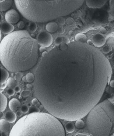
20 Cryoplunge 3 and Solarus 950 systems Frozen hydrated lipid vesicles prepared using Cryoplunge 3 system. Image was recorded at TEM magnification of 25kx at 200 kev and electron dose of ~20 e - /Å 2 using model 910 Multi-specimen single tilt cryo transfer holder and UltraScan 4000 camera. Sample was prepared on Quantifoil specimen support, using Solarus 950 advanced plasma cleaner prior to freezing. Image courtesy of Jessica Goodwin and Htet Khant, National Center for Macromolecular Imaging, Baylor College of Medicine, Houston, TX, USA.

 specimen support, using Solarus 950 advanced plasma cleaning system prior to freezing.")
21 Cryoplunge 3 and Solarus 950 systems Frozen hydrated influenza virus prepared using Cryoplunge 3 system. Gold particles were added as fiducial markers for cryoelectron tomography. Zero loss image was recorded at TEM magnification of 50kx and electron dose of ~20 e - /Å 2 at 300 kev using UltraScan 4000 camera. Sample was prepared on Quantifoil (2/2) specimen support, using Solarus 950 advanced plasma cleaning system prior to freezing. Image courtesy of Dr. Ruben Diaz-Avalos, New York Structural Biology Center, NY, USA.

22 Cryoplunge 3 and Solarus 950 Systems Frozen-hydrated image of the Ndc80 complex decorated microtubules: magnification 30kx, electron dose 40 e - /Å 2. Specimens were prepared on CFlat Protochips, Inc. CF-2/1-4C-50 holey carbon grids which were plasma cleaned using the Solarus 950 advanced plasma cleaning. Frozen hydrated preparations produced using Cryoplunge 3 system. Image recorded at 100 kv on a TEM equipped with a model Single tilt liquid nitrogen cryotransfer holder. Image courtesy of Dr. Elizabeth Wilson-Kubalek, Cell Biology, The Scripps Research Institute, La Jolla, CA, USA.
Phase Contrast Cryo Electron Tomography of Ice embedded Biological Specimens. 1. Introduction.
 Phase Contrast Cryo Electron Tomography of Ice embedded Biological Specimens Radostin Danev, Yoshiyuki Fukuda, Kuniaki Nagayama 1. Introduction. This course is aimed at introducing beginner to intermediate
New Approaches to Specimen Preparation for Molecular TEM
 New Approaches to Specimen Preparation for Molecular TEM Clint Potter National Resource for Automated Molecular Microscopy The Scripps Research Institute NRAMM Workshop 10 November 2014 The overall mission
TED PELLA, INC. Microscopy Products for Science and Industry
 PELCO SILICON NITRIDE, SILICON DIOXIDE, BLANK SILICON SUBSTRATES & APERTURES FOR TEM Clean, Debris-free with Exact 3mm TEM Frame and EasyGrip Edges PELCO Silicon Nitride Support Films for TEM Hydrophilic
Monday: Y42 G53 Tuesday: Y42 G53 Wednesday: Y42 J11
 Locations: Irchel building 42, Level H and F Locations: Irchel building 42, Level H and F Self-study sessions: Monday: Y42 G53 Tuesday: Y42 G53 Wednesday: Y42 J11 1 Center for Microscopy and Image Analysis
Preparing 3D protein nano-crystals for electron diffraction studies
 Preparing 3D protein nano-crystals for electron diffraction studies Abstract Single crystal X-ray diffraction is still the leading technique for structure determination in the solid state. However, one
Supporting Online Material for
 www.sciencemag.org/cgi/content/full/1164346/dc1 Supporting Online Material for Electron Cryomicroscopy of E. coli Reveals Filament Bundles Involved in Plasmid DNA Segregation Jeanne Salje,* Benoit Zuber,
Introduction to Electron Microscopy Andres Kaech
 Center for Microscopy and Image Analysis Introduction to Electron Microscopy Andres Kaech The types of electron microscopes Transmission electron microscope (TEM) Scanning electron microscope (SEM) 1 The
Molecular Surveillance of Viral Processes Using Silicon Nitride Membranes
 Micromachines 2013, 4, 90-102; doi:10.3390/mi4010090 Article OPEN ACCESS micromachines ISSN 2072-666X www.mdpi.com/journal/micromachines Molecular Surveillance of Viral Processes Using Silicon Nitride
Electron Microscopy (EM) Grid
 Grid") Anirban Som 25-01-14 Instrumental technique presentation Electron Microscopy (EM) Grid What I will talk about Some basic topics about EM grid Home-made grid preparation Grid cleaning Carbon coating and
Anirban Som 25-01-14 Instrumental technique presentation Electron Microscopy (EM) Grid What I will talk about Some basic topics about EM grid Home-made grid preparation Grid cleaning Carbon coating and
Highly Concentrated Aqueous Dispersions of Carbon Nanotubes for Flexible and Conductive Fibers
 Highly Concentrated Aqueous Dispersions of Carbon Nanotubes for Flexible and Conductive Fibers Laurent Maillaud 1, Robert J. Headrick 1, Vida Jamali 1, Julien Maillaud 1, Dmitri E. Tsentalovich 1, Wilfrid
Real-time observation of protein aggregates in pharmaceutical formulations using liquid cell electron microscopy
 Electronic Supplementary Material (ESI) for Lab on a Chip. This journal is The Royal Society of Chemistry 2016 Real-time observation of protein aggregates in pharmaceutical formulations using liquid cell
Microscopy AND Microanalysis MICROSCOPY SOCIETY OF AMERICA 2008
 Microsc. Microanal. 14, 375 379, 2008 doi:10.1017/s1431927608080781 Microscopy AND Microanalysis MICROSCOPY SOCIETY OF AMERICA 2008 An Improved Cryogen for Plunge Freezing William F. Tivol,* Ariane Briegel,
Correlative Microscopy - From living cells to 3D electron microscopy A comprehensive workflow approach
 Correlative Microscopy - From living cells to 3D electron microscopy A comprehensive workflow approach Maria Marosvoelgyi, Liesbeth Hekking, Ben Lich, Matthias Geisbauer and Alex de Marco (FEI) Maja Guenthert
Supporting Information
 Supporting Information Photoinitiated Growth of Sub-7 nm Silver Nanowires within a Chemically Active Organic Nanotubular Template D. M. Eisele 1, H. v. Berlepsch 2, C. Böttcher 2, K. J. Stevenson 3, D.
Supplemental Information. Site-Specific Cryo-focused Ion Beam Sample Preparation Guided by 3D. Correlative Microscopy
 Biophysical Journal, Volume 110 Supplemental Information Site-Specific Cryo-focused Ion Beam Sample Preparation Guided by 3D Correlative Microscopy Jan Arnold, Julia Mahamid, Vladan Lucic, Alex de Marco,
Workflow of Cryo-Electron Microscopy and Status of Domestic Infrastructure
 pissn 2287-5123 eissn 2287-4445 https://doi.org/10.9729/am.2018.48.1.6 Review Article Workflow of Cryo-Electron Microscopy and Status of Domestic Infrastructure Ki Ju Choi, Jae In Shin, Sung Hun Lee* Division
MODEL NanoMill TEM Specimen Preparation System. Ultra-low-energy, inert-gas ion source. Concentrated ion beam with scanning capabilities
 MODEL 1040 NanoMill TEM Specimen Preparation System The NanoMill system uses an ultra-low energy, concentrated ion beam to produce the highest quality specimens for transmission electron microscopy. Ultra-low-energy,
PP3010T Cryo preparation system for SEM, FE-SEM and FIB/SEM
 PP3010T Cryo preparation system for SEM, FE-SEM and FIB/SEM Recipe driven touch-screen interface Fully automated processes and start up Gas cooled aquilo preparation chamber Superb specimen visibility
Plunge-Freezing (Holey Carbon Method)
") This article was downloaded by: 10.3.98.93 On: 07 Apr 2019 Access details: subscription number Publisher: CRC Press Informa Ltd Registered in England and Wales Registered Number: 1072954 Registered office:
This article was downloaded by: 10.3.98.93 On: 07 Apr 2019 Access details: subscription number Publisher: CRC Press Informa Ltd Registered in England and Wales Registered Number: 1072954 Registered office:
Electron Microscopy Sciences Industry Road. P.O. Box 550. Hatfield, PA Introduction. DuraSiNTM
 DuraSiNTM Electron Microscopy Sciences Introduction DuraSiN TM Film and Mesh products have revolutionized the way samples are prepared for and analyzed in the transmission electron microscope. DuraSiN
EM Sample Preparation High Pressure Freezing. May 2013
 EM Sample Preparation High Pressure Freezing May 2013 Mouse embryonic fibroblast grown on sapphire disc. Soazig Lelay, Jana Maentler and Paul Verkade, MPI-CBG, Dresden, Germany Chapter Subchapter 4 High
Collection of continuous rotation MicroED Data from Ion Beam Milled Crystals of Any Size
 Collection of continuous rotation MicroED Data from Ion Beam Milled Crystals of Any Size Michael W. Martynowycz, 1,2 Wei Zhao, 3 Johan Hattne, 1,2 Grant J. Jensen, 3,4 and Tamir Gonen, 1,2,5,* 1 Howard
Visualizing Cells Molecular Biology of the Cell - Chapter 9
 Visualizing Cells Molecular Biology of the Cell - Chapter 9 Resolution, Detection Magnification Interaction of Light with matter: Absorbtion, Refraction, Reflection, Fluorescence Light Microscopy Absorbtion
MATERIALS. Sample Preparation & Grids for SEM & TEM... L 17 L 01. MATERIALS / Inorganics & thin films guide
 MATERIAS Sample Preparation & CHARACTERISATION Grids for SEM & TEM... 17 01 Grids for SEM & TEM regular mesh grids for tem All grids are 3.05 mm diameter, but the thickness varies with mesh size. We offer
ECE 541/ME 541 Microelectronic Fabrication Techniques
 ECE 541/ME 541 Microelectronic Fabrication Techniques MW 4:00-5:15 pm Introduction to Vacuum Technology Zheng Yang ERF 3017, email: yangzhen@uic.edu ECE541/ME541 Microelectronic Fabrication Techniques
From Eye to Insight. Leica EM ACE. Coater Family
 From Eye to Insight Leica EM ACE Coater Family EM ACE COATERS A Coater Family to cover all your needs. Developed in cooperation with leading scientists, the EM ACE coaters cover all the requirements for
MiniTEM. Designed for nanoparticle characterization
 MiniTEM Designed for nanoparticle characterization MiniTEM revolutionizes access to transmission Transmission electron microscopy (TEM) is unmatched in providing high resolution images that allow visual
Electronic supplementary information
 Electronic Supplementary Material (ESI) for ChemComm. This journal is The Royal Society of Chemistry 2014 Electronic supplementary information An electrochemical sensor for selective TNT sensing based
Grids for Applications in High-Temperature High- Resolution Transmission Electron Microscopy
 Grids for Applications in High-Temperature High- Resolution Transmission Electron Microscopy The MIT Faculty has made this article openly available. Please share how this access benefits you. Your story
Grids for Applications in High-Temperature High- Resolution Transmission Electron Microscopy
 Grids for Applications in High-Temperature High- Resolution Transmission Electron Microscopy The MIT Faculty has made this article openly available. Please share how this access benefits you. Your story
Human Myoglobin ELISA Kit
 Human Myoglobin ELISA Kit Catalog No: IHMYGKT-1 Lot No: SAMPLE INTENDED USE The Myoglobin test kits are a highly sensitive two-site enzyme linked immunoassay (ELISA) for measuring Myoglobin in biological
3-DIMENSIONAL ELECTRON MICROSCOPY OF BIOLOGICAL SPECIMENS. Korrinn M. Strunk. BS, Carnegie Mellon University, 2009
 3-DIMENSIONAL ELECTRON MICROSCOPY OF BIOLOGICAL SPECIMENS by Korrinn M. Strunk BS, Carnegie Mellon University, 2009 Submitted to the Graduate Faculty of The Swanson School of Engineering in partial fulfillment
BIOLOGICAL SAMPLE PREPARATION FOR TEM OBSERVATION. TEM Seminar Nov 16, 2017 Astari Dwiranti, Ph.D
 BIOLOGICAL SAMPLE PREPARATION FOR TEM OBSERVATION TEM Seminar Nov 16, 2017 Astari Dwiranti, Ph.D Why do we need EM for biological samples? (O'Connor and Adams, 2010) Why do we need EM for biological samples?
MODEL 1051 TEM Mill ION MILLING. Ion milling is used on physical science. specimens to reduce thickness to electron
 MODEL 1051 TEM Mill A state-of-the-art ion milling and polishing system offering reliable, high performance specimen preparation. It is compact, precise, and consistently produces high-quality transmission
METHOD FOR IMPROVING FIB PREPARED TEM SAMPLES BY VERY LOW ENERGY Ar + ION MILL POLISHING
 METHOD FOR IMPROVING FIB PREPARED TEM SAMPLES BY VERY LOW ENERGY Ar + ION MILL POLISHING Yaron Kauffmann, Tzipi Cohen-Hyams, Michael Kalina, Hila Sadan-Meltzman and Wayne D. Kaplan Dept. of Materials Engineering
Human Beta 2-Microglobulin ELISA Kit
 Human Beta 2-Microglobulin ELISA Kit CATALOG NO: IRKTAH1125 LOT NO: SAMPLE INTENDED USE The Beta 2-Microglobulin test kit is a highly sensitive two-site enzyme linked immunoassay (ELISA) for measuring
Supporting Information
 Supporting Information Structure and Solution Dynamics of Lithium Methyl Carbonate as a Protective Layer For Lithium Metal Haodong Liu, Xuefeng Wang, Hongyao Zhou, Hee-Dae Lim, Xing Xing, Qizhang Yan,
Q150R Series Rotary Pumped Coaters
 Q u o r u m Te c h n ologies Q150R Modular Coating Systems Q Series Rotary Pumped Coating Systems Innovative and versatile sputter High vacuum sputtering and carbon coater and carbonfor evaporator evaporation
Simple method for formation of nanometer scale holes in membranes. E. O. Lawrence Berkeley National Laboratory, Berkeley, CA 94720
 Simple method for formation of nanometer scale holes in membranes T. Schenkel 1, E. A. Stach, V. Radmilovic, S.-J. Park, and A. Persaud E. O. Lawrence Berkeley National Laboratory, Berkeley, CA 94720 When
Immersion Freezing of Suspended Particles and Cells for Cryo-Electron Microscopy
 Protocol Immersion Freezing of Suspended Particles and Cells for Cryo-Electron Microscopy Guenter P. Resch, 1,4 Marlene Brandstetter, 1 Lisa Königsmaier, 2 Edit Urban, 2 and Angela M. Pickl-Herk 3 1 IMP-IMBA-GMI
Supplementary Information. for
 Electronic Supplementary Material (ESI) for ChemComm. This journal is The Royal Society of Chemistry 2014 Supplementary Information for Nanoslitting Phase-separated Block Copolymers by Solvent Swelling
Canine Transferrin ELISA KIT
 Canine Transferrin ELISA KIT Cat. No.:DEIA7622 Pkg.Size:96T Intended use The Canine Transferrin ELISA KIT is a highly sensitive two-site enzyme linked immunoassay (ELISA) for measuring dog Transferrin
JSM-7800F Field Emission Scanning Electron Microscope
 JSM-7800F catalogue JSM-7800F Field Emission Scanning Electron Microscope We provide high performance The Ultimate Research Tool for Multi-Disciplinary Research Institutions Extreme resolution The super
Isolation of Protein
 Isolation of Protein Ultra-centrifugation http://irfanchemist.wordpress.com/2009/04/19/isolation-of-protein / Protein solutions of various masses or densities may separated based on the time it takes to
FUSION IN SITU HEATING AND ELECTRICAL. Protochips Quantifiably Better
 FUSION IN SITU HEATING AND ELECTRICAL Protochips Quantifiably Better PtCo Nanoparticle 700 C Image courtesy Oak Ridge National Lab Fullerene STUDY MATERIALS 1200 C Image courtesy CIC nanogune WITH THERMAL
Application Note. Sample Protection prior to FIB Processing. related instruments: Leica EM ACE600. Material Research. Life Science Research
 Material Life Science Application Note Sample Protection prior to FIB Processing related instruments: Leica EM ACE600 Medical Industrial Manufacturing Natural Resources 2 Each Atom Really Counts: Protect
Fibrinogen ELISA. For the quantitative determination of fibrinogen in biological fluids, serum, and plasma.
 Fibrinogen ELISA For the quantitative determination of fibrinogen in biological fluids, serum, and plasma. Please read carefully due to Critical Changes, e.g., Changes to preparation of standard volume
Pig Fibrinogen ELISA
 K-ASSAY Pig Fibrinogen ELISA For the quantitative determination of Fibrinogen in pig biological samples Cat. No. KT-484 For Research Use Only. 1 Rev. 9925484 K-ASSAY PRODUCT INFORMATION Pig Fibrinogen
SUPPORTING INFORMATION
 Electronic Supplementary Material (ESI) for New Journal of Chemistry. This journal is The Royal Society of Chemistry and the Centre National de la Recherche Scientifique 2017 SUPPORTING INFORMATION Luminescence
For the quantitative determination of Complement Factor 3 in guinea pig serum and plasma
 For the quantitative determination of Complement Factor 3 in guinea pig serum and plasma For Research Use Only. Not For Use In Diagnostic Procedures. Catalog Number: 41-CO3GP-E01 Size: 96 wells Version:
Light Sensitization of DNA Nanostructures via Incorporation of Photo-Cleavable Spacers
 Electronic Supplementary Material (ESI) for Chemical Communications. This journal is The Royal Society of Chemistry 2016 Light Sensitization of DNA Nanostructures via Incorporation of Photo-Cleavable Spacers
Transferrin ELISA. For the quantitative determination of transferrin in human biological samples
 Transferrin ELISA For the quantitative determination of transferrin in human biological samples Please see Appendix A for Reference Serum information. For Research Use Only. Not For Use In Diagnostic Procedures.
Differentially pumped quadrupole SIMS probe on FIBbased and two-beam microscopes
 Differentially pumped quadrupole SIMS probe on FIBbased and two-beam microscopes Richard J Chater (1), Barbara Shollock (1), David McPhail (1), Alan J Smith (2) and Graham Cooke (2) (1) Department of Materials,
Lecture Day 2 Deposition
 Deposition Lecture Day 2 Deposition PVD - Physical Vapor Deposition E-beam Evaporation Thermal Evaporation (wire feed vs boat) Sputtering CVD - Chemical Vapor Deposition PECVD LPCVD MVD ALD MBE Plating
MODEL TEM Mill. Two independently adjustable TrueFocus ion sources
 MODEL 1050 TEM Mill A state-of-the-art ion milling and polishing system. It is compact, precise, and consistently produces high-quality transmission electron microscopy (TEM) specimens with large electron
Model TEM Mill. Tabletop precision preparation for producing high-quality TEM specimens from a wide variety of materials EXCELLENCE MAGNIFIED
 Model 1050 TEM Mill Tabletop precision preparation for producing high-quality TEM specimens from a wide variety of materials EXCELLENCE MAGNIFIED Modular design for basic instrument operation or fully
Dog Fibrinogen ELISA
 K-ASSAY Dog Fibrinogen ELISA For the quantitative determination of Fibrinogen in dog biological samples Cat. No. KT-457 For Research Use Only. 1 Rev. 010209 K-ASSAY PRODUCT INFORMATION Dog Fibrinogen ELISA
Model Plasma Cleaner. Effectively cleans specimens for electron microscopy EXCELLENCE MAGNIFIED
 Model 1020 Plasma Cleaner Effectively cleans specimens for electron microscopy EXCELLENCE MAGNIFIED Model 1020 Plasma Cleaner Simultaneously cleans specimen and specimen holder. Cleans highly contaminated
The Evolution of Size, Shape, and Surface Morphology. of Gold Nanorods
 Electronic Supplementary Material (ESI) for Chemical Communications. This journal is The Royal Society of Chemistry 2018 Supporting Information The Evolution of Size, Shape, and Surface Morphology of Gold
Immuno-Labelling Cryosections
 Thin sections of biological material, mounted on nickel or gold grids, can be labelled by floating them, section-side down, on small, 10 µl, droplets of antibody. This process is conveniently carried out
Using Monolithic Chromatographic Columns to Improve the Efficiency and Robustness of Biopharmaceutical Manufacturing
 Using Monolithic Chromatographic Columns to Improve the Efficiency and Robustness of Biopharmaceutical Manufacturing IFPAC, Arlington, VA, January 25th 28th, 2015 Nika Lendero Krajnc, PhD; nika.lendero.krajnc@monoliths.com
Transmission Electron Microscopy (TEM) Prof.Dr.Figen KAYA
 Prof.Dr.Figen KAYA") Transmission Electron Microscopy (TEM) Prof.Dr.Figen KAYA Transmission Electron Microscope A transmission electron microscope, similar to a transmission light microscope, has the following components along
Transmission Electron Microscopy (TEM) Prof.Dr.Figen KAYA Transmission Electron Microscope A transmission electron microscope, similar to a transmission light microscope, has the following components along
Supplementary Information
 Supplementary Information Atmospheric microplasma-functionalized 3D microfluidic strips within dense carbon nanotube arrays confine Au nanodots for SERS sensing Samuel Yick, Zhao Jun Han and Kostya (Ken)
IgY (Chicken) ELISA Kit
 ELISA Kit") IgY (Chicken) ELISA Kit Catalog Number KA1957 96 assays Version: 03 Intended for research use only www.abnova.com Table of Contents Introduction... 3 Intended Use... 3 Principle of the Assay... 3 General
IgY (Chicken) ELISA Kit Catalog Number KA1957 96 assays Version: 03 Intended for research use only www.abnova.com Table of Contents Introduction... 3 Intended Use... 3 Principle of the Assay... 3 General
TF (Bovine) ELISA Kit
 ELISA Kit") TF (Bovine) ELISA Kit Catalog Number KA2047 96 assays Version: 03 Intended for research use only www.abnova.com Table of Contents Introduction... 3 Intended Use... 3 Background... 3 Principle of the Assay...
TF (Bovine) ELISA Kit Catalog Number KA2047 96 assays Version: 03 Intended for research use only www.abnova.com Table of Contents Introduction... 3 Intended Use... 3 Background... 3 Principle of the Assay...
NANOMETER AND HIGH ASPECT RATIO PATTERNING BY ELECTRON BEAM LITHOGRAPHY USING A SIMPLE DUV NEGATIVE TONE RESIST
 NANOMETER AND HIGH ASPECT RATIO PATTERNING BY ELECTRON BEAM LITHOGRAPHY USING A SIMPLE DUV NEGATIVE TONE RESIST H. Elsner and H.-G. Meyer Institute for Physical High Technology (IPHT), Dept. of Cryoelectronics,
Monkey Albumin ELISA
 Monkey Albumin ELISA For the quantitative determination of Albumin in monkey biological fluid For Research Use Only. Not For Use In Diagnostic Procedures. Catalog Number: 41-ALBMK-E01 Size: 96 wells Version:
Chicken IgY ELISA. Cat. No. KT-456 K-ASSAY. For the quantitative determination of IgY in chicken serum or plasma. For Research Use Only. 1 Rev.
 K-ASSAY Chicken IgY ELISA For the quantitative determination of IgY in chicken serum or plasma Cat. No. KT-456 For Research Use Only. 1 Rev. 010209 K-ASSAY PRODUCT INFORMATION Chicken IgY ELISA Cat. No.
Mouse Clusterin ELISA
 Mouse Clusterin ELISA For the quantitative determination of clusterin LQ PRXVH serum and plasma For Research Use Only. Not For Use In Diagnostic Procedures. Catalog Number: Size: Version: 41-CLUMS-E01
Supplementary Figure 1 Two distinct conformational states of the HNH domain in crystal structures. a
 Supplementary Figure 1 Two distinct conformational states of the HNH domain in crystal structures. a HNH-state 1 in PDB 4OO8, in which the distance from the C atom of the HNH catalytic residue 840 to the
Surface Micromachining
 Surface Micromachining Outline Introduction Material often used in surface micromachining Material selection criteria in surface micromachining Case study: Fabrication of electrostatic motor Major issues
The principles and practice of electron microscopy
 The principles and practice of electron microscopy Second Edition Ian M. Watt CAMBRIDGE UNIVERSITY PRESS Contents Preface tofirstedition page ix Preface to second edition xi 1 Microscopy with light and
Supporting Information
 Supporting Information Wiley-VCH 2006 69451 Weinheim, Germany Single Crystal Nanoflowers with Different Chemical Composition and Physical Properties Grown by Limited Ligand Protection Arun Narayanaswamy,
Linear Broad Beam Ion Sources ACC-30x150 IS, ACC-40x300 IS and ACC-40 x 600 IS
 Dr. Hermann Schlemm Ion Beam- and Surface Technology Saalbahnhofstraße 6 D - 07743 JENA, Germany Tel.: ++ 49 3641 22 73 29 Fax: ++ 49 3641 22 87 60 email: hermann.schlemm@jenion.de http://www.jenion.de
Supporting Information. Octopods vs. Concave Nanocrystals: Control of Morphology by Manipulating the. Kinetics of Seeded Growth via Co-reduction
 Supporting Information Octopods vs. Concave Nanocrystals: Control of Morphology by Manipulating the Kinetics of Seeded Growth via Co-reduction Christopher J. DeSantis, Angela A. Peverly, Dennis G. Peters,
HUMAN IgG. Immunoperoxidase Assay for Determination of IgG in Human Samples
 HUMAN IgG Immunoperoxidase Assay for Determination of IgG in Human Samples DIRECTIONS FOR USE Version3 L30A 31 For Research Use Only, NOT for Diagnostic Purposes Please Read this Package Insert Completely
Sheep IgG ELISA Kit. Innovative Research, Inc. Catalog No: ISHIGGKT. Lot No: SAMPLE
 Catalog No: ISHIGGKT Sheep IgG ELISA Kit Lot No: SAMPLE INTENDED USE The total Sheep IgG test kits are a highly sensitive two-site enzyme linked immunoassay (ELISA) for measuring IgG in sheep biological
RAT KIM-1 ELISA. For the quantitative determination of Kidney Injury Molecule-1 in rat serum, plasma, and urine.
 RAT KIM-1 ELISA For the quantitative determination of Kidney Injury Molecule-1 in rat serum, plasma, and urine. For Research Use Only. Not For Use In Diagnostic Procedures. Catalog Number: 41-KIMRT-E01
1 The role of scanning electron microscopy in cell and molecular biology: SEM basics, past accomplishments, and new frontiers
 1 The role of scanning electron microscopy in cell and molecular biology: SEM basics, past accomplishments, and new frontiers 1.1 Introduction New developments in scanning electron microscopy (SEM) have
Roman Chistyakov and Bassam Abraham Zond Inc/Zpulser LLC, Mansfield, MA
 HIPIMS Arc-Free Reactive Sputtering of Non-conductive Films Using the ENDURA 200 mm Cluster Tool: Direct Comparison Between Pulsed DC Pinnacle Plus and HIPIMS Cyprium Roman Chistyakov and Bassam Abraham
INDIAN INSTITUTE OF SCIENCE EDUCATION AND RESEARCH PUNE
 INDIAN INSTITUTE OF SCIENCE EDUCATION AND RESEARCH PUNE CLARIFICATION ON TENDER NUMBER - IISER-PUR-0458-16 ITEM DESCRIPTION- PROCUREMENT OF HIGH RESOLUTION TRANSMISSION ELECTRON MICROSCOPE Refer our Press
Cleaning samples. Options available. » Do not use organic. » Never, never, use squeeze. » Use detergents instead e.g. » Carbon Dioxide snow
 Sample Preparation Cleaning samples» Do not use organic solvents as these are always contaminated, even when fresh electronic grade» Never, never, use squeeze or spray bottles» Use detergents instead e.g.
Advanced SEM: ESEM and Cryo-SEM
 Advanced SEM: ESEM and Cryo-SEM Peter Harris www.reading.ac.uk/emlab Electron Microscopy Scanning electron microscope Transmission electron microscope Electron gun Condenser lens Objective lens Specimen
Scanning Electron Microscope & Surface Analysis. Wageningen EM Centre Marcel Giesbers
 Scanning Electron Microscope & Surface Analysis Wageningen EM Centre Marcel Giesbers Scanning Electron Microscope & Surface Analysis SEM vs Light Microscope and Transmission EM Secondary Electron Imaging
E! 6143 MiniTEM Development of benchtop equipment for automated characterization of viruses and other biological nanoparticles
 E! 6143 MiniTEM Development of benchtop equipment for automated characterization of viruses and other biological nanoparticles 3 years from 7.2011 to 6.2014 Brno - Palackého tř. headquarters, R&D, special
Specimen configuration
 APPLICATIONNOTE Model 1040 NanoMill TEM specimen preparation system Specimen configuration Preparing focused ion beam (FIB) milled specimens for submission to Fischione Instruments. The Model 1040 NanoMill
Journal of Structural Biology
 Journal of Structural Biology 181 (2013) 223 233 Contents lists available at SciVerse ScienceDirect Journal of Structural Biology journal homepage: www.elsevier.com/locate/yjsbi Construction and organization
Mouse Fibrinogen ELISA
 K-ASSAY Mouse Fibrinogen ELISA For the quantitative determination of fibrinogen in mouse biological samples Cat. No. KT-397 For research use only. 1 Rev. 9994397 K-ASSAY PRODUCT INFORMATION Mouse Fibrinogen
Bending Gold Nanorods with Light
 Supporting Information - Bending Gold Nanorods with Light Anastasia Babynina 1, Michael Fedoruk 1, Paul Kühler 1, Alexander Meledin 3, Markus Döblinger 4, Theobald Lohmüller 1,2 * 1 Photonics and Optoelectronics
SunMaxxTM Information Guide: Evacuated Tube Solar Collectors
 SunMaxxTM Information Guide: Evacuated Tube Solar Collectors P: 877.SUNMAXX / 888.SOLAR.11 www.siliconsolar.com / www.sunmaxxsolar.com Silicon Solar Inc Innovative Solar Solutions SunMaxxTM Information
MODEL 1061 SEM Mill ION MILLING. Ion milling is used in the physical. sciences to enhance the sample s surface. characteristics. Inert gas, typically
 MODEL 1061 SEM Mill A state-of-the-art ion milling and polishing system. It is compact, precise, and consistently produces high-quality scanning electron microscopy (SEM) samples in the shortest amount
CANINE HAPTOGLOBIN ELISA KIT
 CANINE HAPTOGLOBIN ELISA KIT CATALOG NO: IRKTAH1113 LOT NO: Sample INTENDED USE The Haptoglobin test kits are a highly sensitive two-site enzyme linked immunoassay (ELISA) for measuring Haptoglobin in
Vitrification after multiple rounds of sample application and blotting improves particle density on cryo-electron microscopy grids.
 Vitrification after multiple rounds of sample application and blotting improves particle density on cryo-electron microscopy grids. Joost Snijder 1, Andrew J. Borst 1, Annie Dosey 1, Alexandra C. Walls
Sodium dodecyl sulphate polyacrylamide gel electrophoresis (SDS-PAGE)
") Sodium dodecyl sulphate polyacrylamide gel electrophoresis (SDS-PAGE) mini-gel type A. Preparation of separating gel 1. Mix distilled water, SDS, acrylamide-bis stock and 1.5 M Tris-HCl to give
Sodium dodecyl sulphate polyacrylamide gel electrophoresis (SDS-PAGE) mini-gel type A. Preparation of separating gel 1. Mix distilled water, SDS, acrylamide-bis stock and 1.5 M Tris-HCl to give
Probing Battery Chemistry with Liquid Cell Electron Energy Loss Spectroscopy
 Electronic Supplementary Material (ESI) for ChemComm. This journal is The Royal Society of Chemistry 2015 Electronic Supplementary Information (ESI ). Probing Battery Chemistry with Liquid Cell Electron
Heteroepitaxy of Monolayer MoS 2 and WS 2
 Supporting Information Seed Crystal Homogeneity Controls Lateral and Vertical Heteroepitaxy of Monolayer MoS 2 and WS 2 Youngdong Yoo, Zachary P. Degregorio, James E. Johns* Department of Chemistry, University
MODEL Plasma Cleaner. Simultaneously cleans specimen and specimen holder. Cleans highly contaminated specimens in 2 minutes or less
 MODEL 1020 Plasma Cleaner Cleans specimens immediately before they are inserted into the electron microscope; removes existing carbonaceous debris from the specimen and prevents contamination from occurring
HUMAN APOLIPOPROTEIN H (APO-H) ELISA Assay Kit
 ELISA Assay Kit") HUMAN APOLIPOPROTEIN H (APO-H) ELISA Assay Kit Immunoperoxidase Assay for Determination of Apolipoprotein H in Human Samples INTENDED USE DIRECTIONS FOR USE For Research Use Only, NOT for Diagnostic Purposes
HUMAN APOLIPOPROTEIN H (APO-H) ELISA Assay Kit Immunoperoxidase Assay for Determination of Apolipoprotein H in Human Samples INTENDED USE DIRECTIONS FOR USE For Research Use Only, NOT for Diagnostic Purposes
A STUDY OF THE EFFECTIVENESS OF THE REMOVAL OF HYDROCARBON CONTAMINATION BY OXIDATIVE CLEANING INSIDE THE SEM.
 A STUDY OF THE EFFECTIVENESS OF THE REMOVAL OF HYDROCARBON CONTAMINATION BY OXIDATIVE CLEANING INSIDE THE SEM. Neal Sullivan, Tung Mai, Scott Bowdoin* and Ronald Vane** A poster paper presented at Microscopy
Biomimetic synthesis of gold nanocrystals using a reducing amphiphile. Ferdinand Gonzaga, Sherdeep Singh and Michael A. Brook. Department of Chemistry
 Biomimetic synthesis of gold nanocrystals using a reducing amphiphile. Ferdinand Gonzaga, Sherdeep Singh and Michael A. Brook Department of Chemistry 1280 Main St. W. Hamilton ON L8S 4M1 Canada Outline