CHARACTERIZATION OF SELECTIVE INHIBITION OF ADENYLYL CYCLASE ACTIVITY BY SMALL MOLECULE INHIBITOR NKY80
|
|
|
- Hortense Short
- 6 years ago
- Views:
Transcription

. UT GSBS Dissertations and Theses (Open Access). 323. http://digitalcommons.library.")
1 Texas Medical Center Library UT GSBS Dissertations and Theses (Open Access) Graduate School of Biomedical Sciences CHARACTERIZATION OF SELECTIVE INHIBITION OF ADENYLYL CYCLASE ACTIVITY BY SMALL MOLECULE INHIBITOR NKY80 Cameron S. Brand Follow this and additional works at: Part of the Medicine and Health Sciences Commons Recommended Citation Brand, Cameron S., "CHARACTERIZATION OF SELECTIVE INHIBITION OF ADENYLYL CYCLASE ACTIVITY BY SMALL MOLECULE INHIBITOR NKY80" (2012). UT GSBS Dissertations and Theses (Open Access) This Thesis (MS) is brought to you for free and open access by the Graduate School of Biomedical Sciences at It has been accepted for inclusion in UT GSBS Dissertations and Theses (Open Access) by an authorized administrator of For more information, please contact
2 CHARACTERIZATION OF SELECTIVE INHIBITION OF ADENYLYL CYCLASE ACTIVITY BY SMALL MOLECULE INHIBITOR NKY80 by Cameron S. Brand, B.S. APPROVED: Carmen Dessauer, PhD Supervisory Committee Chair Jeffrey Frost, PhD Richard Clark, PhD Alemayehu Gorfe, PhD John McMurray, PhD APPROVED: Dean, The University of Texas Graduate School of Biomedical Sciences at Houston
3 CHARACTERIZATION OF SELECTIVE INHIBITION OF ADENYLYL CYCLASE ACTIVITY BY SMALL MOLECULE INHIBITOR NKY80 A THESIS Presented to the Faculty of The University of Texas Health Science Center at Houston and The University of Texas MD Anderson Cancer Center Graduate School of Biomedical Sciences in Partial Fulfillment of the Requirements for the Degree of MASTER OF SCIENCE by Cameron Servetus Brand, B.S. Houston, Texas December 2012
4 Acknowledgements I would like to thank Dr. Carmen Dessauer for her invaluable mentorship. My adjustment at a young age from undergraduate student to graduate researcher was trickier than it is for most, and I cannot express enough gratitude for the expert advice and direction that has been given. I also would like to thank the faculty who served on committees. Dr. Terry Walters, Dr. Zhizhong Pan, Dr. Andy Bean, Dr. Alex Gorfe, and Dr. John McMurray are all greatly appreciated for their critiques and advice. Particularly with regards to seeing the big picture, Dr. Richard Clark and Dr. Jeff Frost deserve special mention for, along with Dr. Dessauer, sitting as members on my committees and giving feedback on my projects during inter-lab meetings and spur-of-the-moment conversations. Fellow members of the Dessauer Lab both past and present also deserve recognition. Kathryn Hassell was of great help in getting acclimated to the lab. Dr. Riad Efendi and Dr. Yong Li have been good friends and role models as researchers. There are too many CRB members of both past and present to list here, but they and the program in general have my profound thanks. My fellow CRB students have been a fantastic support system, and I simply could not have completed this thesis without all of them for practice talks, throwing research ideas back and forth, etc. Lastly, I would like to thank my parents, Anita and Gary Brand. For 24 years, they have raised, taught, and encouraged me, and have served as a grounding and constant source of support. iii
5 CHARACTERIZATION OF SELECTIVE INHIBITION OF ADENYLYL CYCLASE ACTIVITY BY SMALL MOLECULE INHIBITOR NKY80 Publication No. Cameron Servetus Brand, Ph.D. Supervisory Professor: Carmen W. Dessauer, Ph.D. The nine membrane-bound isoforms of adenylyl cyclase (AC), via synthesis of the signaling molecule cyclic AMP (camp), are involved in many isoform specific physiological functions. Decreasing AC5 activity has been shown to have potential therapeutic benefit, including reduced stress on the heart, pain relief, and attenuation of morphine dependence and withdrawal behaviors. However, AC structure is well conserved, and there are currently no isoform selective AC inhibitors in clinical use. P-site inhibitors inhibit AC directly at the catalytic site, but with an uncompetitive or noncompetitive mechanism. Due to this mechanism and nanomolar potency in cell-free systems, attempts at ligand-based drug design of novel AC inhibitors frequently use P-site inhibitors as a starting template. One small molecule inhibitor designed through this process, NKY80, is described as an AC5 selective inhibitor with low micromolar potency in vitro. P-site inhibitors reveal important ligand binding pockets in the AC catalytic site, but specific interactions that give NKY80 selectivity are unclear. Identifying and characterizing unique interactions between NKY80 and AC isoforms would significantly aid the development of isoform selective AC inhibitors. I hypothesized that NKY80 s selective inhibition is conferred by AC isoform specific interactions with the compound within the catalytic site. iv
6 A structure-based virtual screen of the AC catalytic site was used to identify novel small molecule AC inhibitors. Identified novel inhibitors are isoform selective, supporting the catalytic site as a region capable of more potent isoform selective inhibition. Although NKY80 is touted commercially as an AC5 selective inhibitor, its characterization suggests strong inhibition of both AC5 and the closely related AC6. NKY80 was also virtually docked to AC to determine how NKY80 binds to the catalytic site. My results show a difference between NKY80 binding and the conformation of classic P-site inhibitors. The selectivity and notable differences in NKY80 binding to the AC catalytic site suggest a catalytic subregion more flexible in AC5 and AC6 that can be targeted by selective small molecule inhibitors. v
7 Lay Abstract Adenylyl cyclase (AC) is a protein that produces the signaling molecule cyclic AMP (camp). There are nine AC isoforms at cell membranes that, despite being structurally similar and found in various tissue types, have many isoform specific physiological roles. AC5 activity, for example, has been implicated in pain sensation, effects of morphine and other opioids, and heart function. This makes AC5 inhibition desirable as a potential treatment to block chronic pain, lessen the dependence and withdrawal effects of opioids, or protect against chronic heart failure. However, isoform specific inhibition would be important for AC inhibiting drugs in order to limit the number of potential side effects. There are AC inhibitors used in research that are not suitable for drug development because they are not potent enough, specific enough, and/or are unable to enter cells. This project aimed to characterize isoform selective AC inhibition with a known isoform selective inhibitor and predict how it binds to the catalytic site of AC differently from nonselective inhibitors. Novel AC inhibitors with some isoform selectivity were identified in a virtual screen predicting the binding of small drug-like molecules to the AC catalytic site. Additionally, although being commercially sold as an AC5 specific inhibitor, the small molecule NKY80 was shown to inhibit AC5 as well as the closely related AC6 more potently than the other seven isoforms. Virtual binding of NKY80 to AC predicted that NKY80 has a unique interaction within catalytic site that classical nonselective AC inhibitors do not have so consistently. We propose this region is more flexible in AC5 and AC6, explaining NKY80 s selectivity and allowing for targeting of this region to develop more selective AC inhibitors. vi
8 Table of Contents Approval Page... i Title Page... ii Acknowledgements... iii Abstract... iv Lay Abstract... vi Table of Contents... vii List of Illustrations... x List of Tables... xi Abbreviations... xii Chapter 1: Introduction... 1 Adenylyl Cyclase Function and Structure... 2 AC Regulation... 2 AC Roles in Physiology... 4 History of AC Inhibitors... 8 Significance of Proposed Research Chapter 2: Materials and Methods Virtual Ligand Docking AC Sequence Alignment Cell Line Maintenance Sf9 AC Expression Sf9 Membrane Preparation HEK AC Transfection vii
9 HEK Membrane Preparation Adenylyl Cyclase Activity Assays Chapter 3: Virtual Docking of AC Catalytic Site Derivation of AC Docking Target Validation of Docking to AC Target Docking of P-Site Inhibitors to AC Target Summary Chapter 4: Screening for Novel Inhibitors at AC Catalytic Site Current Progress on Small Molecule AC Inhibitor Development High Throughput Virtual Screening for Novel AC Inhibitors Screening Candidates for Ability to Inhibit AC Characterizing Selectivity of Novel AC Inhibitors Summary Chapter 5: NKY80 Docking and Full Characterization of AC Isoform Selectivity History of NKY80 Inhibitor NKY80 Docking to AC Comparison of NKY80 Inhibition of AC5 and AC NKY80 Inhibition Curves for AC Summary Chapter 6: Discussion AC Isoform Groups and NKY80 Selectivity AC Catalytic Site Residues and Inhibitor Selectivity Chemical Groups Important for NKY80 Inhibition of AC viii
10 Comparison of NKY80 to Other AC Inhibitors Cardiac Complexes of AC5/AC6 and Inhibitor Selectivity Future Directions Summary Bibliography Vita ix
11 List of Illustrations Chapter 1 Figure 1: Schematic of AC Structure Figure 2: Notable Small Molecule AC Inhibitors Chapter 3 Figure 3: Alignment of Catalytic Site of Crystal AC Structure and Human AC Figure 4: Replication of Binding Conformation of 2',3'-dd-ATP Figure 5: P-Site Inhibitors Accurately Docked to Catalytic Site Chapter 4 Figure 6: Histogram of Top 10% of Scores for ChemBridge Drug-Like Small Molecules.. 29 Figure 7: Predicted Binding Conformation and Characterized Isoform Selectivity of Novel AC Inhibitor CB Figure 8: Predicted Binding Conformation and Characterized Isoform Selectivity of Novel AC Inhibitor CB Figure 9: Predicted Binding Conformation and Characterized Isoform Selectivity of Novel AC Inhibitor CB Chapter 5 Figure 10: Predicted Binding Conformation of NKY Figure 11: Full AC Isoform Selectivity Characterization of NKY Chapter 6 Figure 12: Structural Flexibility in the AC Catalytic Site x
12 List of Tables Chapter 1 Table 1: AC Isoform Expression and Function Chapter 5 Table 2: Predicted and Experimental IC 50 for NKY80 Inhibition of Adenylyl Cyclase xi
13 Abbreviations AC ADP AKAP ATP BLAST Adenylyl Cyclase Adenosine Diphosphate A-Kinase Anchoring Protein Adenosine Triphosphate Basic Local Alignment Search Tool C1 Cytosolic Domain 1 C2 Cytosolic Domain 2 camp Cl DMEM DNA DTT EDTA FBS Fsk Gβγ GDP Giα GPCR Goα Gsα HEK Cyclic Adenosine Monophosphate Chlorine Dulbecco s Modified Eagle Medium Deoxyribonucleic Acid Dithiothreitol Ethylenediaminetetraacetic Acid Fetal Bovine Serum Forskolin Heterotrimeric G Protein Beta-Gamma Subunit Guanosine Diphosphate Heterotrimeric G Protein Inhibitory Alpha Subunit G Protein Coupled Receptor Heterotrimeric G Protein Olfactory Alpha Subunit Heterotrimeric G Protein Stimulatory Alpha Subunit Human Embryonic Kidney xii
14 HEPES MANT Mg Na NT PBS PDB PKA PKC PSI sac SDS Sf9 VMD 4-(2-hydroxyethyl)-1-piperazineethanesulfonic Acid Methylanthraniloyl Magnesium Sodium Amino Terminus Phosphate Buffered Saline Protein Data Bank Protein Kinase A Protein Kinase C Pounds per Square Inch Soluble Adenylyl Cyclase Sodium Dodecyl Sulfate Spodoptera frugiperda (clonal isolate) Visual Molecular Dynamics xiii
15 Chapter 1 Introduction 1
16 Adenylyl Cyclase Function and Structure The primary function of adenylyl cyclase (AC) is to produce the signaling molecule cyclic AMP (camp), an important 2 nd messenger in cell signaling[1]. Production of camp occurs through conversion of the cellular energy storage molecule adenosine triphosphate (ATP) into camp and pyrophosphate, with camp then being free to target downstream signaling proteins. There are nine mammalian membrane-bound isoforms of AC (AC1-9), as well as a soluble mammalian form (sac). This sac is similar to the bacterial cyclase predecessor of mammalian AC[2]. Global structure of the membrane-bound AC isoforms is well conserved. Basic organization involves a cytosolic N-terminus (NT) and two other cytosolic domains (C1 and C2) separated by a pair of 6-transmembrane helices[3](figure 1). The NT domain has sequence and length that varies widely amongst the isoforms. The NT variability allows for some mechanisms of isoform specific regulation. This includes scaffolding actions that may facilitate signal transduction. For example, it has been shown that the AC5 NT anchors heterotrimeric G proteins[4]. The two C1/C2 cytoplasmic domains are 40% similar in sequence, pseudosymmetrical, and form the catalytic area where ATP binds and synthesis of camp takes place[5]. These C1/C2 domains are also where various intracellular regulators such as heterotrimeric G proteins interact with AC[6, 7]. AC Regulation Signaling proteins that similarly regulate all AC isoforms are rare; one notable exception is the heterotrimeric G protein subunit Gsα, which stimulates all AC isoforms[8-10]. AC isoform specific function is due to differences in tissue expression patterns (Table 1), 2

17 Figure 1: Schematic of AC Structure. regulation, and organization of signaling by scaffolds such as A-kinase anchoring proteins (AKAPs). Some notable differences in regulation allow for organization of AC1-9 into groups. AC 1, 3, and 8 are noted for their stimulation by calcium-stimulated calmodulin; AC 2, 4, and 7 are characterized by considerable conditional stimulation by Gβγ; AC 5 and 6 both have sensitive, micromolar level inhibition by free calcium; and AC9 is in a unique class by itself, including insensitivity to the AC activator forskolin[11]. Since C1 and C2 AC domains form the ATP binding site where camp production takes place, most regulators of AC directly or indirectly modify this catalytic cleft in order to modulate AC activity. The same signaling protein can also regulate AC isoforms differentially; for example, Gβγ stimulates AC 2/4/5/6/7, but inhibits AC 1/3/8[12, 13]. Metal ions such as Mn 2+, Ca 2+, and Mg 2+ also have significant effects at the catalytic site on camp production[14]. AC catalyzes phosphoryl transfer by a metal ion mechanism involving Mn 2+ or Mg 2+. This metal requirement is similar to that for DNA polymerase function. Conversely, Ca 2+ and Zn 2+ can inhibit AC[15, 16]. 3
18 Forskolin is a diterpene derived from the root of the plant Coleus forskohlii[17]. It is able to clearly stimulate activity of all the membrane-bound AC isoforms except AC9[11, 18]. A single forskolin molecule binds at the interface of the C1 and C2 domains[19]. Due to the C1/C2 pseudosymmetrical structure, the forskolin binding pocket is structurally related to the ATP-binding active site. AC9 is missing a key residue within this forskolin-binding pocket that when mutated can elevate forskolin sensitivity to the level exhibited by the other AC isoforms[20]. Gsα and forskolin are also capable of simultaneous stimulation of AC; whether this stimulation is synergistic or additive is isoform specific[18]. AC5 and 6 are very closely related AC isoforms. They are both stimulated by Gsα and Gβγ[12] and inhibited by Ca 2+ [21] and PKA[22]. Neither is directly inhibited by Goα, although it has been shown that there is a mechanism of indirect inhibition of AC5 or AC6 by Goα signaling that could be blocked by antibodies targeting Goα[23]. The known differences between regulation of AC5 and AC6 are typically subtle. For example, Gsα stimulated activity of either isoform is inhibited by Giα, but basal activity of AC5 is also inhibited by Giα while AC6 basal activity is not[24]. Also, AC5 is stimulated by PKC-α and -ζ[25], while AC6 is inhibited by PKC-δ and -ε[26, 27]. Gqα signaling can enhance AC6 activity, but not that of AC5, within intact cells by a calcium/calmodulin dependent mechanism[28]. Both isoforms thus have numerous stimulatory and inhibitory inputs to control camp levels. AC Roles in Physiology The various AC isoforms are involved in many physiological phenomena, with isoform specific roles that are sometimes unclear. Physiological roles of AC cannot solely be explained by tissue distribution. Each AC isoform has a unique tissue expression pattern, but multiple 4
19 isoforms are found in nearly all cell types[29]. Originally, the AC/cAMP pathway was characterized by its role in glycolysis and glucose metabolism[30]. Over time, other physiological roles have been identified as well; reviews have been produced that catalogue evidence for various isoform specific functions[6], while a somewhat briefer summary will be given here (Table 1). Learning, memory, and synaptic plasticity involve the closely related isoforms AC1 and AC8[31-36]. AC2, AC4, and AC7 have few clear physiological roles at this point, although evidence suggests AC7 s involvement in ethanol dependency[37] and immune cell function[38]. AC3 is involved in olfactory function[39, 40], as well as regulation of renal filtration[41]. AC3 and sac both appear to have roles in the testes for sperm production and function[42-44]. AC9 has suggested roles in ion channel mediated repolarization of the heart[45] and immune cell migration[46]. Of the AC isoforms, AC5 has by far the widest variety of identified physiological functions, typically being described by AC5 knockout mouse models. AC1 and AC5 have been implicated in different aspects of pain response, with AC5 mouse knockouts having attenuated responses to both mechanical and inflammatory pain[47]. These mice are characterized by limited mechanical and thermal response to normal stimuli in neuropathic pain models. Conversely, pharmacological stimulation of the AC/cAMP pathway increased sensitivity to the inflammatory pain response[48]. AC5 also has been shown, along with AC1, AC6, and AC8, to have an ability for sensitized stimulation. Sensitized stimulation of AC5 occurs through receptor-coupled Gsα stimulation following prolonged inhibition by receptor-coupled Giα protein. Such sensitized activation has been shown with prolonged stimulation of various Giαcoupled GPCRs, including D2 dopamine and μ-opioid receptors[49-52]. Tolerance, reward and physiological dependence, and withdrawal symptoms of the opiates are functions where these 5

20 receptors have been strongly suggested to play roles. For AC5, this sensitized stimulation appears to also be involved in opiate dependency and withdrawal behaviors, as the effects of morphine were attenuated in AC5 knockout mice[53]. AC5 has also been shown to be involved in motor coordination, with AC5 mouse knockouts developing Parkinson s-like tremors and other motor dysfunctions[54]. All three of these physiological functions are regulated to some degree in the brain by the striatum, where AC5 expression is strongly enriched and is the principal signaling node for D1 and D2 dopamine receptors[55]. 6
21 Additionally, although multiple AC isoforms are expressed in the heart, AC5 and AC6 are most predominantly expressed[56] and have the most clearly demonstrated cardiac functions at this time. A role of the AC/cAMP signaling pathway in heart failure has been established by models of increased expression of signaling proteins upstream and downstream of AC resulting in cardiomyopathy[57-59]. The primary treatment of choice for individuals that are at high risk for chronic heart failture is β-blockers, consistent with significant involvement of camp signaling pathways in cardiac dysfunction[60]. Both AC5 and AC6 are expressed equally in the neonatal heart but AC5 is dominant in the adult heart[61]. Other work has shown AC5 expression mainly in the ventricle while AC6 was in both the ventricle and atria[62]. Thus, it is likely AC5 and AC6 roles in cardiac contraction differ. Disruption of AC5 under thoracic banding simulated stress conditions is protective against heart failure[63]. However, the critical functions of AC5 in the heart are murky as both sympathetic and parasympathetic cardiac regulations are affected in AC5 knockout mice[64]. This includes a complete loss of acetylcholine-mediated Giα inhibition and a significant reduction in Ca 2+ -mediated inhibition of camp production consistent with reduced effects of muscarinic signaling. AC5 loss also desensitizes cardiomyocytes to catecholamine stress, preventing cardiac apoptosis that would otherwise result[65]. This apoptotic reduction may underlie the reduced cardiac stress following aortic banding in knockout mice[63]. AC5 knockout mice also have an increased lifespan and are protected against age-induced cardiomyopathy[66]. This phenotype includes cardiac hypertrophy, apoptosis, and reduced function. Some mouse models for AC5 loss also show increased basal left ventricular function, but decreased responsiveness to beta-adrenergic stimulation that suggests a higher threshold for increasing camp signaling over basal levels[67]. 7
22 History of AC Inhibitors With all of these physiological functions associated with the AC/cAMP signaling pathway, it is not surprising that the pathway is an attractive target for drugs to modulate these functions. As a result, many drugs have been identified that target camp signaling[68]. Salmeterol is a partial agonist of the β2-adrenergic receptor, increasing camp production to increase circulation useful in the treatment of asthma. Theophylline is a phosphodiesterase inhibitor and adenosine receptor antagonist also used as an asthmatic treatment by maintaining elevated camp, albeit as a less frequently used treatment. Beta-blockers decrease camp and are used as treatments for various heart conditions, such as metoprolol for hypertension and heart failure. Morphine is a µ-opioid receptor agonist that decreases camp and, in turn, inhibits pain sensation. Various dopamine receptor agonists and antagonists modulate camp activity downstream as therapeutics for schizophrenia, Parkinson s disease, and nausea. However, these therapeutics target upstream GPCRs or downstream regulators of camp concentration. Direct control of camp production by targeting AC activity has been a more difficult target. The structural similarity and overlapping tissue expression of AC isoforms provide great risk for nonspecific effects. Since AC is an enzymatic protein with transmembrane and cytoplasmic domains, the lack of extracellular domains such as those targeted by ligands on GPCRs mean an AC drug must be capable of passing through the plasma membrane, which has severely limited the kind of molecules that progress past cell-based systems and preclinical animal models. Some AC inhibitor prodrug packages have been attempted to circumvent this issue[69, 70]. However, the current state is that there are no isoform specific AC inhibitors in clinical use. 8
23 That being said, there has been progress in identifying AC isoforms that would be desirable as targets if they could be selectively inhibited. Since it has been shown that AC5, AC1, and AC8 are involved in inflammatory/neuropathic pain, but only AC5 does not cause any major defects in knockout mouse models, AC5 inhibitors have promise as analgesics. Since negative side effects due to compensatory mechanisms are induced by long-term opioid use in patients with chronic pain, AC5 selective inhibitors for patients could also be more viable for long-term treatment. Based on AC5 knockout models, AC5 inhibitors could also act similarly to beta-blockers in the treatment of heart failure. The predominant expression of AC5 and closely related AC6 in heart failure combined with their differing effects on cardiac function requires a selective ligand that would inhibit AC5 but not AC6. The drastic AC5 activity differences in chronic versus acute heart failure also has to be taken into account for prospective drug development. It has been suggested that increased camp from AC5 activity is beneficial as a rescue function in acute heart failure, but is clearly detrimental in chronic failure models. Colforsin daropate hydrochloride is a direct activator of AC5 (with AC6 potency unknown)[71]. Its effects include coronary vasodilation[72], and the activator is an approved treatment in Japan for acute heart failure[73]. In research, two distinct classes of small molecule AC inhibitor are commonly among those utilized. Examples of these inhibitor classes and some notable inhibitors derived from them are shown in Figure 2. One of these classes is P-site inhibitors, which bind in the AC catalytic site and decrease activity through uncompetitive inhibition[74, 75]. The original P-site inhibitors, which we will call classic P-site inhibitors, are adenosine nucleotide analogs with a phosphate or polyphosphate group at the 3 position of a ribose ring. These inhibitors are more potent on activated forms of AC versus the basal state, forming a product-like transition state 9
24 Figure 2: Notable Small Molecule AC Inhibitors. with pyrophosphate to prevent further ATP binding[76]. Although most of the classic P-site inhibitors were synthetically made, some are naturally occurring variants of ADP and ATP[77]. More potent inhibitors, such as adenosine 3 -polyphosphates (i.e. 2,5 -dideoxy-3 -ATP), bind in the absence of pyrophosphate with the aid of the 3 -phosphates, increasing the affinity of these 10
25 inhibitors by 100- to 1,000-fold[78, 79]. This conformation is also produced by the noncompetitive ATP analog 2,3 -dideoxy-5 -ATP, currently the most potent AC inhibitor at a K i of 16nM for rat brain AC[80]. Although most classic P-site inhibitors lack isoform selectivity, there are some exceptions. It has been shown that 2 -deoxy-3 -AMP is mildly selective for AC1 over AC2 and AC6[81]. Also, AC9 is insensitive to 2 -deoxy-3 -AMP [81]. In general, however, P-site inhibitors that are not products of inhibitor optimization experiments are not greatly selective against certain isoforms. P-site based inhibitors nonetheless are extremely direct and potent inhibitors; these compounds typically have affinities of nanomolar concentration. The other predominant class of direct small molecule AC inhibitors is the MANTnucleotide inhibitors. One such example is MANT-GTP, which has a K i of 53nM in Sf9 cyclymphoma cell membranes[82]. MANT-GTP binds in the ATP-binding site with a nucleotide in reverse orientation compared to ATP analogs such as P-site inhibitors[83, 84]. Interestingly, published research suggests that MANT-GTP and similar MANT-nucleotides show potential for designing isoform selective compounds that target the AC catalytic site despite the structural similarity of AC isoforms[83, 85]. These MANT-GTP screens suggested inhibitor affinity was more dependent on the ribose and polyphosphate components of such small molecule AC inhibitors, not necessarily the base component. Thus, non-nucleotide based inhibitors able to modulate AC activity directly at the catalytic site are theoretically possible at therapeutically desirable potency levels. The crystal structure of MANT-GTP with AC also suggests catalytic site flexibility, with the movement of helices to accommodate the MANT group in a C pocket that can accommodate hydrophobic interactions. Crystallized P-site inhibitors show only two subregions of the ATP binding site; an A pocket where hydrogen 11
26 bonding to the purine group takes place, and a B pocket including the two metal ion binding sites and typically containing metal-phosphate interactions. MANT-GTPγS introduced into whole-cell mouse cardiomyocytes by patch pipette has been used to attenuate L-type calcium channel currents that was presumably through inhibition of coupled AC5[86]. Significance of Proposed Research Validation of isoform selective small molecule AC inhibitors in preclinical animal models is a stage of therapeutic development rarely reached. The most significant obstacle is generating small molecule inhibitors with sufficient potency, selectivity, and therapeutic potential combined. One small molecule inhibitor that resulted from searching for these characteristics in a compound derived from P-site inhibitors, NKY80, is described as an AC5 selective inhibitor with low micromolar potency in vitro. Classic small molecule AC inhibitors have revealed important ligand-binding pockets in the AC catalytic site, but what specific interactions in these regions give NKY80 selectivity is currently unclear. Identifying and characterizing unique interactions between NKY80 and AC isoforms could be used to determine isoform specific differences in the interaction of the well conserved catalytic site with regulatory molecules, and as a result significantly aid the development of isoform selective AC inhibitors with therapeutic potential. I hypothesize that unique isoform specific interactions between the AC catalytic site and NKY80 explain the small molecule s selective inhibition profile. 12
27 Chapter 2 Materials and Methods 13
28 Virtual Ligand Docking Searching for novel directly binding AC inhibitors was performed by using Glide docking software from Schrdinger to dock ligands specifically to the catalytic ATP binding site of AC. Both known P-site inhibitors and 35,000 compounds from the generic ChemBridge library of drug-like small molecules that follow Lipinski s rule of five were used as ligand libraries. These libraries were docked to a model of the AC catalytic site adapted from crystal structure of the AC C1/C2 domains bound with the inhibitor 2,3 -dd-atp (PDB ID 1CJT)[16]. This model had waters, forskolin, Gαs protein, Mg 2+ ion, Cl - ion, and buffer molecules removed, leaving only the AC C1 and C2 domains. Ligand 3D structures were prepared, including unprotonated state and creation of stereoisomers, using Schrdinger s LigPrep software. Docking was restricted to the ATP binding region of the catalytic cleft. Hydrogen bonds were defined by a donor-acceptor distance cutoff of 3.5 Å and a donor-hydrogenacceptor angle between 150 and 180. ChemBridge ligands were ranked based on the Glide predicted energy score in kcal/mol and most energetically favorable binding pose. AutoDock 4.2 was used to dock known P-site inhibitors and NKY80 to the entire structure of the AC catalytic site model from crystal structure 1CJT. Docking to the entire AC structure was chosne to test if AutoDock correctly predicts P-site inhibitor binding to the catalytic site in an unbiased manner, as well as to provide a comprehensive prediction of the NKY80 binding site. The value of such a blind docking procedure has been demonstrated previously[87]. Briefly, small molecule inhibitor ligands were prepared with AutoDock tools, creating a nonpolar atomic charge sum and calculating the Gasteiger charges and torsions[88, 89]. AutoDock 4.2 was then used to dock the ligands onto a grid large enough to encompass the entire surface of the AC catalytic site from crystal structure 1CJT with Å grid spacing. A 14
29 population size of 150, with 2 Å clustering cutoff, 10,000 LGA hybrid with 256 runs was used. Predicted poses were analyzed in terms of their energetic binding potential. Hydrogen bonds were defined as described for Glide docking. Van der Waals contacts were defined by a carboncarbon distance cutoff of 5.0 Å. Potential poses were predicted and ranked based on the predicted binding affinities. The free ICM-Browser available from Molsoft L.L.C. was used to visually inspect predicted conformations of ChemBridge small molecules during virtual screening. In addition, VMD Viewer 1.9 from the Theoretical and Computational Biophysics Group at the University of Illinois at Urbana-Champaign was used for visual analysis of the known inhibitor docking results, and for image rendering. AC Sequence Alignment Alignment of two or more AC sequences using BLAST ( was utilized to compare C1 and C2 domains of various AC isoforms. The primary sequences of the C1/C2 domains used in a crystallized AC structure (PDB ID 1CJT) were aligned with the corresponding C1/C2 sequences of human AC1-9. Cell Line Maintenance Sf9 insect cells were maintained with SF-900 II SFM media contained in cell suspension flasks. Cells were passaged to maintain a concentration of log phase cells between 0.5 X 10 6 and 5.5 X 10 6 cells/ml. Cell incubation was in a 27 C shaker at 150rpm. 15
30 HEK293 cells were maintained in Dulbecco s Modified Eagle Medium (DMEM) with 10% fetal bovine serum and 1% penicillin/streptomycin on 10cm dishes in a 37 C, 5% CO 2 incubator. Sf9 AC Expression Baculoviruses were amplified for 5-7 days using 500µl of storage virus stock with 50ml of Sf9 cells in log phase in SF-900 II SFM media with 50 μg/ml gentamicin at a concentration of X 10 6 cells/ml to produce a baculovirus working stock with a minimum titer of 1 X 10 7 pfu/ml. 25ml of the appropriate AC isoform baculovirus was used to infect 1 liter of Sf9 cells at a concentration of X 106 cells/ml. After 48 hours of infection, Sf9 cells were harvested by centrifugation at 1000 g for 10 minutes at 4 C. Sf9 Membrane Preparation Harvested cells were suspended in 600ml of cold lysis buffer consisting of 50 mm NaHEPES (ph 8.0), 0.1 mm EDTA, 3 mm MgCl 2, 10 mm β-mercaptoethanol, 100 mm NaCl, 10 μm GDP, 0.02 mg/ml phenylmethylsulfonyl fluoride, 0.03 mg/ml leupeptin, 0.02 mg/ml 1- chloro-3-tosylamido-7-amino-2-heptanone, 0.02 mg/ml L-1-tosylamido-2-phenylethyl chloromethyl ketone, and 0.03 mg/ml lima bean trypsin inhibitor. Cells were lysed by nitrogen cavitation at 500 PSI, 4 C for 30 min. Cell lysates were centrifuged at 750g for 10 min at 4 C to remove intact cells and nuclei. The supernatants were centrifuged at 100,000g for 30 min at 4 C. Resulting pellets were suspended in 300 ml of buffer consisting of 50 mm NaHEPES (ph 8.0), 3 mm MgCl 2, 10 mm β-mercaptoethanol, 50 mm NaCl, 10 μm GDP, and proteinase inhibitors as above, dounce homogenized, and centrifuged again at 100,000g for 30 min at 4 C. 16
31 Resulting pellets were then resuspended. Resuspended Sf9 membrane pellets had concentration determined by Bradford assay, with membranes then immediately frozen in liquid nitrogen and stored at -80 C in one-use aliquots for future adenylyl cyclase activity assays (see below). HEK AC Transfection HEK293 cells were seeded at 3 X 10 6 cells per 10cm dish in DMEM with 10% FBS approximately 24 hours before transfection. On the next day, medium was replaced with fresh DMEM (no FBS or penicillin/streptomycin). The appropriate plasmids (10ug DNA total per plate) and Lipofectamine 2000 transfection reaction (3µl per 1ug DNA, or 30µl per plate) were separately incubated in OPTI-MEM medium for 5 minutes, and then in combination for an additional 20 minutes. The transfection solution was then added dropwise to the appropriate cell dishes. Cells were incubated at 37 C for 4-6 hours. The media was then replaced with DMEM + 10% FBS and returned to the incubator. In total, the cells were incubated for approximately 40 hours before harvesting. HEK Membrane Preparation Following 40 hour cell transfections (see above), HEK293 cells were rinsed with cold PBS and scraped from 10cm dishes with cold PBS + protease inhibitors. Cells were then pelleted by centrifugation at 3000g, 4 C for 5 minutes. Cell pellets were aspirated and resuspended in 20mM HEPES, 1mM EDTA, 2mM MgCl2, 1mM DTT, 250mM sucrose, and protease inhibitors. Cells were incubated on ice for minutes and then homogenized with a 2ml dounce homogenizer. Cell homogenates were centrifuged at 1800g, at 4 C for 5 minutes to pellet nuclei. The supernatants were transferred to firm-walled microfuge tubes and 17
32 centrifuged at 60,000g, 4 C for 20 minutes. The resulting membrane pellet was resuspended in 20mM HEPES, 1mM EDTA, 2mM MgCl2, 1mM DTT, and 250mM sucrose. HEK membrane concentrations were determined by Bradford assay. Membranes were immediately assayed in adenylyl cyclase activity assays (see below) or frozen in liquid nitrogen and stored at -80 C in one-use aliquots for future assays. Adenylyl Cyclase Activity Assays As performed and described previously[90], Sf9 or HEK membrane preparations (see above) were incubated for 10 minutes at 30 C with an AC mix containing {α- 32 P}ATP and appropriate activators. The activators used were 50µM forskolin for Sf9 membranes of AC1-7, 100µM calcium and 300µM calmodulin for HEK membranes of AC8, and 300nM Gsα for HEK membranes of AC9. Reactions were stopped with a solution of 2.5% SDS, 50mM ATP, and 1.75mM camp. Nucleotides in each reaction sample were then separated by column chromatography to isolate { 32 P}cAMP product produced by AC membranes during the reaction period, using { 3 H}cAMP to monitor column recovery rates. Separated samples were collected in scintillation vials with scintillation fluid. Production of camp was measured by scintillation counting using a dual dpm program to count { 3 H}cAMP and { 32 P}cAMP isolated from each sample. 18
33 Chapter 3 Virtual Docking of AC Catalytic Site 19
34 Derivation of AC Docking Target Crystal structures of AC have been obtained from purified C1/C2 catalytic domains of AC, as the N-terminus and membrane-spanning domains are too unstable for crystallization. These purified C1/C2 domains were originally produced to have a soluble, membrane-free setting for characterizing C1/C2 domain interactions with each other and/or AC regulators[91]. In solution, these domains are capable of reproducing conversion of ATP into camp[92, 93]. The original crystallization producing an AC structure model used a pair of rat AC2 C2 domains with the activator forskolin[94], but this cytosolic domain combination was catalytically dead. The first active crystallized structure, consisting of canine AC5 C1 and rat AC2 C2 domains, was stable enough to form a crystallized structure with bound activators forskolin and Gsα[5]. This also clarified the pseudosymmetrical nature of AC catalytic structure, including its mechanism of regulation by heterotrimeric G protein α subunits. Exploration of the method of metal catalysis of AC activity by crystallization of the AC catalytic core followed[16]. These 5C1/2C2/Fsk/Gsα catalytic structures showed variants of open conformations conducive to active AC, but not the inactive closed conformations that would be expected to occur with a bound inhibitor. Subsequent crystallization of AC catalytic cores aimed to identify such inactive conformations. Such crystallizations included the conformations of P-site inhibitors binding to the ATP-binding site despite their typically uncompetitive inhibitory mechanism[76], the competitive MANT-nucleotide inhibitors binding at the same site[84, 85], and the binding of free calcium ions to inhibit AC5 and AC6[95]. 20
35 In order to perform a virtual screen against AC, we used PDB 1CJT, which is an AC structure comprised of 5C1/2C2/Fsk/Gsα co-crystallized with one Mg 2+ and one Mn 2+ in the metal ion binding sites, and the ATP analog 2,3 -dideoxy-5 -ATP[16]. Modification of this crystal structure into a computational target began with the removal of all waters and the Gsα protein in order to focus on interactions between ligands and the C1/C2 domains of AC. Two metal ions are typically seen in crystallized structures, but it is questionable that both are required for catalysis; experiments suggest complexes of P-site inhibitors with one metal[96]. Additionally, in terms of computational docking, metal ions are typically a major charge-based energetic demand that is favored over other interactions that may have more importance for ligand-target specificity. Thus, to minimize false positives only binding to the metal ions, only the Mn 2+ metal that interacts with the γ-phosphate of 2,3 -dd-atp was kept. The crystal structure in the area within 5 Å of this inhibitor s bound conformation was confirmed by sequence alignment to be identical to human AC5 (Figure 3). This is despite the C2 domain being from AC2, as the catalytic region is so homologous. Figure 3: Alignment of Catalytic Site of Crystal AC Structure and Human AC5. Residues within 5 Å of the crystallized conformation of 2',3'-dd-ATP in PDB ID 1CJT are indicated in red. 21


36 Validation of Docking to AC Target It had to be confirmed that it was possible to replicate the known crystal binding position of 2,3 -dd-atp with the binding position predicted by virtual docking. In order to justify use of the modified version of crystallized AC structure as a computational docking target, 2,3 -dd-atp was thus redocked with Glide and AutoDock. Results show that the predicted docking pose that was produced for 2,3 -dd-atp closely replicates the crystallized binding conformation (Figure 4). This demonstrates the ability to replicate the known bound conformation of a small molecule ligand, in this case the inhibitor 2,3 -dd-atp, through virtual docking. Figure 4: Replication of Binding Conformation of 2',3'-dd-ATP. (A) Chemical structure of 2',3'-dd-ATP. (B) Overlay of predicted (cyan carbons) and crystallized (grey) binding conformations of 2',3'-dd-ATP by AutoDock 4.2 docking program. AC C1 domain is yellow, AC C2 domain is silver, manganese metal ion is orange. 22
37 Docking of P-Site Inhibitors to AC Target Classic P-site inhibitors all bind to AC similarly, with an adenine or adenine-like group in the A pocket, and a multi-phosphate group in the metal-containing B pocket. Other crystallized structures of AC C1/C2/Gsα/Fsk with classic P-site inhibitors include the conformations of 2 -deoxy-3 -AMP with pyrophosphate (PDB ID 1CS4) and 2,5 -dideoxy-3 - AMP with triphosphate (PDB ID 1CUL). These P-site inhibitors were previously used in other crystallized structures of AC to explore the mechanism of P-site regulation[76]. It was of interest to determine if the developed AC catalytic site target was capable of producing realistic binding positions for other small molecule inhibitors. Thus, as ligands known to bind to the AC catalytic site but not included in the 1CJT crystallization, these P-site inhibitors were docked to the AC target derived from 1CJT. As mentioned, the 1CS4 and 1CUL crystal structures used pyrophosphate and triphosphate respectively and our docks were with one ligand. Thus we docked 2 -d-amp without the pyrophosphate molecule, and docked 2 5 -dd-atp as an estimation of 2,5 -dd-amp with triphosphate. Again, the ligand docking produced conformations with adenine-like groups bound to the A pocket and phosphates predictably interacting with the manganese ion in the B pocket (Figure 5). 2 -d-amp s highest scoring predicted pose from docking to the 1CJT structure model overlapped significantly with the crystallized pose for 2 -d-amp and pyrophosphate from 1CS4. The largest deviations were the small molecule s single phosphate attempting to compensate for the missing pyrophosphate s metal interactions. Although the highest scoring pose when docking 2 5 -dd-atp to the 1CJT-derived model did not have significant overlap with the crystallized conformation from 1CUL (Figure 5D), the P-site 23
38 inhibitor was targeted to the ATP-binding area of the catalytic domains. Furthermore, the second highest scored pose did show significant replication of the crystallized position (Figure 5E). The difference in predicted affinity for these two conformations was 0.43 kcal/mol, or approximately 6.13 attomolar. This is a fairly small difference between two predicted conformations; thus, the accurately predicted pose with the second highest score can be considered as a co-highest scored pose. As P-site inhibitors bind similarly, it is worth noting that all three P-site inhibitors tested have overlap in the same binding pockets across crystal structures. Thus, predicted binding conformations of analogs for crystallized P-site inhibitors were able to closely simulate their crystal structure counterparts despite being docked to a slightly different representation of the AC catalytic region, albeit one with little variation. This suggests the computational AC catalytic target developed is useful for docking of any P-site inhibitor like compounds. Summary Using the structure of the AC catalytic domains crystallized with the inhibitor 2,3 -dd- ATP (PDB ID 1CJT), a target for computational docking to the ATP-binding region of AC was developed. Redocking of this inhibitor replicated the actual crystallized conformation. Additionally, attempted docking of P-site inhibitors to the same site closely replicated their crystallized positions, supporting the developed AC target as a reliable model of the catalytic site for docking P-site inhibitors and similar compounds. 24
 Overlay of top scoring predicted conformation of 2 -d-amp (cyan carbons) and crystallized binding conformation of 2 -d-amp with")
 Overlay of top scoring predicted conformation of 2,5 -dd-atp (cyan carbons) and crystallized binding conformation of 2,5 -dd-amp")
 Overlay of second highest scoring predicted conformation of 2,5 - dd-atp (cyan) and crystallized binding conformation of 2,5")
39 Figure 5: P-Site Inhibitors Accurately Docked to Catalytic Site. (A) Chemical structure of 2'-d-AMP. (B) Overlay of top scoring predicted conformation of 2 -d-amp (cyan carbons) and crystallized binding conformation of 2 -d-amp with pyrophosphate (grey) by AutoDock 4.2 docking program. (C) Chemical structure of 2',5 dd-amp. (D) Overlay of top scoring predicted conformation of 2,5 -dd-atp (cyan carbons) and crystallized binding conformation of 2,5 -dd-amp with triphosphate (grey) by AutoDock 4.2 docking program. (E) Overlay of second highest scoring predicted conformation of 2,5 - dd-atp (cyan) and crystallized binding conformation of 2,5 -dd-amp with triphosphate (grey) by AutoDock 4.2 docking program. For (B), (D), and (E), AC C1 domain is yellow, AC C2 domain is silver, manganese ion is orange. 25
40 Chapter 4 Screening for Novel Inhibitors at AC Catalytic Site 26
41 Current Progress on Small Molecule AC Inhibitor Development Due to an uncompetitive or noncompetitive inhibitory mechanism and a nanomolar range of potency in cell-free systems, attempts at ligand-based drug design of novel AC inhibitors have frequently used P-site inhibitors as a starting point. This is despite their typically nonselective inhibition of AC isoforms, providing risk for off-target effects. A main concern regarding P-site inhibitors is the requirement of adenine group and ribose ring[97]. Such a requirement could cause inhibition of enzymes such as DNA polymerases, kinases or ATP synthases[70]. However, small molecule compounds subsequently developed do not necessarily have these chemical groups. A notable group of compounds derived from P-site inhibitors contain an additional metal chelating group and exhibit enhanced AC isoform selectivity, most notably the AC5 selective PMC-6[98-100]. There is also limited development of small molecules that inhibit AC at the C1/C2 catalytic cleft but do not bind in the ATP-binding site pockets. Examples of these are the tyrphostins, catechol oestrogens, and calmidazolium, which like PMC-6 chelate metal ions necessary for catalysis of ATP conversion to camp[101, 102]. Only a few in vivo studies have been conducted with small molecules developed from classic P-site inhibitors. One success story is HTS09836, a patented AC1 inhibitor and orally available drug. HTS09836 decreases camp in human neuronal slices and decreases painrelated behaviors in rat pain models, demonstrating the therapeutic potential of compounds based on P-site inhibitors[103]. As most P-site inhibitors cannot cross cell membranes and/or are degraded by adenosine deaminase in intact cells, HTS09836 is a notable exception to the rule of P-site inhibitors performing poorly in cultured cells or animals. 27
42 High Throughput Virtual Screening for Novel AC Inhibitors The validated AC docking target derived from PDB 1CJT was used to identify novel AC inhibitor candidates that lack the issues for therapeutic development that classic P-site inhibitors do, such as similarity to ATP. Flexible drug-like small molecules from ChemBridge were docked at the ATP binding site of AC in a structure-based virtual screen. The library consisted of approximately 35,000 small drug-like ligands. Higher scoring compounds consistently had interactions in the previously mentioned A and B pockets of the ATP binding site. As these compounds were from a generic drug-like molecule library, predicted hits rarely had chemical groups resembling adenine in the A pocket and never had phosphates in the B pocket. Screening Candidates for Ability to Inhibit AC A randomly produced list of 300 ligands of the 3500 that scored in the 90 th percentile or above was visually inspected for realism of predicted binding conformation and the existence of interactions beyond those between the ligand and Mn 2+. Based on this subjective inspection and subsequent clustering of the acceptable predicted hits, 58 highly distinct ligands were selected for in vitro testing. These compounds were then tested with an AC activity assay to determine their ability to inhibit AC. Of the compounds that were able to inhibit AC5 at a concentration of 1mM, an inhibitor concentration of 100µM was tested against AC1, AC2, AC5, and AC6. Three compounds showed isoform selective inhibition at 100µM; CB , CB , and CB All of the compounds selected for testing turned out to have scored near or above 99 th percentile of the structure-based virtual screen (Figure 6). 28
43 Figure 6: Histogram of Top 10% of Scores for ChemBridge Drug-Like Small Molecules. Top 1% of scores to the left of red line. Glide docking scores from the virtual docking screen for ChemBridge small drug-like molecules identified as isoform selective AC inhibitors in vitro are indicated by blue arrows. Characterizing Selectivity of Novel AC Inhibitors These three novel inhibitors all decreased AC5 activity by at least 50% at a concentration of 1mM (Figures 7B, 8B, 9B). While 1mM is a relatively high concentration for a desirable AC inhibitor, all three compounds had chemical groups not closely related to adenine or triphopshate groups. Additionally, the requirement of a ribose or ribose-like ring was not found in these small molecule inhibitors. With the inhibition of AC5 demonstrated by 29
44 these novel molecules, it was then determined whether these inhibitors also had any AC isoform selectivity properties. CB was able to inhibit AC1 much more strongly than the other AC isoforms at 100µM (Figure 7). Its binding conformation puts a large number of AC residues in close proximity, but no unique interactions are apparent. CB binds in the catalytic cleft, inserting its pyrazole rings to the A pocket. However, the placement of its benzene ring in free space where presumably there would be water in a biological setting suggests a slightly different actual conformation for either the inhibitor or the AC structure. CB has a unique binding conformation that makes hydrogen bonds that are typically seen by ligands in the A pocket (Figure 8). The ligand structure is not near any AC residues while the other end blocks ATP binding via A pocket interactions. This appears to be a shared property of the predicted conformations for CB and CB Interactions with AC residues in close proximity to these ligands are not provided outside the A pocket despite the presence of large chemical groups for both ligands that are outside this pocket. For CB , this non-pocket chemical structure includes a chlorine atom. Regarding inhibitory potential, CB has isoform selective inhibition of AC2. Thus, the novel AC isoform inhibitors identified differ in their selectivity profiles. Lastly, CB selectively inhibits AC1 and has a more conventional predicted binding conformation (Figure 9). Hydrogen bonds are formed in the A pocket between CB and Asp1018 in the as well as Ile1019 of AC. The carboxyl group on the other end of the ligand is stabilized by close proximity to the Mn 2+ ion in the B pocket, and to a lesser extent by a possible interaction with Thr
45 The AC1 and AC2 selective inhibition by these novel small molecules support different binding conformations in the AC catalytic site conferring different isoform selectivity profiles. Comparison of interacting residues with both CB (Figure 7D) and CB (Figure 9D) shows significant interaction overlap. However, the majority of this overlap is in the A pocket where any direct inhibitor must bind, and no residues previously characterized as important for ligand binding are closer than they would be to a typical P-site inhibitor. Thus, there is no novel interaction that clearly is conferring the observed AC1 selectivity to these inhibitors. AC residue Thr401 has an oxygen within 3.08 Å of CB , and its backbone amide is within 2.89 Å of CB Although not yet confirmed, the catalytic site subregion containing Thr401 may provide the AC1 selectivity seen in these molecules. We were able to find isoform selective novel AC inhibitors as predicted, although it is surprising that none of the three had the intended selectivity for AC5 inhibition. Summary A large-scale structure-based virtual screen docked small drug-like molecules at the AC catalytic site with Glide. Using this approach, novel small molecule inhibitors from the ChemBridge library of drug-like compounds were identified that have little to no similarity in chemical structure to other known AC inhibitors. Three novel inhibitors identified show varying isoform selective inhibition of AC. 31
 Isoform selective AC inhibition by CB-6673567. 15µg of membranes of indicated AC isoform stimulated by 50µM forskolin, incubated with indicated concentration of inhibitor. p < 0.05 for *, p < 0.")
46 Figure 7: Predicted Binding Conformation and Characterized Isoform Selectivity of Novel AC Inhibitor CB (A) Chemical structure of novel AC inhibitor CB (B) Isoform selective AC inhibition by CB µg of membranes of indicated AC isoform stimulated by 50µM forskolin, incubated with indicated concentration of inhibitor. p < 0.05 for *, p < 0.01 for **, p < for ***. (C) Predicted binding conformation of CB (cyan carbons) in AC catalytic site by Glide docking program. C1 domain is yellow, C2 domain is silver, manganese metal ion is orange. (D) Predicted binding conformation of CB (cyan carbons) with AC catalytic site residues within 3.5 angstroms displayed (grey carbons). 32
 Isoform selective AC inhibition by CB-7833407. 15µg of membranes of indicated AC isoform stimulated by 50µM forskolin, incubated with indicated concentration of inhibitor. p < 0.05 for *, p < 0.")
47 Figure 8: Predicted Binding Conformation and Characterized Isoform Selectivity of Novel AC Inhibitor CB (A) Chemical structure of novel AC inhibitor CB (B) Isoform selective AC inhibition by CB µg of membranes of indicated AC isoform stimulated by 50µM forskolin, incubated with indicated concentration of inhibitor. p < 0.05 for *, p < 0.01 for **, p < for ***. (C) Predicted binding conformation of CB (cyan carbons) in AC catalytic site by Glide docking program. C1 domain is yellow, C2 domain is silver, manganese metal ion is orange. (D) Predicted binding conformation of CB (cyan carbons) with AC catalytic site residues within 3.5 angstroms displayed (grey carbons). 33
 Isoform selective AC inhibition by CB-7921220. 15µg of membranes of indicated AC isoform stimulated by 50µM forskolin, incubated with indicated concentration of inhibitor. p < 0.05 for *, p < 0.")
48 Figure 9: Predicted Binding Conformation and Characterized Isoform Selectivity of Novel AC Inhibitor CB (A) Chemical structure of novel AC inhibitor CB (B) Isoform selective AC inhibition by CB µg of membranes of indicated AC isoform stimulated by 50µM forskolin, incubated with indicated concentration of inhibitor. p < 0.05 for *, p < 0.01 for **, p < for ***. (C) Predicted binding conformation of CB (cyan carbons) in AC catalytic site by Glide docking program. C1 domain is yellow, C2 domain is silver, manganese metal ion is orange. (D) Predicted binding conformation of CB (cyan carbons) with AC catalytic site residues within 3.5 angstroms displayed (grey carbons). 34
49 Chapter 5 NKY80 Docking and Full Characterization of AC Isoform Selectivity 35
50 History of NKY80 Inhibitor Based on the structure of the cell permeable, AC5 selective P-site inhibitor SQ22536, NKY80 was developed in order to produce a similarly selective inhibitor with a structure that did not contain an intact adenine ring. This is due to concern over off-target effects involving inhibition of DNA polymerases, kinases, and ATP synthases. NKY80 maintains an adeninelike motif at the C2 and C6 carbons that was accurately predicted to be essential for AC5 inhibitors[104]. However, the adenine ring in NKY80 is modified elsewhere, eliminating DNA synthesis inhibition as a potential off-target effect. NKY80 shows inhibition for AC5 with a K i in cell-free assays of 8.3µM[104]. This inhibition is more potent compared to the ability of NKY80 to inhibit AC3 (16-fold) and AC2 (200-fold) [104], and thus may serve as a good template for the development of other AC5 inhibitors. Like its P-site inhibitor predecessors, NKY80 is also not a competitive inhibitor[104]. However, while it is derived from a cell permeable AC inhibitor, NKY80 was not effective when tested in a cell-based assay[105]. Reasons for membrane impermeability based on the chemical structure of NKY80 are not apparent. Additionally, its potential for inhibiting isoforms other than AC2, AC3, or AC5 has not been characterized. Regardless, for research NKY80 is currently considered a standard for selective small molecule inhibition of AC5. NKY80 Docking to AC Classic P-site inhibitors bind with an adenine group in the A pocket, a multiphosphate group in the metal-containing B pocket, and a connecting ribose or ribose-like ring group. This binding position supported in the literature has been replicated as part of validating the computational structure of the AC catalytic site that was developed as a docking target for 36
51 small molecule AC inhibitors. NKY80 is an optimized small molecule inhibitor originally derived from the structure of a P-site inhibitor, and has been shown previously to have a selectivity characteristic that classic P-site inhibitors do not have. In order to determine the AC structural interactions that could explain this unique selectivity, NKY80 was docked to the AC structure with AutoDock (Figure 10). The adenine-modified chemical group of NKY80 bound to the A pocket. However, NKY80 has no phosphates to interact with metal ions in the B pocket as shown by P-site inhibitors, either directly or indirectly with pyrophosphate. The clearest difference of NKY80 to 2,3 -dd-atp is in the ring region between the A and B pockets. 2,3 -dd-atp has a ribose ring that, while having some potential for intermolecular interaction, more notably serves as a connector between high affinity phosphate-metal interactions and A pocket interactions that block off the binding region for an adenine base. The furan ring of NKY80 instead shows a substantial shift towards Asn1025 on the C2 domain of AC. This shift brings a likely potential hydrogen bond between NKY80 and Asn1025 within a bond distance of 2.9 Å. Although its sole importance cannot be verified by this method, this result does suggest a strong role for Asn1025 in AC inhibition by NKY80. It is unclear if this interaction explains the selectivity profile of NKY80, or how it is able to inhibit at the catalytic pocket at nanomolar concentrations despite lacking a phosphate or polyphosphate group. Comparison of NKY80 Inhibition of AC5 and AC6 In previous NKY80 characterizations, NKY80 had its inhibition potency for AC5 compared to AC2 and AC3[104]. In order to determine how truly selective the NKY80 molecule is, the inhibition capability of AC5 was compared to AC6, the two most closely related AC isoforms, in an AC activity assay (Figure 11A). AC5 was marginally more potently 37
 as reference. C1 domain is yellow, C2 domain is silver, manganese metal ion is orange.")
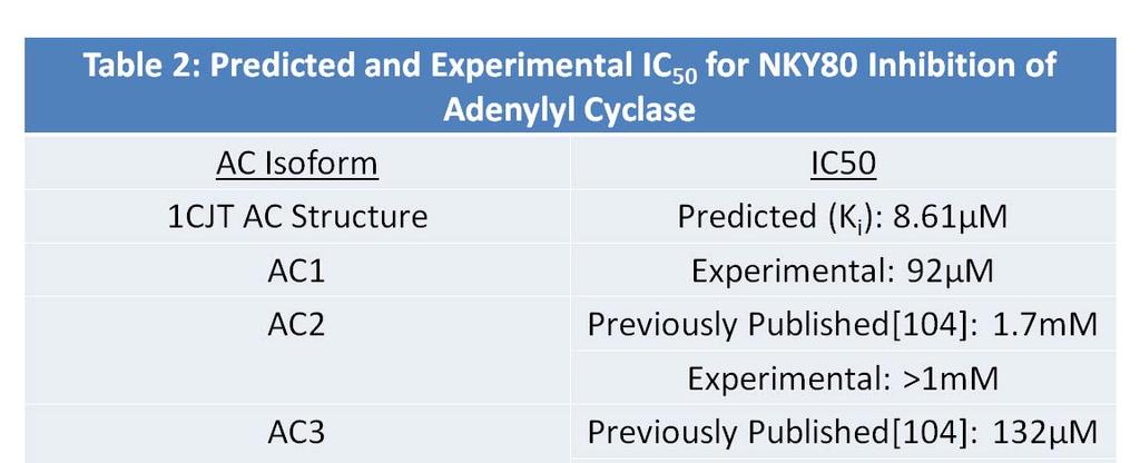
52 Figure 10: Predicted Binding Conformation of NKY80. (A) Chemical structure of AC inhibitor NKY80. (B) Predicted binding conformation of NKY80 (cyan carbons) in AC catalytic site by AutoDock 4.2. NKY80 conformation is an overlay with of 2,3 dd-atp conformation (grey carbons) as reference. C1 domain is yellow, C2 domain is silver, manganese metal ion is orange. (C) Predicted binding conformation of NKY80 (cyan carbons) with AC catalytic site residues within 3.5 angstroms displayed (grey carbons). inhibited than AC6 at each concentration of NKY80 used. This is shown by observed IC 50 of 7.1µM for AC5 compared to 17µM for AC6, an approximately 2.5 fold difference. This potency shift was generally not significant at equivalent concentrations of NKY80, and it can be concluded that AC5 and AC6 are similarly inhibited by NKY80. 38
53 NKY80 Inhibition Curves for AC 1-9 With NKY80 shown to not have perfect AC5 isoform specificity, all 9 AC isoforms were tested in AC activity assays with NKY80 to determine how truly selective the small molecule inhibitor was (Figure 11B). AC1-7 were expressed in Sf9 membranes; as AC8 and AC9 do not express well in Sf9 membranes, expression in HEK membranes was instead used for these two isoforms. Results show that NKY80 inhibition of AC5 and AC6 is much more potent than inhibition of all other isoforms. There appears to be an intermediate potency group of AC1, AC3, and AC4, as well as a low potency group including AC2, AC7, AC8, and AC9. Unlike the more strongly inhibited AC5 and AC6 isoform group, these two other groupings do not follow the typically categorized AC isoform families with evolutionary similarity and higher overlap in regulators. Observed IC 50 for AC2, AC3, and AC5 agree with previously published results[104], and interestingly the docking score for NKY80 to the 1CJT AC derived structure predicted affinity of NKY80 for AC was very close to the inhibition potency of NKY80 for AC5 (Table 2). Summary The small molecule AC inhibitor NKY80 was docked to AC structure with AutoDock 4.2, and its concentration-dependent ability to inhibit all 9 AC isoforms was characterized experimentally. Despite being advertised as an AC5 selective inhibitor, NKY80 also strongly inhibits the closely related isoform AC6. When compared to nonselective P-site inhibitors, the ring structure of NKY80 moves towards a predicted hydrogen bond with AC residue Asn
54 Figure 11: Full AC Isoform Selectivity Characterization of NKY80. (A) Concentration curves of AC5 and AC6 inhibition by NKY80 from a representative experiment. 15µg of membranes of indicated AC isoform stimulated by 50µM forskolin, incubated with indicated concentration of inhibitor. p < 0.05 for *. (B) Concentration curves of NKY80 inhibition of transmembrane AC isoforms. 15µg of membranes of indicated AC isoform stimulated by 50µM forskolin, incubated with indicated concentration of inhibitor. AC isoform family is indicated by color of the inhibition curve; Group I AC s are red (AC1 = red, AC3 = dark red, AC8 = light red), Group II AC s are green (AC2 = green, AC4 = dark green, AC7 = light green), Group III AC s are blue (AC5 = blue, AC6 = cyan), AC9 is grey. 40
55 41
56 Chapter 6 Discussion 42
57 AC Isoform Groups and NKY80 Selectivity It was expected that NKY80 selectivity for inhibiting AC isoforms would follow classic AC groups that are phylogenetically related and have closer patterns of regulation. These are Group 1 (AC1, AC3, and AC8), Group 2 (AC2, AC4, and AC7), Group 3 (AC5 and AC6), and Group 4 (AC9)[6]. Consistent with this prediction, NKY80 inhibited AC5 and AC6 much better than the other isoforms. However, the other inhibition curves surprisingly did not show separation of Group 1 and Group 2 AC isoforms. AC1, AC3, and AC4 had NKY80 IC 50 in the high micromolar range, consistent with the previously published AC3 IC 50 of approximately 132µM. AC2, AC7, AC8, and AC9 had NKY80 IC 50 somewhere beyond the 1mM concentration tested. The IC 50 of AC2 has previously been published to be about 1.2 mm; we assume that AC7, AC8, and AC9 remain on a similar inhibition curve beyond 1mM as AC2. These groupings also do not follow known similarities in AC isoform regulation. Lack of known isoform similarity for these groupings combined with the observed 10-fold differences in NKY80 inhibitory potential between AC isoforms being possible from minor changes in AC spatial conformation, the prospect of explaining the catalytic structure differences between the AC1/3/4 and AC2/7/8/9 groups seems unlikely. Determining why NKY80 inhibits AC5 and AC6 better than the remainder of AC isoforms is more probable, considering the greater similarity of these two AC isoforms. Although NKY80 is described as an AC5 selective inhibitor, it strongly inhibits both AC5 and AC6. Supposed AC5 selectivity issues are not just restricted to AC inhibitors. The AC catalytic site is pseudosymmetrical, and the binding pocket for the AC activator forskolin is found opposite to the ATP binding site. Novel AC selective small molecule activators derived from forskolin have been developed that show selectivity for cardiac AC5 over AC2 and AC3, 43
58 but were not tested against the closely related AC6[106]. One of the activators developed is clinically approved for acute heart failure, colforsin daropate[71]. Considering the beneficial effects from AC6 activation for chronic heart failure, it seems reasonable to consider if colforsin daropate stimulates AC6 as well as AC5. AC Catalytic Site Residues and Inhibitor Selectivity Asn1025 is a residue in the catalytic site of AC that virtual docking of NKY80 suggests is involved in the ability of NKY80 to bind at the AC catalytic pocket. Asn1025 is conserved in all 9 isoforms, so why it would confer isoform selectivity to NKY80 is not immediately clear by a difference in isoform sequence. Thus, it is unclear if the cause for this residue s interaction with NKY80 is directly due to the residue or residue type, or if the interaction is induced by an isoform specific allosteric effect. However, it is clear that Asn1025 location or placement is important for NKY80 binding. It is known that NKY80 is somewhat isoform selective, with a preference for inhibition of AC5[104] and, apparently, AC6. It is noteworthy that P-site inhibitors are one of two larger families of classic AC small molecule inhibitors. The other is MANT-nucleotide analogs of ATP. MANT-GTP and similar inhibitors share an increased inhibitory potency for AC5 and AC6, although with a less profound difference between these isoforms and others. The MANT group on these molecules, for example with MANT-GTP, binds to AC based on a crystal structure where there is structural helix movement to allow a new pocket that we shall call a C pocket in addition to the A and B inhibitor binding pockets used by P-site inhibitors[84]. The beginning of the opening to this extra C pocket, consisting of the α1 helix of the C1 domain and the α4 helix of the C2 domain, lies just beyond Asn1025. The large 44
59 movement of these two helices accommodates the hydrophobic region which interacts with the MANT group, as seen when comparing the 1CJT and 1TL7 AC structures (Figure 12A). Hydrophobic region opening also includes significant movement of the sidechains for Thr401 and Asn1025, the residues noticed as possible causes for the selectivity of the novel AC1 inhibitors and NKY80 respectively. This is also supported by the α1 helix having isoform specific residue variability that is rare to find in the AC catalytic site (Figure 12B). More flexibility for AC5 and AC6 in this subregion would increase entropy, and thus make interaction of the subregion residues with inhibitors more energetically favorable. Thus, NKY80 and MANT-attached nucleotides may similarly interact with AC in this mobile subregion of the catalytic site conferring similar AC5 and AC6 selectivity. Sites unique to the C1/C2 domains of AC5 and AC6 are not without precedence. The AC5/6 C1 domains both have a binding site for Giα, for example[107]. However, such regulatory sites can usually be identified in the AC primary sequence. Instead, in the case of NKY80 and MANT-GTP, it is possible that the AC5/6 isoform family is more flexible in this catalytic subregion bracketed by the α1 and α4 helices, and thus more accommodating to stable conformations with interactions by these small molecule inhibitors. NKY80 is similar in selectivity to the competitive inhibitor MANT-GTP, but is derived from uncompetitive P-site inhibitors. As NKY80 is also not a competitive inhibitor, this supports the development of novel isoform selective inhibitors that also do not have a competitive mechanism. As previously mentioned, one of the justifications for using P-site inhibitors as a template for a desired AC isoform selective therapeutic as opposed to MANTnucleotides is the desire for an uncompetitive inhibitory mechanism. Uncompetitive inhibition causing decreased maximal camp concentrations capable by cardiac AC5 as a chronic heart 45
 Overlay of crystal structures of AC catalytic site from 2,3 -dd-atp (PDB ID 1CJT; C1 = yellow, C2 = silver) and MANT-GTP (PDB ID 1TL7; C1 = tan, C2 = black).")
60 Figure 12: Structural Flexibility in the AC Catalytic Site. (A) Overlay of crystal structures of AC catalytic site from 2,3 -dd-atp (PDB ID 1CJT; C1 = yellow, C2 = silver) and MANT-GTP (PDB ID 1TL7; C1 = tan, C2 = black). Manganese metal ion is orange and labeled. Positions of Thr401 and Asn1025 indicated for 1CJT with cyan carbons, for 1TL7 with grey carbons. The α1 and α4 helices of AC are also labeled. (B) Primary sequence alignments for α1 and α4 helix domains of crystal AC structure, human AC s 1-9. Residues that differ from the primary sequence of the 1CJT crystal structure C1/C2 domains are red. Residue Thr401 in the AC C1 sequence alignment is blue. Residue Asn1025 in the AC C2 sequence alignment is green. failure treatment, for example, would still allow lower levels of camp activity. Thus, such a mechanism would likely make long term treatment easier by not shutting down AC5/cAMP 46
Gα i Activation Assay Kit
 A helping hand for your research Product Manual Configuration-specific Monoclonal Antibody Based Gα i Activation Assay Kit Catalog Number 80301 20 assays NewEast Biosciences, Inc 1 Table of Content Product
Gα 13 Activation Assay Kit
 A helping hand for your research Product Manual Configuration-specific Monoclonal Antibody Based Gα 13 Activation Assay Kit Catalog Number: 80401 20 assays NewEast Biosciences 1 Table of Content Product
ab G alpha i Activation Assay Kit
 ab173234 G alpha i Activation Assay Kit Instructions for Use For the simple and fast measurement of G alpha i activation. This product is for research use only and is not intended for diagnostic use. Version
Cdc42 Activation Assay Kit
 A helping hand for your research Product Manual Configuration-specific Monoclonal Antibody Based Cdc42 Activation Assay Kit Catalog Number: 80701 20 assays 1 Table of Content Product Description 3 Assay
Accelerating Scientific Discovery
 Elite TM Fluorescent Membrane Potential Dye Kit CATALOG NUMBER: CA-M165 Description camp is a key second messenger involved extensively in cellular signal transduction pathways associated with the majority
1. QUANTITY OF LYSATE 2. LYSIS BUFFER
 SAMPLE PREPARATION 1. QUANTITY OF LYSATE The amount of protein requested for the Kinex KAM-880 Antibody Microarray service is 100 µg per sample at an approximate concentration of 2 mg/ml. If your samples
Data Sheet Adenosine A2A Receptor Functional Recombinant Stable Cell Line Catalog # 79381
 Data Sheet Adenosine A2A Receptor Functional Recombinant Stable Cell Line Catalog # 79381 Product Description Adenosine A2a receptor (A2aR or ADORA2A) stably expressed in HEK-293 reporter cells with a
Data Sheet. camp/pka Signaling Pathway CRE/CREB Reporter (Luc) HEK293 Cell Line Catalog #: 60515
 HEK293 Cell Line Catalog #: 60515") Data Sheet camp/pka Signaling Pathway CRE/CREB Reporter (Luc) HEK293 Cell Line Catalog #: 60515 Background The camp/pka Signaling Pathway CRE/CREB Reporter (Luc) HEK293 Cell Line is designed for monitoring
Data Sheet camp/pka Signaling Pathway CRE/CREB Reporter (Luc) HEK293 Cell Line Catalog #: 60515 Background The camp/pka Signaling Pathway CRE/CREB Reporter (Luc) HEK293 Cell Line is designed for monitoring
Assay ID Assay name Description Components of the assay SYS-A115 DRD5/ARRB2 targetscreener
 DRD5/ARRB2 targetscreener Assay SYS-A115 SYS-A115C4 systasy bioscience GmbH Adams-Lehmann-Str. 56 80797 München Tel. +49 (0) 89 2155 3085 Fax. +49 (0) 89 4400 55853 E-mail: support@systasy.de Product Description
NANYANG TECHNOLOGICAL UNIVERSITY SEMESTER I EXAMINATION CBC922 Medicinal Chemistry. NOVEMBER TIME ALLOWED: 120 min
 AYAG TECLGICAL UIVERSITY SEMESTER I EXAMIATI 2006-2007 CBC922 Medicinal Chemistry VEMBER 2006 - TIME ALLWED 120 min ISTRUCTIS T CADIDATES 1. This examination paper contains TW (2) parts and comprises SIX
Mitochondria/Cytosol Fractionation Kit
 Mitochondria/Cytosol Fractionation Kit Sufficient for analysis of 50 samples Cat. No. MIT1000 FOR RESEARCH USE ONLY Not for use in diagnostic procedures. USA & Canada Phone: +1(800) 437-7500 Fax: +1 (951)
Arf6 Activation Assay Kit
 A helping hand for your research Product Manual Configuration-specific Monoclonal Antibody Based Arf6 Activation Assay Kit Catalog Number: 82401 20 assays NewEast Biosciences 1 Table of Content Product
PROCEDURE FOR USE NICKEL NTA Magnetic Agarose Beads (5%)
") 1 AFFINITY HIS-TAG PURIFICATION PROCEDURE FOR USE NICKEL NTA Magnetic Agarose Beads (5%) DESCRIPTION Nickel NTA Magnetic Agarose Beads are products that allow rapid and easy small-scale purification of
1 AFFINITY HIS-TAG PURIFICATION PROCEDURE FOR USE NICKEL NTA Magnetic Agarose Beads (5%) DESCRIPTION Nickel NTA Magnetic Agarose Beads are products that allow rapid and easy small-scale purification of
RheB Activation Assay Kit
 A helping hand for your research Product Manual Configuration-specific Monoclonal Antibody Based RheB Activation Assay Kit Catalog Number: 81201 20 assays NewEast Biosciences 1 FAX: 610-945-2008 Table
10X ACTOne Membrane Potential Dye Solution, 10 ml each bottle, 10 bottles 10X ACTOne Dye Dilution Buffer, 100 ml
 Codex Technical Data Sheet Codex ACTOne TM Membrane Potential Dye Bulk Kit Product Information Catalog Number: Components: CB-80500-211 10X ACTOne Membrane Potential Dye Solution, 10 ml each bottle, 10
Rab5 Activation Assay Kit
 A helping hand for your research Product Manual Configuration-specific Monoclonal Antibody Based Rab5 Activation Assay Kit Catalog Number: 83701 20 assays 24 Whitewoods Lane 1 Table of Content Product
Data Sheet. CRE/CREB Reporter Assay Kit (camp/pka Cell Signaling Pathway) Catalog #: 60611
 Catalog #: 60611") Data Sheet CRE/CREB Reporter Assay Kit (camp/pka Cell Signaling Pathway) Catalog #: 60611 Background The main role of the camp response element, or CRE, is mediating the effects of Protein Kinase A (PKA)
Data Sheet CRE/CREB Reporter Assay Kit (camp/pka Cell Signaling Pathway) Catalog #: 60611 Background The main role of the camp response element, or CRE, is mediating the effects of Protein Kinase A (PKA)
Ni-NTA Agarose. User Manual. 320 Harbor Way South San Francisco, CA Phone: 1 (888) MCLAB-88 Fax: 1 (650)
 MCLAB-88 Fax: 1 (650)") Ni-NTA Agarose User Manual 320 Harbor Way South San Francisco, CA 94080 Phone: 1 (888) MCLAB-88 Fax: 1 (650) 871-8796 www. Contents Introduction -----------------------------------------------------------------------
Ni-NTA Agarose User Manual 320 Harbor Way South San Francisco, CA 94080 Phone: 1 (888) MCLAB-88 Fax: 1 (650) 871-8796 www. Contents Introduction -----------------------------------------------------------------------
Data Sheet. Hedgehog Signaling Pathway Gli Reporter NIH3T3 Cell Line Catalog #: 60409
 Data Sheet Hedgehog Signaling Pathway Gli Reporter NIH3T3 Cell Line Catalog #: 60409 Product Description The Gli Reporter NIH3T3 Cell Line is designed for monitoring the activity of the hedgehog signaling
Immunoprecipitation Protocol
 Immunoprecipitation Protocol Immunoprecipitation is a general method to obtain the enrichment of a specific protein from tissue lysate and cell lysate. It can be used to purify a specific protein, to identify
Data Sheet IDO2 - HEK293 Recombinant Cell Line Cat #: 60533
 Data Sheet IDO2 - HEK293 Recombinant Cell Line Cat #: 60533 Description Recombinant HEK293 cell line expressing tetracycline-inducible human indoleamine 2,3- dioxygenase (IDO2), Genbank accession number
Assay ID Assay name Description Components of the assay SYS-A124 ADRB2/ARRB2 targetscreener
 ADRB2/ARRB2 targetscreener Assay SYS-A124 SYS-A124C5 systasy bioscience GmbH Adams-Lehmann-Str. 56 80797 München Tel. +49 (0) 89 2155 3085 Fax. +49 (0) 89 4400 55853 E-mail: support@systasy.de Product
Assay ID Assay name Description Components of the assay. 1. SYS-V441, phtr2a-v2r-ntev-tevs-gv. Cells 1. SYS-C10, PC12 (only SYS-A102C10)
") HTR2A/ARRB2 targetscreener Assay SYS-A102 SYS-A102C10 systasy bioscience GmbH Adams-Lehmann-Str. 56 80797 München Tel. +49 (0) 89 2155 3085 Fax. +49 (0) 89 4400 55853 E-mail: support@systasy.de Product
HTR2A/ARRB2 targetscreener Assay SYS-A102 SYS-A102C10 systasy bioscience GmbH Adams-Lehmann-Str. 56 80797 München Tel. +49 (0) 89 2155 3085 Fax. +49 (0) 89 4400 55853 E-mail: support@systasy.de Product
ProteoEnrich ATP-Binders Kit 1 kit
 Novagen User Protocol TB413 Rev. C 0805 1 of 6 ProteoEnrich TM ATP-Binders TM Kit About the Kit ProteoEnrich ATP-Binders Kit 1 kit 71438-3 Description The ProteoEnrich ATP-Binders Kit is designed for enrichment
CRE/CREB Reporter Assay Kit camp/pka Cell Signaling Pathway Catalog #: 60611
 Data Sheet CRE/CREB Reporter Assay Kit camp/pka Cell Signaling Pathway Catalog #: 60611 Background The main role of the camp response element, or CRE, is mediating the effects of Protein Kinase A (PKA)
Data Sheet CD137/NF-κB Reporter - HEK293 Recombinant Cell Line Catalog # 79289
 Data Sheet CD137/NF-κB Reporter - HEK293 Recombinant Cell Line Catalog # 79289 Background Human CD137 (4-1BB; TNFRS9) is an inducible co-stimulatory molecule that activates T cells. CD137:CD137L-mediated
SensoLyte Homogeneous AFC Caspase-8 Assay Kit *Fluorimetric*
 SensoLyte Homogeneous AFC Caspase-8 Assay Kit *Fluorimetric* Catalog # 72088-100 Kit Size 100 Assays (96-well plate) Optimized Performance: This kit is optimized to detect caspase-8 activity. Enhanced
SensoLyte Anti-alpha-Synuclein Quantitative ELISA Kit (Human/Mouse/Rat) *Colorimetric*
 *Colorimetric*") Catalog # Kit Size SensoLyte Anti-alpha-Synuclein Quantitative ELISA Kit (Human/Mouse/Rat) *Colorimetric* AS-55550 One 96-well strip plate This kit is optimized to detect human/mouse/rat alpha-synuclein
Catalog # Kit Size SensoLyte Anti-alpha-Synuclein Quantitative ELISA Kit (Human/Mouse/Rat) *Colorimetric* AS-55550 One 96-well strip plate This kit is optimized to detect human/mouse/rat alpha-synuclein
Data Sheet. Glucocorticoid Receptor Pathway GAL4 Reporter (Luc)-HEK293 cell Line Catalog #: w70666
-HEK293 cell Line Catalog #: w70666") Data Sheet Glucocorticoid Receptor Pathway GAL4 Reporter (Luc)-HEK293 cell Line Catalog #: w70666 Background The glucocorticoid signaling pathway plays an important role in development, fluid homeostasis,
Data Sheet Glucocorticoid Receptor Pathway GAL4 Reporter (Luc)-HEK293 cell Line Catalog #: w70666 Background The glucocorticoid signaling pathway plays an important role in development, fluid homeostasis,
Faster Chemiluminescent camp detection by HitHunter camp XS+ on the SpectraMax L Luminescence Microplate Reader
 SpectraMax Systems Faster Chemiluminescent camp detection by HitHunter camp XS+ on the SpectraMax L Luminescence Microplate Reader By Rajini Bompelli and Sunitha Ambikapathi, DiscoveRx Corporation, 42501
SERVA Ni-NTA Magnetic Beads
 INSTRUCTION MANUAL SERVA Ni-NTA Magnetic Beads Magnetic beads for Affinity Purification of His-Tag Fusion Proteins (Cat. No. 42179) SERVA Electrophoresis GmbH - Carl-Benz-Str. 7-69115 Heidelberg Phone
EpiQuik Global Histone H3 Phosphorylation (Ser28) Assay Kit (Colorimetric)
 Assay Kit (Colorimetric)") EpiQuik Global Histone H3 Phosphorylation (Ser28) Assay Kit (Colorimetric) Base Catalog # PLEASE READ THIS ENTIRE USER GUIDE BEFORE USE The EpiQuik Global Histone H3 Phosphorylation (Ser28) Assay Kit (Colorimetric)
EpiQuik Global Histone H3 Phosphorylation (Ser28) Assay Kit (Colorimetric) Base Catalog # PLEASE READ THIS ENTIRE USER GUIDE BEFORE USE The EpiQuik Global Histone H3 Phosphorylation (Ser28) Assay Kit (Colorimetric)
Automated Imaging Assay for Characterizing Ca 2+ Flux with R-GECO Biosensor
 A p p l i c a t i o n N o t e Automated Imaging Assay for Characterizing Ca 2+ Flux with R-GECO Biosensor Joe Clayton, Ph.D., and Peter Banks, Ph.D., BioTek Instruments, Inc., Winooski, VT USA Abstract
Solutions to 7.02 Quiz II 10/27/05
 Solutions to 7.02 Quiz II 10/27/05 Class Average = 83 Standard Deviation = 9 Range Grade % 87-100 A 43 74-86 B 39 55-73 C 17 > 54 D 1 Question 1 (56 points) While studying deep sea bacteria, you discover
Data Sheet. Glucocorticoid Receptor Pathway GR-GAL4 Reporter (Luc)-HEK293 cell Line Catalog #: 60655
-HEK293 cell Line Catalog #: 60655") Data Sheet Glucocorticoid Receptor Pathway GR-GAL4 Reporter (Luc)-HEK293 cell Line Catalog #: 60655 Background The glucocorticoid signaling pathway plays an important role in development, fluid homeostasis,
Data Sheet Glucocorticoid Receptor Pathway GR-GAL4 Reporter (Luc)-HEK293 cell Line Catalog #: 60655 Background The glucocorticoid signaling pathway plays an important role in development, fluid homeostasis,
SensoLyte Homogeneous AFC Caspase-3/7 Assay Kit
 SensoLyte Homogeneous AFC Caspase-3/7 Assay Kit Revision Number:1.1 Last Revised: October 2014 Catalog # Kit Size AS-71114 500 Assays (96-well) or 1250 Assays (384-well) Convenient Format: All essential
Data Sheet. Hedgehog Signaling Pathway Gli Reporter NIH3T3 Cell Line Catalog #: 60409
 Data Sheet Hedgehog Signaling Pathway Gli Reporter NIH3T3 Cell Line Catalog #: 60409 Product Description The Gli Reporter NIH3T3 Cell Line is designed for monitoring the activity of the hedgehog signaling
TECHNICAL BULLETIN NUCLEI EZ PREP NUCLEI ISOLATION KIT. Product Number NUC-101 Store at 2-8 C
 NUCLEI EZ PREP NUCLEI ISOLATION KIT Product Number NUC-101 Store at 2-8 C TECHNICAL BULLETIN Product Description Sigma s Nuclei EZ Prep Kit is designed for the rapid isolation of nuclei from mammalian
Mitochondrial DNA Isolation Kit
 Mitochondrial DNA Isolation Kit Catalog Number KA0895 50 assays Version: 03 Intended for research use only www.abnova.com Table of Contents Introduction... 3 Background... 3 General Information... 4 Materials
Data Sheet. TGFβ/SMAD Signaling Pathway SBE Reporter HEK293 Cell Line Catalog #: 60653
 Data Sheet TGFβ/SMAD Signaling Pathway SBE Reporter HEK293 Cell Line Catalog #: 60653 Background The SBE Reporter HEK293 Cell Line is designed for monitoring the activity of the TGFβ/SMAD signaling pathway.
PROTOCOL 1850 Millrace Drive, Suite 3A Eugene, Oregon
 PROTOCOL ATP Synthase Protein Quantity Microplate Assay Kit 1850 Millrace Drive, Suite 3A Eugene, Oregon 97403 Rev.0 DESCRIPTION ATP Synthase Protein Quantity Microplate Assay Kit Sufficient materials
+ M III. IMAP Screening Express Kit Product #8073 Quantity: 8000, 20 µl reactions (80 µl final volumes) Low FP. M III High FP.
 Low FP. M III High FP.") Product Insert IMAP Screening Express Kit Product #8073 Quantity: 8000, 20 µl reactions (80 µl final volumes) Introduction About the IMAP Screening Express Kit The IMAP Screening Express Kit is used to
Product Insert IMAP Screening Express Kit Product #8073 Quantity: 8000, 20 µl reactions (80 µl final volumes) Introduction About the IMAP Screening Express Kit The IMAP Screening Express Kit is used to
FlashPlate File #15. High Throughput Screening. J. Watson SmithKline Beecham Pharmaceuticals, UK.
 Drug Discovery Research Clinical Screening High Throughput Screening FlashPlate File #15 Use of Novel FlashPlate Technology to Measure camp Accumulation in Chinese Hamster Ovary Cells Expressing Human
Supplementary information for Small molecule inhibitors block Gas6-inducible TAM activation and tumorigenicity
 Supplementary information for Small molecule inhibitors block Gas6-inducible TAM activation and tumorigenicity Stanley G. Kimani 1 *, Sushil Kumar 1 *, Nitu Bansal 2 *, Kamalendra Singh 3, Vladyslav Kholodovych
How Targets Are Chosen. Chris Wayman 12 th April 2012
 How Targets Are Chosen Chris Wayman 12 th April 2012 A few questions How many ideas does it take to make a medicine? 10 20 20-50 50-100 A few questions How long does it take to bring a product from bench
SensoLyte Anti-alpha-Synuclein Quantitative ELISA Kit (Rat) *Colorimetric*
 *Colorimetric*") SensoLyte Anti-alpha-Synuclein Quantitative ELISA Kit (Rat) *Colorimetric* Revision number: 1.3 Last updated: 1/15/18 Catalog # AS-55550-R Kit Size One 96-well strip plate This kit is optimized to detect
SensoLyte Anti-alpha-Synuclein Quantitative ELISA Kit (Rat) *Colorimetric* Revision number: 1.3 Last updated: 1/15/18 Catalog # AS-55550-R Kit Size One 96-well strip plate This kit is optimized to detect
TaKaRa MiniBEST Universal RNA Extraction Kit
 Cat. # 9767 For Research Use TaKaRa MiniBEST Universal RNA Extraction Kit Product Manual Table of Contents I. Description...3 II. III. IV. Kit Components...3 Storage and Shipping...4 Precautions for Preventing
of the Triphosphate of ATP
 A Small Aptamer with Strong and Specific Recognition of the Triphosphate of Peter L. Sazani, Rosa Larralde and Jack W. Szostak Howard Hughes Medical Institute, and Department of Molecular Biology, Massachusetts
A hybrid structural approach to analyze ligand binding by the 5-HT4. receptor
 Supplement A hybrid structural approach to analyze ligand binding by the 5-HT4 receptor Pius S. Padayatti 1**, Liwen Wang 2**, Sayan Gupta 3, Tivadar Orban 4, Wenyu Sun 1, David Salom 1, Steve Jordan 5,
Instructions For Research Use Only. Not For Use In Diagnostic Procedures
 Instructions For Research Use Only. Not For Use In Diagnostic Procedures 3D Culture Cell Harvesting Kit For harvesting and lysate preparation from 3D Culture for Western analysis Sufficient reagents for
Chapter 3 Nucleic Acids, Proteins, and Enzymes
 3 Nucleic Acids, Proteins, and Enzymes Chapter 3 Nucleic Acids, Proteins, and Enzymes Key Concepts 3.1 Nucleic Acids Are Informational Macromolecules 3.2 Proteins Are Polymers with Important Structural
6 pts. B) State two mechanisms to explain the effect shown in the figure.
 State two mechanisms to explain the effect shown in the figure.") Question 1. (10 points) The rate of uptake of glucose into rat adipocytes is increased by addition of insulin to the medium. The increase reaches a maximum within 30 seconds at 37 C and is energy dependent.
Question 1. (10 points) The rate of uptake of glucose into rat adipocytes is increased by addition of insulin to the medium. The increase reaches a maximum within 30 seconds at 37 C and is energy dependent.
The implementation of laboratory investigations for diagnosing. pyruvate kinase deficiency at the Johannesburg Hospital
 The implementation of laboratory investigations for diagnosing pyruvate kinase deficiency at the Johannesburg Hospital Pierre Durand (Student no: 8604833/H) A report submitted to the Faculty of Health
SUPPLEMENTARY INFORMATION. The nucleotide binding dynamics of MSH2/MSH3 are lesion-dependent.
 SUPPLEMENTARY INFORMATION The nucleotide binding dynamics of /MSH are lesion-dependent. Barbara A. L. Owen, Walter H. Lang, and Cynthia T. McMurray* mau 1 8 6 4 2 mau 8 6 4 2 mau 8 6 4 2 mau 8 7 6 5 4
Insect Cell PE LB. Insect Cell Protein Extraction & Lysis Buffer. (Cat. # ) think proteins! think G-Biosciences
 think proteins! think G-Biosciences") 240PR G-Biosciences 1-800-628-7730 1-314-991-6034 technical@gbiosciences.com A Geno Technology, Inc. (USA) brand name Insect Cell PE LB Insect Cell Protein Extraction & Lysis Buffer (Cat. # 786 411) think
240PR G-Biosciences 1-800-628-7730 1-314-991-6034 technical@gbiosciences.com A Geno Technology, Inc. (USA) brand name Insect Cell PE LB Insect Cell Protein Extraction & Lysis Buffer (Cat. # 786 411) think
Kit Components (Included) Cat # # of vials Product Name Quantity Storage Human Renal Cortical Epithelial Cells (HRCEpiC)
 Cat # # of vials Product Name Quantity Storage Human Renal Cortical Epithelial Cells (HRCEpiC)") 3D Renal Tubule Formation Kit 3D-RTF Cat # 3D-4110 Product Description The human kidney is frequently exposed to drugs and toxic compounds, which can result in nephrotoxicity. A drug s uptake and clearance
3D Renal Tubule Formation Kit 3D-RTF Cat # 3D-4110 Product Description The human kidney is frequently exposed to drugs and toxic compounds, which can result in nephrotoxicity. A drug s uptake and clearance
EPIGENTEK. EpiQuik Tissue Chromatin Immunoprecipitation Kit. Base Catalog # P-2003 PLEASE READ THIS ENTIRE USER GUIDE BEFORE USE
 EpiQuik Tissue Chromatin Immunoprecipitation Kit Base Catalog # P-2003 PLEASE READ THIS ENTIRE USER GUIDE BEFORE USE The EpiQuik Tissue Chromatin Immunoprecipitation Kit is suitable for combining the specificity
OPPF-UK Standard Protocols: Mammalian Expression
 OPPF-UK Standard Protocols: Mammalian Expression Joanne Nettleship joanne@strubi.ox.ac.uk Table of Contents 1. Materials... 3 2. Cell Maintenance... 4 3. 24-Well Transient Expression Screen... 5 4. DNA
SideStep Lysis and Stabilization Buffer
 SideStep Lysis and Stabilization Buffer INSTRUCTION MANUAL Catalog #400900 Revision B.0 For Research Use Only. Not for use in diagnostic procedures. 400900-12 LIMITED PRODUCT WARRANTY This warranty limits
The Study of the Effect of Drugs of Abuse on Protein Kinase A Activity in Mouse Brain and Spinal Cord
 Virginia Commonwealth University VCU Scholars Compass Theses and Dissertations Graduate School 2005 The Study of the Effect of Drugs of Abuse on Protein Kinase A Activity in Mouse Brain and Spinal Cord
HOOK 6X His Protein Purification (Yeast)
") G-Biosciences 1-800-628-7730 1-314-991-6034 technical@gbiosciences.com A Geno Technology, Inc. (USA) brand name HOOK 6X His Protein Purification (Yeast) For The Purification of His Tagged Proteins from
G-Biosciences 1-800-628-7730 1-314-991-6034 technical@gbiosciences.com A Geno Technology, Inc. (USA) brand name HOOK 6X His Protein Purification (Yeast) For The Purification of His Tagged Proteins from
AlphaScreen SureFire STAT3 Total Assay Kits. Manual
 AlphaScreen SureFire STAT3 Total Assay Kits Manual Assay Points Catalog # 500 TGRTS3S500 10 000 TGRTS3S10K 50 000 TGRTS3S50K For Research Use Only Research Reagents for Research Purposes Only TGRKVS73.5
Miniaturized Whole Cell-based GPCR camp Assay Using a Novel Detection System
 Miniaturized Whole Cell-based GPCR camp Assay Using a Novel Detection System Geetha Shankar, Ph.D. Associate Director New Lead Discovery Exelixis, Inc South San Francisco, CA Overview GPCR HTS assays ACT:One
RNP-IP (Modified Method)-Getting Majority RNA from RNA Binding Protein in the Cytoplasm Fengzhi Liu *
-Getting Majority RNA from RNA Binding Protein in the Cytoplasm Fengzhi Liu *") RNP-IP (Modified Method)-Getting Majority RNA from RNA Binding Protein in the Cytoplasm Fengzhi Liu * School of Biomedical Sciences, Thomas Jefferson University, Philadelphia, USA *For correspondence:
RNP-IP (Modified Method)-Getting Majority RNA from RNA Binding Protein in the Cytoplasm Fengzhi Liu * School of Biomedical Sciences, Thomas Jefferson University, Philadelphia, USA *For correspondence:
The Need for a PARP Pharmacodynamic Assay
 The Need for a PARP Pharmacodynamic Assay Version 05/07/09 amsbiotechnology(europe)ltd info@amsbio.com www.amsbio.com (UK)+44(0)1235828200 (CH)+41(0)916045522 (DE)+49(0)69779099 DNA Repair Pathways Base
No camp - maximum HRP activity
 Product Insert CatchPoint Cyclic-AMP Fluorescent Assay Kit Product # R8044 Quantity: 768, 60 μl reactions Introduction About the camp Assay The CatchPoint cyclic-amp Fluorescent Assay Kit measures levels
SensoLyte Homogeneous AFC Caspase-8 Assay Kit *Fluorimetric*
 SensoLyte Homogeneous AFC Caspase-8 Assay Kit *Fluorimetric* Catalog # 7288-2 Kit Size 2 Assays (96-well plate) Optimized Performance: This kit is optimized to detect caspase-8 activity. Enhanced Value:
Data Sheet. TCR activator / PD-L1 - CHO Recombinant Cell line Cat. #: 60536
 Data Sheet TCR activator / PD-L1 - CHO Recombinant Cell line Cat. #: 60536 Product Description Recombinant CHO-K1 cells constitutively expressing human PD-L1 (Programmed Cell Death 1 Ligand 1, CD274, B7
Purification of Concentrated, High-Quality RNA from Cells and Tissues
 Purification of Concentrated, High-Quality RNA from Cells and Tissues ReliaPrep RNA Cell and Tissue Miniprep Systems Don Smith, Promega Corporation Products Featured: ReliaPrep RNA Cell Miniprep System
RayBio Human NF-κB p65 Transcription Factor Activity Assay Kit
 RayBio Human NF-κB p65 Transcription Factor Activity Assay Kit Catalog #: TFEH-p65 User Manual Mar 13, 2017 3607 Parkway Lane, Suite 200 Norcross, GA 30092 Tel: 1-888-494-8555 (Toll Free) or 770-729-2992,
Human Caspase-3 FlowCytomix Simplex Kit
 PRODUCT INFORMATION & MANUAL Human Caspase-3 FlowCytomix Simplex Kit BMS82012FF For research use only. Not for diagnostic or therapeutic procedures. Human Caspase-3 FlowCytomix Simplex Kit North America
Total Histone H3 Acetylation Detection Fast Kit (Fluorometric)
") Total Histone H3 Acetylation Detection Fast Kit (Fluorometric) Catalog Number KA1539 48 assays Version: 02 Intended for research use only www.abnova.com Table of Contents Introduction... 3 Intended Use...
Total Histone H3 Acetylation Detection Fast Kit (Fluorometric) Catalog Number KA1539 48 assays Version: 02 Intended for research use only www.abnova.com Table of Contents Introduction... 3 Intended Use...
Attenuation of synaptic toxicity and MARK4/PAR1-mediated Tau phosphorylation by
 Supplementary Methods and Figures Attenuation of synaptic toxicity and MARK4/PAR1-mediated Tau phosphorylation by methylene blue for Alzheimer s disease treatment Wenchao Sun 1, Seongsoo Lee 1,2, Xiaoran
camp ELISA Kit (Colorimetric)
") Product Manual camp ELISA Kit (Colorimetric) Catalog Numbers STA-500 STA-500-5 96 assays 5 x 96 assays FOR RESEARCH USE ONLY Not for use in diagnostic procedures Introduction Adenosine 3,5 -cyclic monophosphate
Product Manual camp ELISA Kit (Colorimetric) Catalog Numbers STA-500 STA-500-5 96 assays 5 x 96 assays FOR RESEARCH USE ONLY Not for use in diagnostic procedures Introduction Adenosine 3,5 -cyclic monophosphate
Assay ID Assay name Description Components of the assay SYS-A044 ERBB2-3/ SH2(GRB2) receptor. readout
 receptor. readout") ERBB2-3/SH2(GRB2) targetscreener Assay SYS-A044 SYS-A044C10 systasy bioscience GmbH Adams-Lehmann-Str. 56 80797 München Tel. +49 (0) 89 2155 3085 Fax. +49 (0) 89 4400 55853 E-mail: support@systasy.de Product
ERBB2-3/SH2(GRB2) targetscreener Assay SYS-A044 SYS-A044C10 systasy bioscience GmbH Adams-Lehmann-Str. 56 80797 München Tel. +49 (0) 89 2155 3085 Fax. +49 (0) 89 4400 55853 E-mail: support@systasy.de Product
EZ-10 SPIN COLUMN GENOMIC DNA MINIPREPS KIT HANDBOOK
 EZ-0 SPIN COLUMN GENOMIC DNA MINIPREPS KIT HANDBOOK (Bacteria, Plant, Animal, Blood) Version 8 Rev 05/0/03 EZ-0 Genomic DNA Kit Handbook Table of Contents Introduction Limitations of Use Features Applications
Science June 3, 1988 v240 n4857 p1310(7) Page 1
 Page 1") Science June 3, 1988 v240 n4857 p1310(7) Page 1 by Brian K. Kobilka, Tong Sun Kobilka, Kiefer Daniel, John W. Regan, Marc G. Caron and Robert J. Lefkowitz COPYRIGHT 1988 American Association for the Advancement
Science June 3, 1988 v240 n4857 p1310(7) Page 1 by Brian K. Kobilka, Tong Sun Kobilka, Kiefer Daniel, John W. Regan, Marc G. Caron and Robert J. Lefkowitz COPYRIGHT 1988 American Association for the Advancement
Use of a Label-Free Platform in a preclinical Contract Research Organization Environment
 Use of a Label-Free Platform in a preclinical Contract Research Organization Environment Scott Perschke Director, Assay Development Contents Introduction 2 Problem Statement 2 Previous Options 2 Solution
5.36 Biochemistry Laboratory Spring 2009
 MIT OpenCourseWare http://ocw.mit.edu 5.36 Biochemistry Laboratory Spring 2009 For information about citing these materials or our Terms of Use, visit: http://ocw.mit.edu/terms. Laboratory Manual for URIECA
Tissue Acetyl-Histone H4 ChIP Kit
 Tissue Acetyl-Histone H4 ChIP Kit Catalog Number KA0672 48 assays Version: 03 Intended for research use only www.abnova.com Table of Contents Introduction... 3 Intended Use... 3 Background... 3 Principle
Z -LYTE fluorescent kinase assay technology. Z -LYTE Kinase Assay Platform
 Z -LYTE fluorescent kinase assay technology Z -LYTE Kinase Assay Platform Z -LYTE Kinase Assay Platform Non-radioactive assay format for screening a diverse collection of over 185 kinase targets Assay
SensoLyte Homogeneous AFC Caspase-3/7 Assay Kit
 SensoLyte Homogeneous AFC Caspase-3/7 Assay Kit Catalog # 71114 Unit Size Kit Size 1 Kit 500 Assays (96-well) or 1250 Assays (384-well) This kit is optimized to detect caspase-3/7 activity in cell culture
College of the Canyon s Introduction to Biotechnology Course, Custom Lab
 College of the Canyon s Introduction to Biotechnology Course, Custom Lab Protein Extraction and Concentration Determination Version 7-22-12 The standard curve that you created in the protein standard curve
SensoLyte AMC Calpain Activity Assay Kit *Fluorimetric*
 SensoLyte AMC Calpain Activity Assay Kit *Fluorimetric* Catalog # 72150 Kit Size 100 Assays (96-well plate) Optimized Performance: This kit is optimized to detect calpain activity Enhanced Value: It provides
TECHNICAL BULLETIN NUCLEI PURE PREP NUCLEI ISOLATION KIT. Product No. NUC-201 Technical Bulletin No. MB-735 Store at 2-8 C
 NUCLEI PURE PREP NUCLEI ISOLATION KIT Product No. NUC-201 Technical Bulletin No. MB-735 Store at 2-8 C TECHNICAL BULLETIN Product Description Sigma s Nuclei PURE Prep Nuclei Isolation Kit is designed for
EPIGENTEK. EpiQuik Chromatin Immunoprecipitation Kit. Base Catalog # P-2002 PLEASE READ THIS ENTIRE USER GUIDE BEFORE USE
 EpiQuik Chromatin Immunoprecipitation Kit Base Catalog # P-2002 PLEASE READ THIS ENTIRE USER GUIDE BEFORE USE The EpiQuik Chromatin Immunoprecipitation Kit is suitable for combining the specificity of
ab65354 Superoxide Dismutase Activity Assay kit (Colorimetric)
") Version 9 Last updated 11 January 2018 ab65354 Superoxide Dismutase Activity Assay kit (Colorimetric) For the measurement of Superoxide Dismutase Activity in various samples. This product is for research
Version 9 Last updated 11 January 2018 ab65354 Superoxide Dismutase Activity Assay kit (Colorimetric) For the measurement of Superoxide Dismutase Activity in various samples. This product is for research
Propagation of H7 hesc From: UW (John Stamatoyannopoulos) ENCODE group Date: 12/17/2009 Prepared By: S. Paige/S. Hansen (UW)
 ENCODE group Date: 12/17/2009 Prepared By: S. Paige/S. Hansen (UW)") Propagation of H7 hesc From: UW (John Stamatoyannopoulos) ENCODE group Date: 12/17/2009 Prepared By: S. Paige/S. Hansen (UW) Growth and Harvest Modifications Addendum to: Propagation of H7 hesc from UW
Propagation of H7 hesc From: UW (John Stamatoyannopoulos) ENCODE group Date: 12/17/2009 Prepared By: S. Paige/S. Hansen (UW) Growth and Harvest Modifications Addendum to: Propagation of H7 hesc from UW
ab Ran Activation Assay Kit
 ab173247 Ran Activation Assay Kit Instructions for Use For the simple and fast measurement of Ran activation. This product is for research use only and is not intended for diagnostic use. Version 1 Last
AP Biology Book Notes Chapter 3 v Nucleic acids Ø Polymers specialized for the storage transmission and use of genetic information Ø Two types DNA
 AP Biology Book Notes Chapter 3 v Nucleic acids Ø Polymers specialized for the storage transmission and use of genetic information Ø Two types DNA Encodes hereditary information Used to specify the amino
NT-proBNP (Human) ELISA Kit
 ELISA Kit") NT-proBNP (Human) ELISA Kit Catalog Number KA2119 96 assays Version: 10 Intended for research use only www.abnova.com Table of Contents Introduction... 3 Intended Use... 3 Background... 3 Principle of
NT-proBNP (Human) ELISA Kit Catalog Number KA2119 96 assays Version: 10 Intended for research use only www.abnova.com Table of Contents Introduction... 3 Intended Use... 3 Background... 3 Principle of
EpiQuik Tissue Methyl-CpG Binding Domain Protein 2 ChIP Kit
 EpiQuik Tissue Methyl-CpG Binding Domain Protein 2 ChIP Kit Base Catalog # PLEASE READ THIS ENTIRE USER GUIDE BEFORE USE The EpiQuik Tissue Methyl-CpG Binding Domain Protein 2 ChIP Kit is suitable for
For Research Use Only. Not for use in diagnostic procedures.
 Printed December 13, 2011 Version 1.0 For Research Use Only. Not for use in diagnostic procedures. DDDDK-tagged Protein PURIFICATION GEL with Elution Peptide (MoAb. clone FLA-1) CODE No. 3326 / 3327 PURIFICATION
1. Cross-linking and cell harvesting
 ChIP is a powerful tool that allows the specific matching of proteins or histone modifications to regions of the genome. Chromatin is isolated and antibodies to the antigen of interest are used to determine
EpiQuik Methyl-CpG Binding Domain Protein 2 ChIP Kit
 EpiQuik Methyl-CpG Binding Domain Protein 2 ChIP Kit Base Catalog # PLEASE READ THIS ENTIRE USER GUIDE BEFORE USE The EpiQuik Methyl-CpG Binding Domain Protein 2 ChIP Kit is suitable for combining the
Yeast Nuclei Isolation Kit
 Yeast Nuclei Isolation Kit Catalog Number KA3951 50 assays Version: 02 Intended for research use only www.abnova.com Table of Contents Introduction... 3 Intended Use... 3 Background... 3 General Information...