cell and tissue imaging by fluorescence microscopy
|
|
|
- Edwin Owens
- 6 years ago
- Views:
Transcription

1 cell and tissue imaging by fluorescence microscopy Steven NEDELLEC Plateforme Micropicell SFR Santé François Bonamy Nantes 1


2 A matter of size Limit of resolution 0.15mm aims: building the image of an object using photons (or electrons) enlarge the image size thanks to lens 2

3 Fluorescence and fluorochromes Jablonski diagram Fluorochrome (also termed fluorophore), is a fluorescent chemical compound that can re-emit light upon light excitation. The emission of light through the fluorescence process is nearly simultaneous with the absorption of the excitation light due to a relatively short time delay between photon absorption and emission, ranging usually less than a microsecond in duration 3

4 What does a fluorescence microscope look like? 4
 Acquisition of in-focus well defined images (solid line) but also out-of-focus blurry (dotted line) images objective Focal")
5 Wide-field fluorescence microscopy Observation tube, camera Emission filter Dichroic beamsplitter Excitation light Excitation filter Lamp (mercury, xenon ) Acquisition of in-focus well defined images (solid line) but also out-of-focus blurry (dotted line) images objective Focal plane 5

6 Optical slicing Confocal microscopy Photomultiplier detector Emission filter The pinhole aperture eliminates out-of-focus interfering events, allowing detection of the only infocus objects Dichroic beamsplitter Confocal imaging approach provides a significant improvement in both axial and lateral resolution objective Focal plane 6


7 Hypothetical specimen under a confocal microscope The initial scan collects an optical slice of the image and then stores that image in the computer Below is a collection of individual slices called a mosaic. The series of slices were taken of a pollen grain. Each slice shows only a portion of the entire sample. The lower right image is a projection image made from the series The stage is moved in the Z direction and another optical section is taken. The digital image is again stored in the computer The final optical slice is scanned and stored in the computer From the image stack an image is produced called a projection Source: Paperproject.org 7
 vs a laser scanning confocal image (right) of a mouse intestine (from Carl")
8 Wide-field vs Confocal imaging Wide field confocal A wide-field fluorescent image (left) vs a laser scanning confocal image (right) of a mouse intestine (from Carl Zeiss). 8
9 Applications of fluorescence microscopy to cellular and tissular biology Most molecules constituting living cells display very low fluorescence levels that can t be quantified Fluorescent staining of sub-cellular compartments needs specific tools 9
10 Fluorescent staining: Probing Cell Structure and functions - High absorption - High fluorescent quantum yield - Water solubility - Cell permeant - Affinity to a particular part of the cell - Chemical and photostability - Stability in cell conditions - Low cytotoxicity Cell structure Nucleus Cytoskeletal proteins Plasma membrane and lipids Organelles (mitochondria, reticulum ) Cell functions Viability, proliferation Endocytosis Ions channels Signal transduction Intracellular ions indicators Intracellular ph Membrane potential 10


11 Probing Cell Structure: Nuclear stains DAPI Λabs=358nm λem=461nm Hoechst Λabs=350nm Λem=461nm HeLa cells Molecular probes handbook 11
 Λabs=540nm λem=580nm Phalloidin alexa 488")
 and the")
12 Probing Cell Structure: Plasma membrane stains Cytoskeleton stains Alexa 488 phalloidin DiL (lipophilic probe) Λabs=540nm λem=580nm Phalloidin alexa 488 Λabs=488nm λem=518nm Gustatory mouse neuron Molecular probes handbook Fibroblast labeled with probes for actin (green-fluorescent Alexa488 phalloidin) and the nucleus (TO-PRO-3) 12


13 Probing Cell Structure: mitochondria MitoTracker Red CMXRos λabs=578nm λem=599nm MitoTracker Green FM λabs=490nm λem=516nm Green: actin, blue: nucleus, red, mitochondria Molecular probes handbook 13

14 Probing Cell Function: Calcium indicators Fura-2 λabs=340/380nm λem=510nm Fluo-4 λabs=480nm λem=520nm Hi Ca 2+ level Lo Ca 2+ level Source: Dr Michele Sweeney, Newcastle University (Andor technology) 14
 In vitro time lapse imaging with a wide-field fluorescence microscope: images were recorded every 15 seconds Lo Ca 2+")
15 Probing Cell Function: Calcium indicators Fura2 loaded T cells Hi Ca 2+ level Non fluorescent infected target cell (dendritic cell) In vitro time lapse imaging with a wide-field fluorescence microscope: images were recorded every 15 seconds Lo Ca 2+ level 15


16 Probing Cell Function: Calcium indicators Tumoral target B cells are co-cultured with fura-2 probed T lymphocytes. On the right, target cells are pre-treated with a blocking antibody, preventing recognition by effector cells. Hi Ca 2+ level In vitro time lapse imaging with a wide-field fluorescence microscope: images were recorded every 15 seconds Dilek N. et al, 2011 Lo Ca 2+ level Such probe is very useful in order to quantify the activation kinetic of an effector cell 16

17 Probing Cell Function: Cytoplasm stains In vitro time lapse imaging with a wide-field fluorescence microscope: images were recorded every 15 seconds Tumoral target cells loaded with calcein-green probe Non fluorescent T cells Loss of target cell fluorescence is linked to cell death 17

18 Probing Cell Function: mitosis Mitosis in pig kidney epithelium Green: tubulin,red: DNA MicroscopyU.com 18

19 Immuno staining: fluorescent antibodies Immunostaining refers to the process of detecting antigens in cells of a tissue section by exploiting the principle of antibodies binding specifically to antigens in biological tissues. It takes its name from the roots "immuno," in reference to antibodies used in the procedure. Specific molecular markers are characteristic of particular cellular events (such as proliferation, cell death, physiologic activity etc ). Fluorescent antibodies are designed to specifically stain those markers and can be used to detect surface molecules on living cells or intracellular molecules on fixed cells. By using this technique, it s possible to identify accurately protein location. Various antibodies tagged with different fluorochromes can be used simultaneously. 19


20 Immuno staining: fluorescent antibodies HeLa cells labeled with nuclear probe and fitc conjugated anti-keratin antibody HeLa cells labeled with nuclear probe (DAPI), FITC conjugated phalloidin (green) and TRITC conjugated anti-vinculin antibody Chatin B et al, 2012 Chatin B et al,
 cells.")
 displays a strong colocalization viewable in yellow. Chen z.")
. Overlay of GM1 and CD5 (red and green) displays a strong colocalization viewable in yellow, whereas CD5 and IgM don t colocalize.")
21 Immuno staining: colocalization A B CD5 GM1 overlay CD5 IgM overlay Colocalization experiment of angiotensinconverting-enzyme (ACE) with beta-2 adrenergic receptors in Chinese hamster ovary (CHO) cells. ACE is stained with an alexa 568 (red) conjugated antibody anti ACE, B2 receptors with an alexa 488 (green) conjugated antibody anti B2. Overlay of both channels (red and green) displays a strong colocalization viewable in yellow. Chen z. et al, 2006 A) Colocalization experiment of membrane receptor CD5 (anti-cd5 FITC staining) and lipid rafts (stained in red with a probe specific of M1 ganglioside). Overlay of GM1 and CD5 (red and green) displays a strong colocalization viewable in yellow, whereas CD5 and IgM don t colocalize. B) Colocalization experiment of membrane receptor CD5 (anti-cd5 FITC staining) and the B Cell Recptor BCR (stained in red with an anti-igm alexa 568). Overlay of both channels reveals an absence of colocalization between these receptors. 21

22 Fluorescent fusion proteins, a tool for live cell imaging Step 1: Fusion X gene GFP gene X protein Plasmid insertion Fusion gene Fusion protein GFP gene GFP: Green Fluorescent Protein Step 2: Transfection 22


23 Fluorescent fusion proteins Mitochondria GFP nucleus Generation of a cell line expressing a mitochondrial fusion protein F1-ATP-synthase GFP. This method allows to generate living cells with intracellular fluorescent proteins. Immunofluorence can reveal intracellular elements only after cell fixation. 23


 interacting with 24 C.")
24 Fixed tissue labelling: in vitro granuloma characterization pathogen Representative movie from microscopy time series showing leukocytes from a healthy donor infected with GFP-tagged C. albicans cells. Representative movie from confocal microscopy showing mature macrophages (red) interacting with 24 C. albicans hyphae (green) inside granuloma.

 were added to a")

 and tumor epithelial")
25 Living tissues labelling: human lung carcinoma biopsy Salmon H et al., 2011 Preactivated T cells (Hoechst; green) were added to a human lung tumor slice that was subsequently stained for fibronectin and EpCAM to respectively identify stromal (red) and tumor epithelial cell (blue) regions 25


26 Fluorescence Recovering After Photobleaching (FRAP) The mobility of a fluorescent protein fusion can be assessed using a technique known as fluorescence recovery after photobleaching (FRAP). In practice, the fluorescently labeled molecules in a small region of the cell are irreversibly photobleached using high laser power, followed by monitoring the subsequent movement of the surrounding non-bleached fluorescent molecules into the photobleached region using low laser power. This technique is used for mobility measurement of membrane or intracellular molecules. control Treated cell HEK cells were transduced to express a GFP tagged of the butyrophilin molecule Control and treated cells display different patterns of BT3 membrane mobility. This differential behaviour is linked to the ability of T lymphocytes to recognize and eliminate cells. 26

27 FLIM/FRET FLIM (Fluorescence Lifetime Imaging Microscopy) is a powerful technique to measure protein-protein interactions, and is based on the FRET (Forster Resonant Energy Transfert) principle, as shown below. X Y The fundamental mechanism of FRET involves a donor fluorophore in an excited electronic state, which may transfer its excitation energy to a nearby acceptor fluorophore. FRET occurs between two appropriately positioned fluorophores only when the distance separating them is 8 to 10 nanometers or less. 27

28 Two photons microscopy Two-photon excitation microscopy is a fluorescence imaging technique that allows imaging of living tissue up to a very high depth, that is up to about one millimeter. The concept of two-photon excitation is based on the idea that two photons of comparably lower energy than needed for one photon excitation can also excite a fluorophore in one quantum event 28

.")
29 Two photons microscopy Radially expanding transglial calcium waves in the intact cerebellum Spontaneous radial expanding calcium wave in the cerebellar molecular layer (xz). A radial expanding calcium wave.. Overlaid color map shows relative fluorescence changes with time (playback is 4 times original speed, diameter of the field of view is 195 m). Hoogland et al., 2009 ATP-triggered calcium wave in the cerebellar molecular layer (xy). Green channel displays raw fluorescence reported by fluo-5f; red channels shows Alexa 594 in pipette and during ATP injection as well as SR101 staining of the molecular layer (playback is 4 times original speed, diameter of the field of view is 286 m). 29

30 Scanner Light Sheet Microscopy Light sheet microscopy is a rapidly emerging technology that combines optical sectioning with multiple-view imaging to observe tissues and living organisms with impressive resolution. the technique can image virtually any plane with multiple views obtained from different angles. Thus, light sheet microscopy is ideal for examining of both large (animals) and small (cells) specimens labeled with fluorescent proteins and other fluorophores. long-term imaging of zebrafish development for 24 h. Keller et al.,


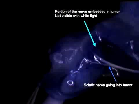

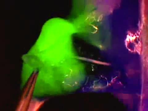
31 Use of molecular fluorescence imaging to guide surgery Source: USCD medical center 31
Fluorescence Light Microscopy for Cell Biology
 Fluorescence Light Microscopy for Cell Biology Why use light microscopy? Traditional questions that light microscopy has addressed: Structure within a cell Locations of specific molecules within a cell
Visualizing Cells Molecular Biology of the Cell - Chapter 9
 Visualizing Cells Molecular Biology of the Cell - Chapter 9 Resolution, Detection Magnification Interaction of Light with matter: Absorbtion, Refraction, Reflection, Fluorescence Light Microscopy Absorbtion
Special Techniques 1. Mark Scott FILM Facility
 Special Techniques 1 Mark Scott FILM Facility SPECIAL TECHNIQUES Multi-photon microscopy Second Harmonic Generation FRAP FRET FLIM In-vivo imaging TWO-PHOTON MICROSCOPY Alternative to confocal and deconvolution
Contents. SCHOOL of FLUORESCENCE. For more information, go to lifetechnologies.com/imagingbasics
 MPSF educator packet This packet contains illustrations and figures from the Molecular Probes School of Fluorescence website. They illustrate concepts from the basic physical properties that underlie fluorescence
MICROSCOPY. "micro" (small) "scopeo" (to watch)
 scopeo (to watch)") MICROSCOPY "micro" (small) "scopeo" (to watch) THE RELATIVE SIZES OF MOLECULES, CELLS AND ORGANISMS THE RELATIVE SIZES OF MOLECULES, CELLS AND ORGANISMS MICROSCOPY 1590 2012 MICROSCOPY THE LIGHT Light:
MICROSCOPY "micro" (small) "scopeo" (to watch) THE RELATIVE SIZES OF MOLECULES, CELLS AND ORGANISMS THE RELATIVE SIZES OF MOLECULES, CELLS AND ORGANISMS MICROSCOPY 1590 2012 MICROSCOPY THE LIGHT Light:
More on fluorescence
 More on fluorescence Last class Fluorescence Absorption emission Jablonski diagrams This class More on fluorescence Common fluorophores Jablonski diagrams to spectra Properties of fluorophores Excitation
Description. Lipodin-Pro TM - Protein Transfection Reagent. 1. Kit Benefits
 Description Lipodin-Pro TM - Protein Transfection Reagent The delivery of proteins inside living cells represents an alternative to nucleic acids transfection and a powerful strategy for functional studies
BIO 315 Lab Exam I. Section #: Name:
 Section #: Name: Also provide this information on the computer grid sheet given to you. (Section # in special code box) BIO 315 Lab Exam I 1. In labeling the parts of a standard compound light microscope
BIO 315 Lab Exam I. Section #: Name:
 Section #: Name: Also provide this information on the computer grid sheet given to you. (Section # in special code box) BIO 315 Lab Exam I 1. In labeling the parts of a standard compound light microscope
Resolution of Microscopes Visible light is nm Dry lens(0.5na), green(530nm light)=0.65µm=650nm for oil lens (1.4NA) UV light (300nm) = 0.13µm f
, green(530nm light)=0.65µm=650nm for oil lens (1.4NA) UV light (300nm) = 0.13µm f") Microscopes and Microscopy MCB 380 Good information sources: Alberts-Molecular Biology of the Cell http://micro.magnet.fsu.edu/primer/ http://www.microscopyu.com/ Approaches to Problems in Cell Biology
Microscopes and Microscopy MCB 380 Good information sources: Alberts-Molecular Biology of the Cell http://micro.magnet.fsu.edu/primer/ http://www.microscopyu.com/ Approaches to Problems in Cell Biology
F* techniques: FRAP, FLIP, FRET, FLIM,
 F* techniques: FRAP, FLIP, FRET, FLIM, FCS Antonia Göhler March 2015 Fluorescence explained in the Bohr model Absorption of light (blue) causes an electron to move to a higher energy orbit. After a particular
Fluorescence Microscopy
 Fluorescence Microscopy Dr. Arne Seitz Swiss Institute of Technology (EPFL) Faculty of Life Sciences Head of BIOIMAGING AND OPTICS BIOP arne.seitz@epfl.ch Fluorescence Microscopy Why do we need fluorescence
Automated Digital Microscopy
 A p p l i c a t i o n G u i d e Peter Banks, Ph.D. and Peter J. Brescia, Applications Department, BioTek Instruments, Inc., Winooski, VT Table of Contents Introduction ----------------------------------------------------------------------------------------------------------------------
Dino-Lite knowledge & education. Fluorescence Microscopes
 Dino-Lite knowledge & education Fluorescence Microscopes Dino-Lite Fluorescence models Smallest fluorescence microscope in the world Revolution to biomedical and educational applications Flexible Easy
Partha Roy
 Fluorescence microscopy http://micro.magnet.fsu.edu/primer/index.html Partha Roy 1 Lecture Outline Definition of fluorescence Common fluorescent reagents Construction ti of a fluorescence microscope Optical
Con-focal and Multi-photon Microscope Experiment Fundamental. Qian Hu, Lab of Laser Scanning Confocal & Two-Photon Microscopy, ION, CAS
 Con-focal and Multi-photon Microscope Experiment Fundamental Qian Hu, Lab of Laser Scanning Confocal & Two-Photon Microscopy, ION, CAS 1. Light is Electromagnetic Wave ν = c / λ 2. Image of a Point Source
Fluorescence Microscopy
 Fluorescence Microscopy Dr. Arne Seitz Swiss Institute of Technology (EPFL) Faculty of Life Sciences Head of BIOIMAGING AND OPTICS BIOP arne.seitz@epfl.ch Fluorescence Microscopy Why do we need fluorescence
Pro-DeliverIN TM - Protein Delivery Reagent Results
 Pro-DeliverIN TM - Protein Delivery Reagent Results OZ Biosciences is delighted to announce the launching of the innovative Pro-DeliverIN - Protein Delivery Reagent. Pro-DeliverIN is a lipid based formulation
Fluorescence Microscopy. Terms and concepts to know: 10/11/2011. Visible spectrum (of light) and energy
 and energy") Fluorescence Microscopy Louisiana Tech University Ruston, Louisiana Microscopy Workshop Dr. Mark DeCoster Associate Professor Biomedical Engineering 1 Terms and concepts to know: Signal to Noise Excitation
Fluorescence Microscopy Louisiana Tech University Ruston, Louisiana Microscopy Workshop Dr. Mark DeCoster Associate Professor Biomedical Engineering 1 Terms and concepts to know: Signal to Noise Excitation
Contact Details. Dr Alexander Galkin. Office: MBC Room 186. Tel: (028) Frequency and wavelength.
 Frequency and wavelength.") Contact Details The electromagnetic spectrum Biological Spectroscopy Dr Alexander Galkin Email: a.galkin@qub.ac.uk Dr Alexander Galkin MSc Biomolecular Function - BBC8045 Office: MBC Room 186 Tel: (028)
Contact Details The electromagnetic spectrum Biological Spectroscopy Dr Alexander Galkin Email: a.galkin@qub.ac.uk Dr Alexander Galkin MSc Biomolecular Function - BBC8045 Office: MBC Room 186 Tel: (028)
Microscopy from Carl Zeiss
 Microscopy from Carl Zeiss LSM 710 In Tune with Your Application Enjoy new freedom in selecting fluorescent dyes with In Tune, the new laser system for the LSM 710. Whatever the wavelength, you can match
Imaging facilities at WUR
 Imaging facilities at WUR Advanced light microscopy facilities at Wageningen UR Programme Thursday 13 June 2013 Lunch meeting organized by Cat-Agro Food 12.00 Welcome and sandwich lunch 12.10 Introduction
Confocal Microscopy Analyzes Cells
 Choosing Filters for Fluorescence A Laurin Publication Photonic Solutions for Biotechnology and Medicine November 2002 Confocal Microscopy Analyzes Cells Reprinted from the November 2002 issue of Biophotonics
11/19/2013. Janine Zankl FACS Core Facility 13. November Cellular Parameters. Cellular Parameters. Monocytes. Granulocytes.
 DEPARTEMENT BIOZENTRUM Janine Zankl FACS Core Facility 13. November 2013 Cellular Parameters Granulocytes Monocytes Basophils Neutrophils Lymphocytes Eosinophils Cellular Parameters 1 What Is Flow Cytometry?
Fuse-It Membrane Fusion
 Fuse-It Membrane Fusion Next Generation Transfection Including: LifeAct Actin Visualization in Living Cells www..com Mechanisms and Methods of Molecular Transfer into Living Cells Fuse-It- Fuse-It-siRNA
Nodes of regulation in cellular systems
 Nodes of regulation in cellular systems cell membrane signal transduction ligands receptors oligomerization transport signal transduction modified protein Golgi transcription factor transport ER transport
Spectral Separation of Multifluorescence Labels with the LSM 510 META
 Microscopy from Carl Zeiss Spectral Separation of Multifluorescence Labels with the LSM 510 META Indians living in the South American rain forest can distinguish between almost 200 hues of green in their
Live cell microscopy
 Live cell microscopy 1. Why do live cell microscopy? 2. Maintaining living cells on a microscope stage. 3. Considerations for imaging living cells. 4. Fluorescence labeling of living cells. 5. Imaging
Confocal Microscopes. Evolution of Imaging
 Confocal Microscopes and Evolution of Imaging Judi Reilly Hans Richter Massachusetts Institute of Technology Environment, Health & Safety Office Radiation Protection What is Confocal? Pinhole diaphragm
Cell Viability and Senescence Detection Kits
 Cell Viability and Senescence Detection Kits Cell viability and proliferation assays Growth factor/cytokine assays Cell culture condition optimization Cell number determination Multiwell and automation
Challenges to measuring intracellular Ca 2+ Calmodulin: nature s Ca 2+ sensor
 Calcium Signals in Biological Systems Lecture 3 (2/9/0) Measuring intracellular Ca 2+ signals II: Genetically encoded Ca 2+ sensors Henry M. Colecraft, Ph.D. Challenges to measuring intracellular Ca 2+
Multiplexed 3D FRET imaging in deep tissue of live embryos Ming Zhao, Xiaoyang Wan, Yu Li, Weibin Zhou and Leilei Peng
 Scientific Reports Multiplexed 3D FRET imaging in deep tissue of live embryos Ming Zhao, Xiaoyang Wan, Yu Li, Weibin Zhou and Leilei Peng 1 Supplementary figures and notes Supplementary Figure S1 Volumetric
A Brief History of Light Microscopy And How It Transformed Biomedical Research
 A Brief History of Light Microscopy And How It Transformed Biomedical Research Suewei Lin Office: Interdisciplinary Research Building 8A08 Email: sueweilin@gate.sinica.edu.tw TEL: 2789-9315 Microscope
Ab-DeliverIN TM - Antibody Delivery Reagent Results
 Ab-DeliverIN TM - Antibody Delivery Reagent Results OZ Biosciences is delighted to announce the launching of the innovative Ab-DeliverIN TM - antibody delivery reagent. Ab-DeliverIN TM is a lipid based
SONOMA STATE UNIVERSITY DEPARTMENT OF BIOLOGY BIOLOGY 344: CELL BIOLOGY Fall 2013
 SONOMA STATE UNIVERSITY DEPARTMENT OF BIOLOGY BIOLOGY 344: CELL BIOLOGY Fall 2013 Instructor Murali C. Pillai, PhD Office 214 Darwin Hall Telephone (707) 664-2981 E-mail pillai@sonoma.edu Website www.sonoma.edu/users/p/pillai
Contents. 11 The Use of Epitope Tags in Histochemistry References... 98
 Contents 1 Antibodies for Immunohistochemistry... 1 1.1 Structure of Antibodies... 2 1.2 Polyclonal Antibodies... 4 1.3 Mouse Monoclonal Antibodies... 4 1.4 Rabbit Monoclonal Antibodies... 5 1.5 Protein
INTRODUCTION TO FLOW CYTOMETRY
 DEPARTEMENT BIOZENTRUM INTRODUCTION TO FLOW CYTOMETRY F ACS C ore F acility Janine Zankl FACS Core Facility 3. Dezember 2015, 4pm Cellular Parameters Granulocytes Monocytes Basophils Lymphocytes Neutrophils
Imaging of Cells using fluorescents dyes. By: Josué A. Benjamín Rivera September 27, 2018
 Imaging of Cells using fluorescents dyes By: Josué A. Benjamín Rivera September 27, 2018 1 History Sir William Henry Perkin BRITISH CHEMIST In 1856, at the age of 18, William Henry Perkin set out with
RaftNote. Imaging and Sorting Living Cells Stained with Vital Dyes on Cell Microsystems CytoSort Array and Automated CellRaft AIR System
 RaftNote Applications and protocols for use with CellRaft Technology Imaging and Sorting Living Cells Stained with Vital Dyes on Cell Microsystems CytoSort Array and Automated CellRaft AIR System Jacquelyn
ALP (alkaline phosphatase) calibrators were analyzed manually in microtiter plates to find the linearity range by following this protocol:
 calibrators were analyzed manually in microtiter plates to find the linearity range by following this protocol:") Exam Mol 3008 May 2009 Subject 1 (15p) ALP (alkaline phosphatase) calibrators were analyzed manually in microtiter plates to find the linearity range by following this protocol: Reaction solutions: 50
Exam Mol 3008 May 2009 Subject 1 (15p) ALP (alkaline phosphatase) calibrators were analyzed manually in microtiter plates to find the linearity range by following this protocol: Reaction solutions: 50
Monitoring and Optimizing the Lipopolysaccharides-plasmid DNA interaction by FLIM-FRET
 Transactions on Science and Technology Vol. 4, No. 3-3, 342-347, 2017 Monitoring and Optimizing the Lipopolysaccharides-plasmid DNA interaction by FLIM-FRET Nur Syahadatain Abdul Razak 1#, Clarence M.
CytoPainter Golgi Staining Kit Green Fluorescence
 ab139483 CytoPainter Golgi Staining Kit Green Fluorescence Instructions for Use Designed for the detection of Golgi bodies by microscopy This product is for research use only and is not intended for diagnostic
ab CytoPainter ER Staining Kit Red Fluorescence
 ab139482 CytoPainter ER Staining Kit Red Fluorescence Instructions for Use Designed to detect Human endoplasmic reticulum by microscopy. This product is for research use only and is not intended for diagnostic
Introduction to Flow Cytometry. -- BD FACSCanto II TM. Daisy Kuo Application Specialist BDBiosciences
 Introduction to Flow Cytometry -- BD FACSCanto II TM Daisy Kuo Application Specialist E-mail: daisy_kuo@bd.com BDBiosciences Outline Basic Concept of Flow Cytometry FACSCanto II System Introduction Application
FLIM Fluorescence Lifetime IMaging
 FLIM Fluorescence Lifetime IMaging Fluorescence lifetime t I(t) = F0 exp( ) τ 1 τ = k f + k nr k nr = k IC + k ISC + k bl Batiaens et al, Trends in Cell Biology, 1999 τ τ = fluorescence lifetime (~ns to
Fluorescence Microscopy: A Biological Perspective
 Fluorescence Microscopy: A Biological Perspective From nanometre to metre: the scale of life Instrumentation and accessible scale limits the questions that can be addressed in biology Why are there limits?
D e c N o. 2 8
 D e c. 2 0 0 7 N o. 2 8 CONFOCAL APPLICATION LETTER resolution FRET Acceptor Photobleaching LAS AF Application Wizard FRET with Leica TCS SP5 LAS AF Version 1.7.0 Introduction Fluorescence Resonance Energy
ab CytoPainter ER Staining Kit Red Fluorescence
 ab139482 CytoPainter ER Staining Kit Red Fluorescence Instructions for Use Designed to detect Human endoplasmic reticulum by microscopy. This product is for research use only and is not intended for diagnostic
Multiphoton Microscopy: Seeing deeper and clearer
 Multiphoton Microscopy: Seeing deeper and clearer Since the invention of simple microscope by Leuwenhoek and Hooke in the 17th century, different types of light microscopy techniques (such as phase contrast,
SUPPLEMENTARY INFORMATION
 doi:10.1038/nature10016 Supplementary discussion on binding site density for protein complexes on the surface: The density of biotin sites on the chip is ~10 3 biotin-peg per µm 2. The biotin sites are
The Basics of Flow Cytometry
 The Basics of Flow Cytometry F ACS C ore F acility Janine Bögli, Biozentrum, 29. January 2018 The functions of the FACS Core Facility Centralization of equipment and expertise Train users Sorter operation
2004 Debye Lecture 4 C. B. Murray. Quantum Dot Applications: Sun Screen. Solar Cells. Bio-tagging. Solid State Lighting?
 2004 Debye Lecture 4 C. B. Murray Quantum Dot Applications: Sun Screen Solar Cells Bio-tagging Solid State Lighting? Quantum Dot Solar cells Nanocrystal Solar Cells Double-labeling of mitochondria
Imaging of endocrine organs
 Imaging of endocrine organs Helen Christian Department of Physiology, Anatomy & Genetics St Anne s College, University of Oxford Diabetesforum, Stockholm 2017 Islets of Langerhan Pituitary gland Renin
Chapter 4. the biological community to assay for protein-protein interactions. FRET describes the
 31 Chapter 4 Determination of nachr stoichiometry using Normalized Försters Resonance Energy Transfer (NFRET) Försters resonance energy transfer (FRET) has become a technique widely used in the biological
Final Exam, 176 points PMB 185: Techniques in Light Microscopy
 Final Exam, 176 points Name PMB 185: Techniques in Light Microscopy Point value is in parentheses at the end of each question. 1) Order the steps in setting up Köhler illumination. It is not necessary
Two-Photon Microscopy for Deep Tissue Imaging of Living Specimens
 for Deep Tissue Imaging of Living Specimens Tilman Franke* and Sebastian Rhode TILL Photonics GmbH, an FEI company, Lochhamer Schlag 21, D-82166 Gräfelfing, Germany *tilman.franke@fei.com Introduction
What to look for in a fluorophore. What to do with a fluorophore. Types of fluorochromes
 What to do with a fluorophore Intracellular localization (ER, Golgi, PM, nuclear, lysosome, MT, actin,...) Dynamic processes (protein synthesis, trafficking, turnover, DNA replication, cytoskeletal remodeling,
Biochemistry. Biochemical Techniques. 18 Spectrofluorimetry
 Description of Module Subject Name Paper Name 12 Module Name/Title 1. Objectives 1.1 To understand technique of Spectrofluorimetry. 1.2 To explain instrumentation design 1.3 What are applications of Spectrofluorimetry?
Fluorescent Imaging in Cell Biology. Invitrogen Cellular Analysis
 Fluorescent Imaging in Cell Biology Invitrogen Cellular Analysis Topics Fluorescent Dyes and Dots Cellular Structure. Multicolor Immuno-Labeling. rganelle Stains Cellular Physiology Intracellular Ions
Organelle Atlas APPENDIX TO CHAPTER 4 4A.1
 APPENDIX TO CHAPTER 4 This atlas contains images of cellular organelles visualized using a wide variety of reagents and techniques and demonstrates the diversity of methods for exploring the cell. The
Supplementary Table-1: List of genes that were identically matched between the ST2 and
 Supplementary data Supplementary Table-1: List of genes that were identically matched between the ST2 and ST3. Supplementary Table-2: List of genes that were differentially expressed in GD2 + cells compared
Introduction to Flow Cytometry. -- BD FACSCanto II TM. Daisy Kuo Assistant Product Manager BDBiosciences
 Introduction to Flow Cytometry -- BD FACSCanto II TM Daisy Kuo Assistant Product Manager E-mail: daisy_kuo@bd.com BDBiosciences Outline Basic Concept of Flow Cytometry FACSCanto II System Introduction
The CQ1 Confocal Quantitative Image Cytometer and its Application to Biological Measurement
 The CQ1 Confocal Quantitative Image Cytometer and its Application to Biological Measurement Hirofumi Sakashita *1 Koji Ohashi *1 Kazuo Ozawa *2 ohei Tsubouchi *1 The CQ1 confocal quantitative image cytometer,
No wash 2 Washes 2 Days ** ** IgG-bead phagocytosis (%)
") Supplementary Figures Supplementary Figure 1. No wash 2 Washes 2 Days Tat Control ** ** 2 4 6 8 IgG-bead phagocytosis (%) Supplementary Figure 1. Reversibility of phagocytosis inhibition by Tat. Human
Supplementary Figures Supplementary Figure 1. No wash 2 Washes 2 Days Tat Control ** ** 2 4 6 8 IgG-bead phagocytosis (%) Supplementary Figure 1. Reversibility of phagocytosis inhibition by Tat. Human
The most extensively used technique for tissue analysis is light microscopy.
 Fluorescence Theory Quantum yield Wavelength shift Ligand interactions Membrane interactions Using quenchning effects Fluorescence in-vivo Localization Distance measurements FRET The most extensively used
Practical light microscopy: an introduction
 Practical light microscopy: an introduction Dr. Mark Leake, Oxford University www.physics.ox.ac.uk/users/leake Aim of today s talk: Explanation of the very (very) basics of how a light microscope works
HYPERSPECTRAL MICROSCOPE PLATFORM FOR HIGHLY MULTIPLEX BIOLOGICAL IMAGING. Marc Verhaegen
 HYPERSPECTRAL MICROSCOPE PLATFORM FOR HIGHLY MULTIPLEX BIOLOGICAL IMAGING Marc Verhaegen CMCS, MONTREAL, MAY 11 th, 2017 OVERVIEW Hyperspectral Imaging Multiplex Biological Imaging Multiplex Single Particle
Confocal Microscopy & Imaging Technology. Yan Wu
 Confocal Microscopy & Imaging Technology Yan Wu Dec. 05, 2014 Cells under the microscope What we use to see the details of the cell? Light and Electron Microscopy - Bright light / fluorescence microscopy
Supplementary material to Alterations in the properties of the cell membrane due to glycosphingolipid accumulation in a model of Gaucher disease
 Supplementary material to Alterations in the properties of the cell membrane due to glycosphingolipid accumulation in a model of Gaucher disease Gyula Batta, Lilla Soltész, Tamás Kovács, Tamás Bozó, Zoltán
Single cell molecular profiling using Quantum Dots. Technical Journal Club Rahel Gerosa
 Single cell molecular profiling using Quantum Dots Technical Journal Club 01.10.2013 Rahel Gerosa Molecular Profiling Powerful technique to study complex molecular networks underlying physiological and
Immunofluorescence Confocal Microscopy of 3D Cultures Grown on Alvetex
 Immunofluorescence Confocal Microscopy of 3D Cultures Grown on Alvetex 1.0. Introduction Immunofluorescence uses the recognition of cellular targets by fluorescent dyes or antigen-specific antibodies coupled
Introduction to N-STORM
 Introduction to N-STORM Dan Metcalf Advanced Imaging Manager Outline Introduction Principles of STORM Applications N-STORM overview Biological Scale Mitochondrion Microtubule Amino Acid 1Å Kinesin 1nm
 Page 1 of 9 Fundamentals and Applications in Multiphoton Excitation Microscopy Two-photon excitation microscopy (also referred to as non-linear, multiphoton, or two-photon laser scanning microscopy) is
λ N -GFP: an RNA reporter system for live-cell imaging
 λ N -GFP: an RNA reporter system for live-cell imaging Nathalie Daigle & Jan Ellenberg Supplementary Figures and Text: Supplementary Figure 1 localization in the cytoplasm. 4 λ N22-3 megfp-m9 serves as
Super Resolution Imaging Solution Provider. Imaging Future
 Super Resolution Imaging Solution Provider Imaging Future Imaging Solution More Than Equipment NanoBioImaging(NBI) is the Industrial Partner of HKUST Super Resolution Imaging Center (SRIC). NBI aims to
Flow Cytometry. Marta Argenti, PhD student. Department of Biomedical Sciences Padua
 Flow Cytometry Marta Argenti, PhD student Department of Biomedical Sciences Padua 14.12.12 Flow ~ cells in motion Cyto ~ cell Metry ~ measure Physical properties: Flow Cytometry is the measurement of cells
[A complex community of T cells, B cells, NK, DC monocytes,neutrophils, etc.] Lymphocyte Communication Does not Obey at least one Aristotelian Ideal:
![[A complex community of T cells, B cells, NK, DC monocytes,neutrophils, etc.] Lymphocyte Communication Does not Obey at least one Aristotelian Ideal:](/thumbs/93/114465749.jpg "[A complex community of T cells, B cells, NK, DC monocytes,neutrophils, etc.] Lymphocyte Communication Does not Obey at least one Aristotelian Ideal:") [A complex community of T cells, B cells, NK, DC monocytes,neutrophils, etc.] Lymphocyte Communication Does not Obey at least one Aristotelian Ideal: Broadcast An entire city should be of a sufficiently
[A complex community of T cells, B cells, NK, DC monocytes,neutrophils, etc.] Lymphocyte Communication Does not Obey at least one Aristotelian Ideal: Broadcast An entire city should be of a sufficiently
Chapter One. Construction of a Fluorescent α5 Subunit. Elucidation of the unique contribution of the α5 subunit is complicated by several factors
 4 Chapter One Construction of a Fluorescent α5 Subunit The significance of the α5 containing nachr receptor (α5* receptor) has been a challenging question for researchers since its characterization by
Workshop advanced light microscopy
 Workshop advanced light microscopy Multi-mode confocal laser scanning microscope Jan Willem Borst Laboratory of Biochemistry Biomolecular Networks www.bic.wur.nl MicroSpectroscopy Centre Wageningen Microspectroscopy
The new LSM 700 from Carl Zeiss
 The new LSM 00 from Carl Zeiss Olaf Selchow, Bernhard Goetze To cite this version: Olaf Selchow, Bernhard Goetze. The new LSM 00 from Carl Zeiss. Biotechnology Journal, Wiley- VCH Verlag, 0, (), pp.. .
SI Appendix. Supporting Materials (references cited here can be found in the reference list in the main text):
:") SI Appendix Supporting Materials (references cited here can be found in the reference list in the main text): SupplementaryText Figs. S1 to S13 Table S1 Captions for Supporting Movies S1 to S5 Other Supporting
SI Appendix Supporting Materials (references cited here can be found in the reference list in the main text): SupplementaryText Figs. S1 to S13 Table S1 Captions for Supporting Movies S1 to S5 Other Supporting
Sapphire. Biomolecular Imager THE NEXT GENERATION OF LASER-BASED IMAGING
 Sapphire Biomolecular Imager THE NEXT GENERATION OF LASER-BASED IMAGING Breakthrough image capture and analysis The Sapphire Biomolecular Imager is a next generation laser scanning system that provides
Basic Principles in Flow Cytometry. Flow Cytometry
 Basic Principles in Flow Cytometry Flow Cytometry» Flow Cytometry is the technological process that allows for the individual measurements of cell fluorescence and light scattering. This process is performed
Cytomics in Action: Cytokine Network Cytometry
 Cytomics in Action: Cytokine Network Cytometry Jonni S. Moore, Ph.D. Director, Clinical and Research Flow Cytometry and PathBioResource Associate Professor of Pathology & Laboratory Medicine University
THE JOURNAL OF CELL BIOLOGY
 Supplemental Material THE JOURNAL OF CELL BIOLOGY Yeung et al., http://www.jcb.org/cgi/content/full/jcb.200903020/dc1 Figure S1. Assessment of the surface charge of maturing phagosomes. (A C and F H) RAW
CD93 and dystroglycan cooperation in human endothelial cell adhesion and migration
 /, Supplementary Advance Publications Materials 2016 CD93 and dystroglycan cooperation in human endothelial cell adhesion and migration Supplementary Materials Supplementary Figure S1: In ECs CD93 silencing
Cell Structure and Function
 Cell Structure and Function Dead White Men Who Discovered (and were made of) Cells: Anton Van Leeuwenhoek Robert Hooke Where the Magic Happened Schleiden Cell Theory All plants are made of cells Schwann
Lecture 4: Fluorescence Microscopy
 Lecture 4: Fluorescence Microscopy Basic concepts Fluorescence: a phenomenon that some molecules absorb energy from light and emit another light of longer wavelength. FL-Microscopy: use fluorescence light
Cell Proliferation and Death
 Cell Proliferation and Death Derek Davies, Cancer Research UK http://www.london-research-institute.org.uk/technologies/120 Proliferation A cell Apoptosis Cell death Proliferation signals Senescence DNA
Biophotonics I W. Petrich
 Biophotonics I W. Petrich Slides of lecture #6 November 20 th, 2017 http://www.kip.uni-heidelberg.de/biophotonik/teaching Lecture Biophotonics I will be credited with 2 CP subject to successfully passing
Beta3 integrin promotes long-lasting activation and polarization of Vascular Endothelial Growth Factor Receptor 2 by immobilized ligand
 SUPPLEMENTAL FIGURES Beta3 integrin promotes long-lasting activation and polarization of Vascular Endothelial Growth Factor Receptor 2 by immobilized ligand C. Ravelli et al. FIGURE S. I Figure S. I: Gremlin
Input. Pulldown GST. GST-Ahi1
 A kd 250 150 100 75 50 37 25 -GST-Ahi1 +GST-Ahi1 kd 148 98 64 50 37 22 GST GST-Ahi1 C kd 250 148 98 64 50 Control Input Hap1A Hap1 Pulldown GST GST-Ahi1 Hap1A Hap1 Hap1A Hap1 Figure S1. (A) Ahi1 western
Lullaby sirna Transfection Reagent - Results
 sirna Transfection Reagent - Results OZ Biosciences is delighted to announce the launching of a new sirna transfection reagent: -sirna. This lipid based transfection reagent is specifically designed for
Microscopy Reagents. for Immunocytochemistry and Immunohistochemistry. World-Class Quality Superior Customer Support Outstanding Value
 Microscopy Reagents for Immunocytochemistry and Immunohistochemistry BioLegend is ISO 9001:2008 and ISO 13485:2003 Certified Toll-Free Tel: (US & Canada): 1.877.BIOLEGEND (246.5343) Tel: 858.768.5800 biolegend.com
ANAT 3231 Cell Biology Lab12 Stem Cell Analysis
 ANAT 3231 Cell Biology Lab12 Stem Cell Analysis 2 June 2010 Dr Antonio Lee Neuromuscular & Regenera9ve Medicine Unit School of Medical Sciences, UNSW Introduction to Flow Cytometry Contributed by Vittoria
Flow Cytometry - The Essentials
 Flow Cytometry - The Essentials Pocket Guide to Flow Cytometry: 1. Know your Cytometer 2. Understanding Fluorescence and Fluorophores 3. Gating Process 4. Controls 5. Optimization 6. Panel Building 7.
Widefield Microscopy Bleed-Through
 In widefield microscopy the excitation wavelengths which illuminate the sample, and the emission wavelengths which reach the CCD camera are selected throughout a filter cube. A filter cube consists of
Imaging & analysis with the LSM780 NLO Discover the secrets beyond the twilight zone
 Imaging & analysis with the LSM780 NLO Discover the secrets beyond the twilight zone Sven Terclavers LSM780 System overview The Scan Module - Core of the LSM 780 1 V/tunable PTC laser ports (405/440, cw/ps;
Tracking Cellular Protein Localization and Movement in Cells with a Flexible Fluorescent Labeling Technology. Chad Zimprich January 2015
 Tracking Cellular Protein Localization and Movement in Cells with a Flexible Fluorescent Labeling Technology Chad Zimprich January 2015 Presentation verview HaloTag Fusion Technology Design Functionality
BEH.462/3.962J Molecular Principles of Biomaterials Spring 2003
 Lecture 17: Drug targeting Last time: Today: Intracellular drug delivery Drug targeting Reading: T.J. Wickham, Ligand-directed targeting of genes to the site of disease, Nat. Med. 9(1) 135-139 (2003) Drug