Dr Karen Whale and Tony Van Galen. Anatomical Pathology Royal Hobart Hospital
|
|
|
- Jemima Thomas
- 5 years ago
- Views:
Transcription


1 Dr Karen Whale and Tony Van Galen Anatomical Pathology Royal Hobart Hospital
2 821 in Australia in in Victorian hospitals Royal Hobart Hospital State referral lab for renal biopsies 49 transplant renal biopsies in 2016
3 Implantation 3 months 12 months Clinical indication Rising serum creatinine Increasing proteinuria
4 Pain Infection Bleeding Clot obstruction Intra-renal AV fistula formation Nephrectomy (1/10000) Death (1/5000) Damage to other organs (bladder, spleen, bowel) Failure


5 16G core, one core for transplants Received fresh and assessed for ancillary studies Light microscopy Immunofluorescence Electron microscopy Light microscopy Walker PD et al. Practice guidelines for the renal biopsy. Mod Pathol Dec;17(12):
6 Light Microscopy Immunohistochemistry Electron Microscopy
7 Fixation Processing Sectioning Staining
8 Enhance staining characteristics Preserve antigenicity Rapid fixation and processing

 60 minutes")
9 10% Neutral Buffered Formalin (ph7.2) C
10 STEP REAGENT TEMPERATURE TIME P/V 1 70% Alcohol 37 C 10 min. Ambient 2 95% Alcohol 37 C 10 min. Ambient 3 Absolute Alcohol 37 C 10 min. Ambient 4 Absolute Alcohol 37 C 10 min. Ambient 5 Absolute Alcohol 37 C 10 min. Ambient 6 Absolute Alcohol 37 C 10 min. Ambient 7 Xylene 37 C 10 min. Ambient 8 Xylene 37 C 15 min. Vacuum 9 Precision Cut Paraffin Wax 62 C 10 min. Vacuum 10 Precision Cut Paraffin Wax 62 C 10 min. Vacuum 11 Precision Cut Paraffin Wax 62 C 15 min. Vacuum
11 Paraffin block edge and surface trimming Pre-cool block using ice water, cold plate, fridge freezer & -80 C freezer Clean disposable knife blade Pre-cool knife holder and blade

12 3 levels of serial 1.0um H&E 3 levels of serial 0.5um PAS 3 levels of serial 0.5um Silver Methenamine/MT 3um Orcein Giemsa 6um Congo Red 4um SV40 & C4d
13 Stains: Celestine blue iron mordant Eosin Y / Phloxine B
14 Results: Nuclei, cytoplasmic RNA, some calcium salts, urates, bacteria (weakly) - Blue Muscle, coarse elastic fibres, fibrin - Bright Red Collagen, reticulin, nerve fibres, amyloid - Pink Red blood cells - Orange
15
16 Schiff s staining time 30 minutes Results: Nuclei - Blue Basement membranes, carbohydrates - Magenta
17
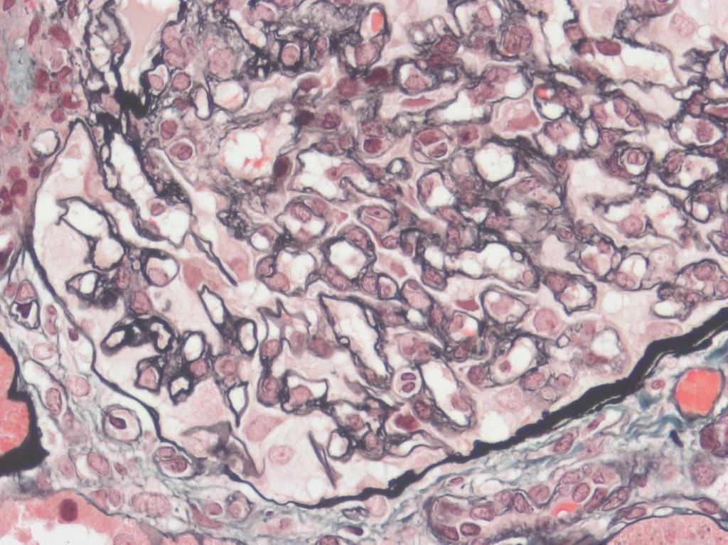
18 Results: Glomerular capillary basement membranes - Black Nuclei - Blue/Black Muscle, red blood cells, fibrin - Red Immunoglobulin - Orange Connective tissue - Green
19
20
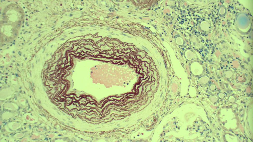
21 Simplicity and Reproducibility Results: Elastic fibres - Brown Nuclei - Blue Background - Pale Pink
22

23 Results: Amyloid - Salmon orange Nuclei - Blue
24
25 Immunofluorescence (frozen sections) Pros: Sensitive, simple and rapid. Cons: Impermanent, fluorescent microscope. Immunoperoxidase (paraffin wax sections) Pros: Localisation of immune deposits, permanent. Cons: More time consuming, costly.
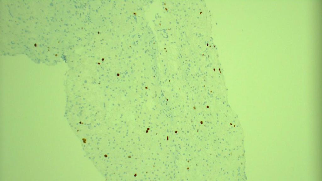
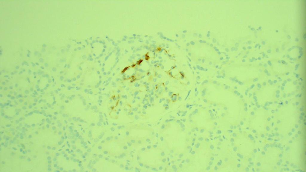
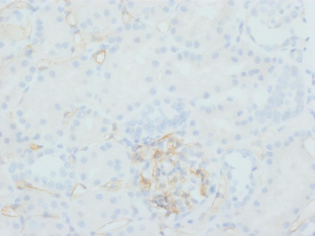
26 SV40 staining Cell Marque SV40 (MRQ-4) Nuclear localisation Simian virus 40 (polyomavirus) C4d staining Roche C4d (SP91) rabbit monoclonal Membranous localisation A complement system protein
27
28
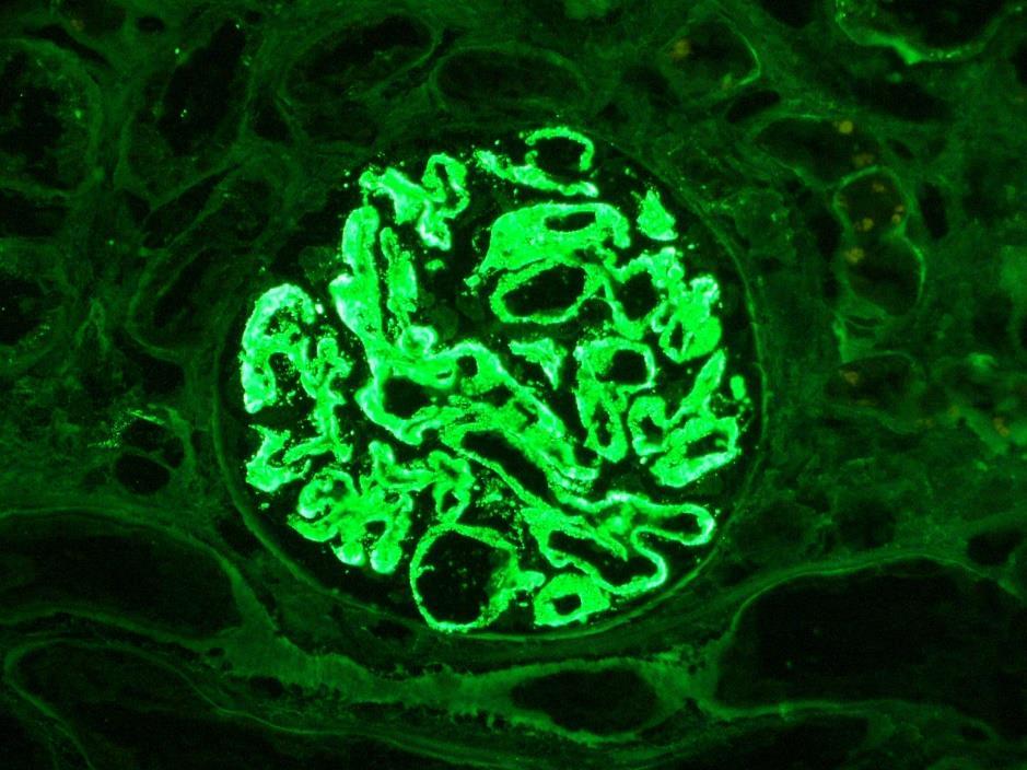
29 Direct IF method for detection of peritubular capillary deposition of Immunoglobulins IgG, IgA & IgM Complement conjugates C3 & C1q Fibrinogen (fibrin) & Albumin Kappa & Lambda light chains
30
31
32
33 Fixed in 2.5% Glutaraldehyde in PIPES 4 C for 24-48hrs. Washed in PIPES buffer. Sent to RCH together with LM/IF report. Processed, photographed and reported. Digital images saved on DVD & IT server.
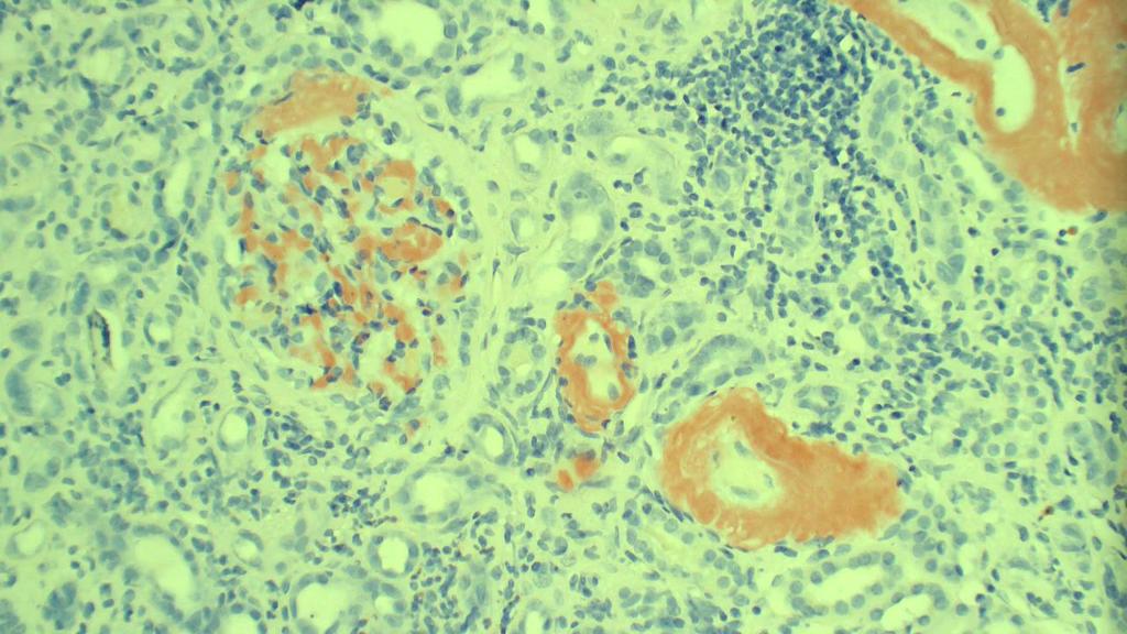
34 Rejection Banff classification Recurrent/de novo disease Infection Drug effect Can have multiple pathologies Comparison to previous biopsies important

 rejection Interstitial fibrosis and")
35 Banff classification Normal Antibody mediated rejection Borderline or suspicious for acute cellular rejection T cell mediated (acute cellular) rejection Interstitial fibrosis and tubular atrophy
36 Interstitial inflammation Tubulitis Vascular inflammation Glomerulitis Interstitial fibrosis Tubular atrophy Arterial fibrointimal thickening Transplant glomerulopathy Mesangial matrix increase Arteriolar hyalinosis Peritubular capillary inflammation C4d Total inflammation
37 For example: Interstitial inflammation i0: <10% of non fibrotic cortex shows mononuclear inflammation i1: 10-25% i2: 26-50% i3: >50% > i1 (interstitial inflammation) + > t1 (tubulitis) or > v0 (vascular inflammation) = T cell mediated rejection
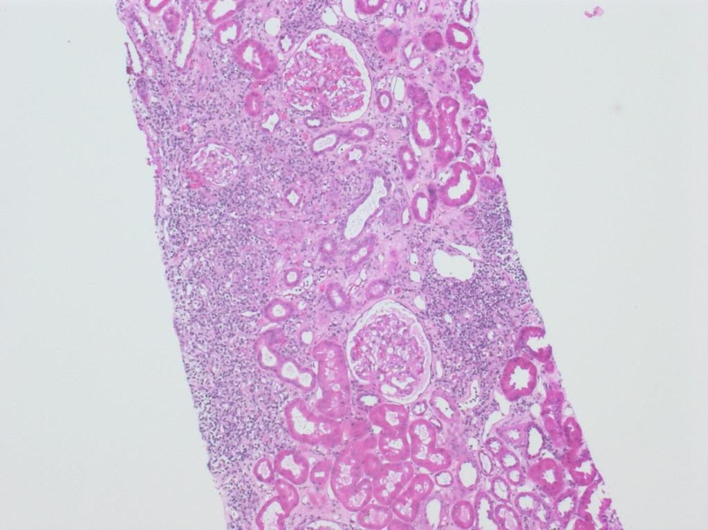
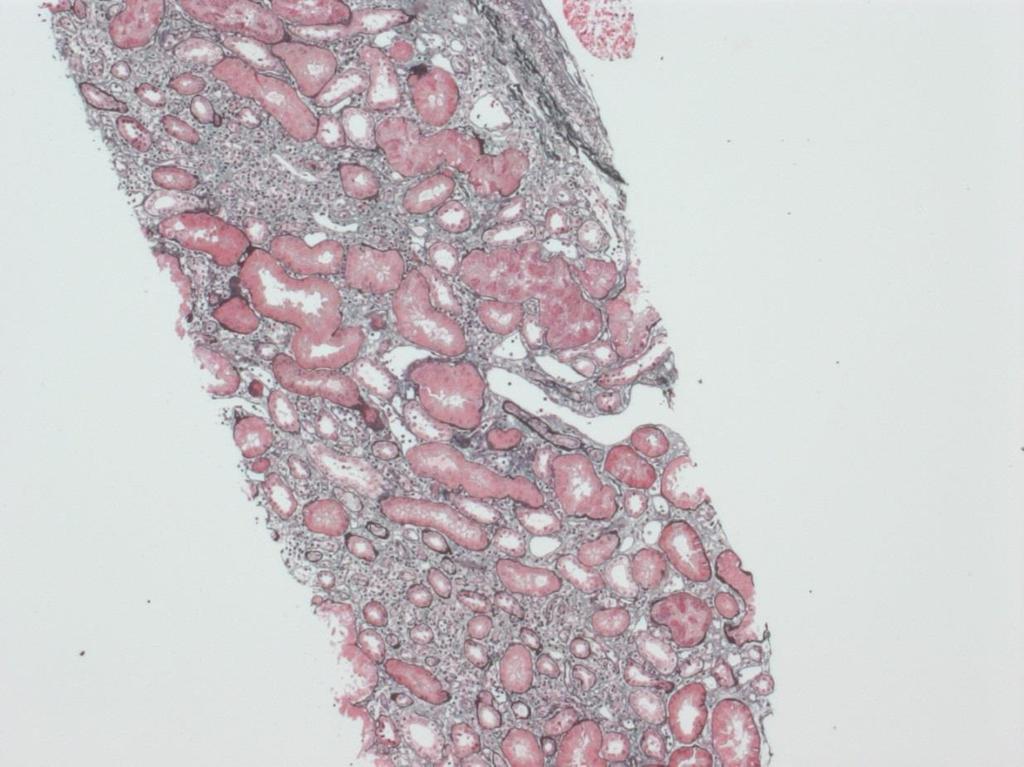
38 H&E 50x


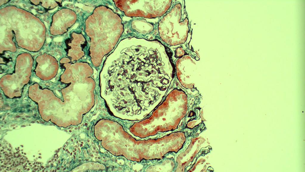
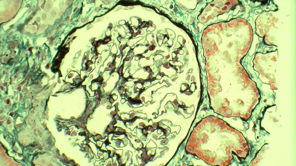
39 PAS 200x Methenamine Silver / Masson Trichrome 200x

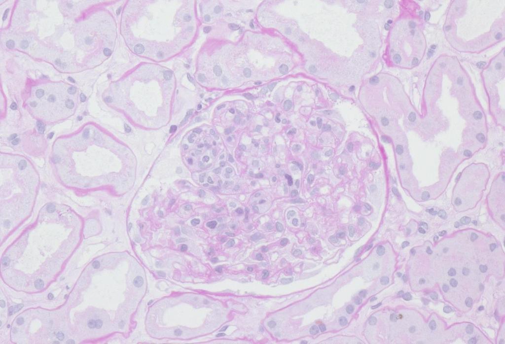
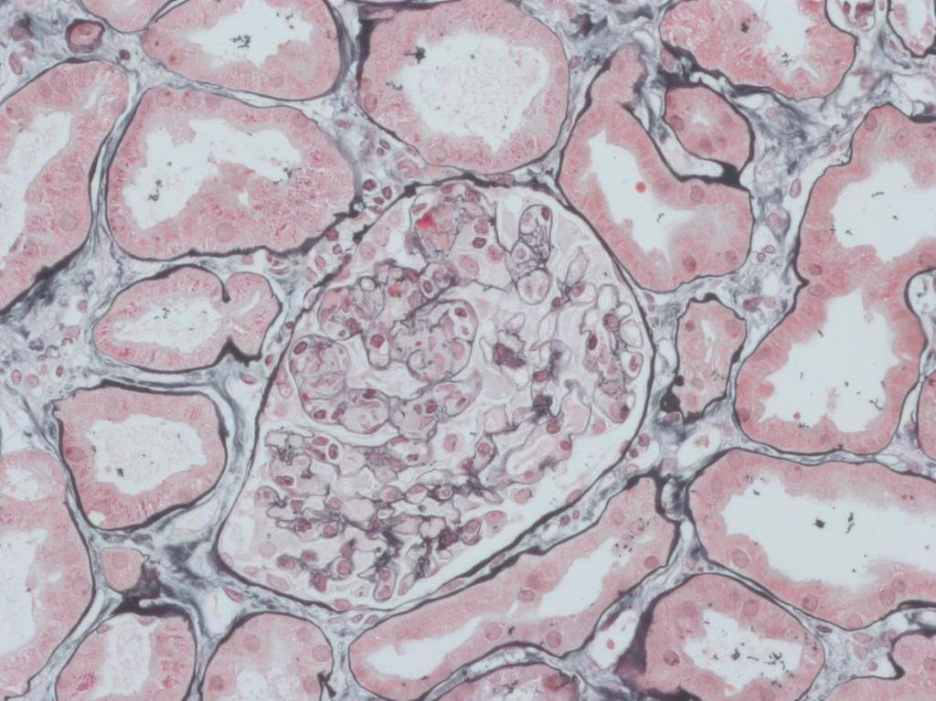
40 H&E 200x PAS 200x Methenamine Silver / Masson Trichrome 200x

41 Methenamine Silver / Masson Trichrome 200x
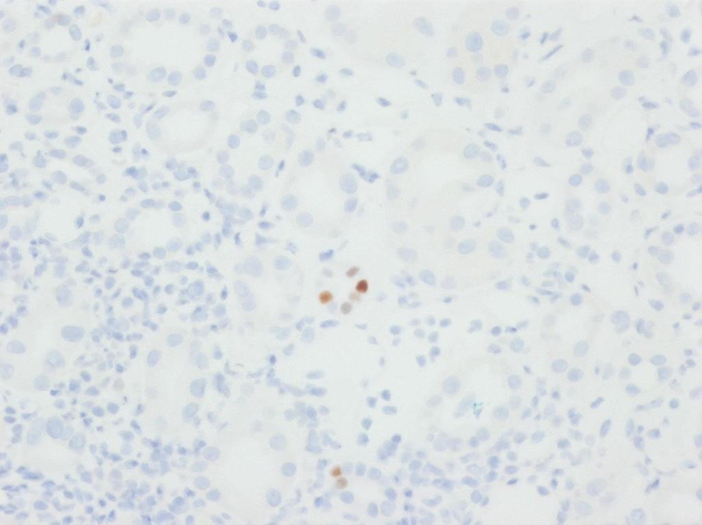
42 SV-40
43


44

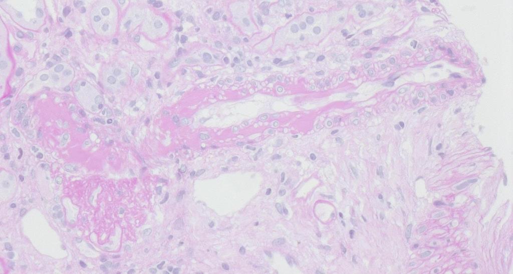
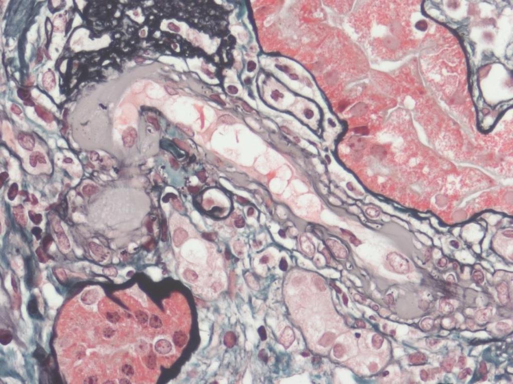
45 PAS 200x Methenamine Silver / Masson Trichrome 200x 50x

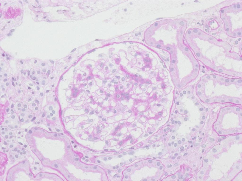
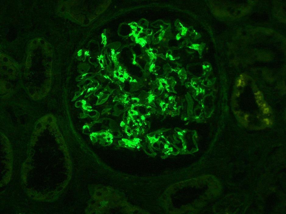
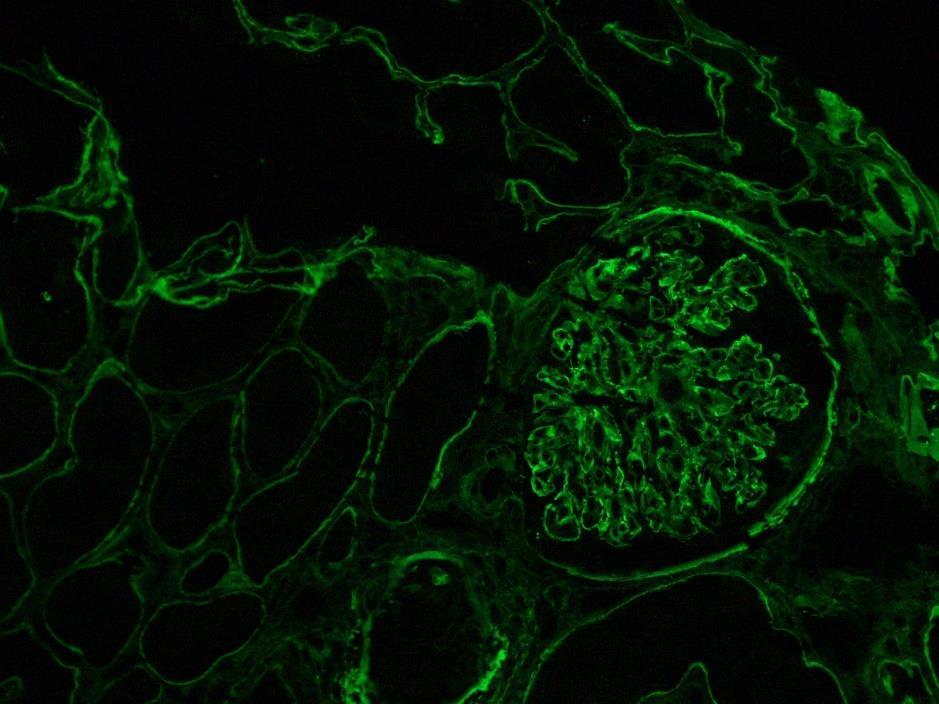
46 PAS 200x IgA immunofluorescence
47 Verity Shugg Clinical Nurse Consultant Organ & Tissue Donation, RHH Dr Ali Graver & Carmel McLeod Clinical Renal Team, RHH Aust. & NZ Dialysis & Transplant Registry Royal Adelaide Hospital, SA
COPYRIGHTED MATERIAL. Tissue Preparation and Microscopy. General Concepts. Chemical Fixation CHAPTER 1
 CHAPTER 1 Tissue Preparation and Microscopy General Concepts I. Biological tissues must undergo a series of treatments to be observed with light and electron microscopes. The process begins by stabilization
UNC DIVISION OF NEPHROPATHOLOGY
 UNC Division of Nephropathology The University of North Carolina School of Medicine Department of Pathology & Laboratory Medicine 09 Brinkhous-Bullitt Building, CB#7 Chapel Hill, NC 799-7 http://www.uncnephropathology.org
PREPARATION OF HISTOLOGICAL SPECIMENS
 PREPARATION OF HISTOLOGICAL SPECIMENS Histo-techniques Preparation of tissue for microscopic examination Series of processes Ultimate aim to make tissue visible as it is Pathology Vs Anatomy Steps vary
Preparation of thin slices for light microscopy
 Preparation of thin slices for light microscopy Optical light microscopy course 23.10.2012 Kirsi Rilla Shortly: Histological sample preparation for microscopy 1. Fixation: To fix the tissue components
Immunohistochemistry guide
 Immunohistochemistry guide overview immunohistochemistry Overview Immunohistochemistry is a laboratory technique utilized for the visual detection of antigens in tissue. When working with cells this technique
Immunological Techniques in Research and Clinical Medicine. Philip L. Cohen, M.D. Chief of Rheumatology, LKSOM 10 March 2016
 Immunological Techniques in Research and Clinical Medicine Philip L. Cohen, M.D. Chief of Rheumatology, LKSOM 10 March 2016 Antibodies Remarkable Tools for Research and Diagnosis You can make an antibody
Methodology for Immunohistochemistry. Learning Objectives:
 Proteomics Methodology for Immunohistochemistry Methodology for Immunohistochemistry A staining process for identifying the proteins location in cells, tissues by using antigen-antibody property. Immuno
TheraLin. Universal Tissue Fixative Enabling Molecular Pathology
 TheraLin Universal Tissue Fixative Enabling Molecular Pathology TheraLin Universal Tissue Fixative Enabling Molecular Pathology Contents Page # TheraLin Universal Tissue Fixative 3 Introduction 5 Easy
Histostaining Artisan Link Pro. Artisan Link Pro. The consistent, safe and easy choice for special stains.
 PR OD U C T I N F O R M A T IO N Histostaining Artisan Link Pro Artisan Link Pro. The consistent, safe and easy choice for special stains. Experience true automation with Artisan Link Pro. Special stains
Introduction to Histology
 Introduction to Histology The name "Histology" is derived from the Greek word for a tissue "Histos", and "-logos" = the study of It is tightly bounded to molecular biology, genetics, immunology and other
Immunoglobulin deposits in Erythema Nodosum Leprosum (ENL)
") Hansen. Int. 3(1), 1978 Immunoglobulin deposits in Erythema Nodosum Leprosum (ENL) JOSEPHINE ANTHONY(*) M. C. VAIDYA (**) A. DASGUPTA (***) SUMMARY Erythema Nodosum Leprosum (ENL) skin lesions observed
Hansen. Int. 3(1), 1978 Immunoglobulin deposits in Erythema Nodosum Leprosum (ENL) JOSEPHINE ANTHONY(*) M. C. VAIDYA (**) A. DASGUPTA (***) SUMMARY Erythema Nodosum Leprosum (ENL) skin lesions observed
Preparation of tissues for study
 Preparation of tissues for study HISTOLOGY : It is the branch of science which deals with the microscopic study of normal tissue HISTOPATHOLOGY : It is the branch of science which deals with the microscopic
THE BASICS OF IMMUNOHISTOCHEMISTRY
 THE BASICS OF IMMUNOHISTOCHEMISTRY Introduction Immunohistochemistry (IHC) identifies specific tissue components by means of a specific antigen/antibody reaction tagged with a visible label. IHC makes
CIHRT Exhibit P-1764 Page 1 IMMUNOHISTOCHEMISTRY ACCURATE LOCALIZATION OF TISSUE OR CELLULAR CONSTITUENTS WITH ANTIBODIES
 CIHRT Exhibit P-1764 Page 1 IMMUNOHISTOCHEMISTRY ACCURATE LOCALIZATION OF TISSUE OR CELLULAR CONSTITUENTS WITH ANTIBODIES CIHRT Exhibit P-1764 Page 2 FUNCTIONAL ROLE OF ANTIBODIES Identify the tissue of
Module IB. Histochemistry. Martin Špaček, MD. (
 Module IB Histochemistry Martin Špaček, MD (E-mail: m.spacek@centrum.cz) http://www.lf3.cuni.cz/histologie What is histochemistry? It is a histological technique used for studying chemistry of tissues
Multiple myeloma (MM) & related disorders
 & related disorders") Multiple myeloma (MM) & related disorders Plasma cell neoplasms Six major variants: (1) Multiple myeloma (2) Solitary plasmacytoma (3) Lymphoplasmacytic lymphoma They secrete a single complete or partial
Multiple myeloma (MM) & related disorders Plasma cell neoplasms Six major variants: (1) Multiple myeloma (2) Solitary plasmacytoma (3) Lymphoplasmacytic lymphoma They secrete a single complete or partial
Sectioning of Paraffin and OCT Embedded Tissue. Signature
 Title SOP Code Effective Date Sectioning of Paraffin and OCT Embedded Tissue SOP117_01 01-Sep-2012 Site Approvals Name and Title (typed or printed) Signature Date dd/mon/yyyy 1.0 PURPOSE This Standard
Materials and Methods Materials Required for Fixing, Embedding and Sectioning. OCT embedding matrix (Thermo Scientific, LAMB/OCT)
") Page 1 Introduction Tissue freezing and sectioning is a rapid method of generating tissue samples (cryosections) for histological analysis, and obviates the need for wax embedding. The method is popular
Page 1 Introduction Tissue freezing and sectioning is a rapid method of generating tissue samples (cryosections) for histological analysis, and obviates the need for wax embedding. The method is popular
HistoMark Double Staining Procedures. Where Better Science Begins.
 HistoMark Double Staining Procedures Where Better Science Begins www.kpl.com HistoMark Double Staining Procedures Researchers often need the ability to visualize multiple proteins in one tissue sample.
Materials and Methods Materials Required for Fixing, Embedding and Sectioning
 Page 1 Introduction Immunofluorescence uses the recognition of cellular targets by fluorescent dyes or antigenspecific antibodies coupled to fluorophores. Depending on the antibody or dye used, proteins,
LAMININ. For Immunohistochemical Demonstration of Laminin in Paraffin-embedded and Frozen Human Tissue Sections Stock No. IMMH-7
 LAMININ For Immunohistochemical Demonstration of Laminin in Paraffin-embedded and Frozen Human Tissue Sections Stock No. IMMH-7 TABLE OF CONTENTS BACKGROUND AND PRINCIPLE... 4 REAGENTS AND EQUIPMENT PROVIDED...
Product datasheet. ARG30119 Pro-B Cell Marker Antibody panel (CD19, CD34, CD38, CD40, CD45)(FACS)
(FACS)") Product datasheet info@arigobio.com Package: 1 kit ARG30119 Pro-B Cell Marker Antibody panel (CD19, CD34, CD38, CD40, CD45)(FACS) Component Cat. No. Component Name ARG62820 anti-cd34 antibody [4H11(APG)]
Product datasheet info@arigobio.com Package: 1 kit ARG30119 Pro-B Cell Marker Antibody panel (CD19, CD34, CD38, CD40, CD45)(FACS) Component Cat. No. Component Name ARG62820 anti-cd34 antibody [4H11(APG)]
NEW STAINS IN TISSUE DIAGNOSIS
 NEW STAINS IN TISSUE DIAGNOSIS CHARLES F. GESCHICKTER, M.D. (Prom the Surgical Pathological Lahoratory of the Johns Hopkins Hospital ami Univel'sily, Baltimore, Maryland) The use of stains has long been
Technical Note. Tissue Section Imaging. Published August The most recent version of this Technical Note is posted at licor.com/bio/support.
 Technical Note Tissue Section Imaging Published August 2017. The most recent version of this Technical Note is posted at licor.com/bio/support. Page 2 - Tissue Section Imaging Table of Contents Page I.
Product Datasheet and Instructions for Use
 Product Datasheet and Instructions for Use Product Code: MP-138-CM01 (0.1ml conc) MP-138-CM05 (0.5ml conc) MP-138-CM1 (1ml conc) Product Description: Biotinylated Bromodeoxyuridine (BrdU) Concentrated
Departments of Electron Microscopy and Neuropathology
 Departments of Electron Microscopy and Neuropathology Method for preserving muscle and nerve biopsies (Procedures 1 and 2) From each biopsy, pieces of tissue should be taken for the following: 1. Frozen
Rapid processing of renal glomeruli for electron microscopy
 516 Rapid processing of renal glomeruli for electron microscopy H. HELIN1, A. PASTERNACK2, and I. RANTALA2 Departments of 1Biomedical and 2Clinical Sciences, University of Tampere, Box 607, SF-33101 Tampere
Introduction to histology and its methods of study
 Introduction to histology and its methods of study Li shulei lishulei@tom.com Department of Histology & Embryology 1 What is histology Definition Cell: smallest units functions in the human body Tissue
Immunofluorescence Confocal Microscopy of 3D Cultures Grown on Alvetex
 Immunofluorescence Confocal Microscopy of 3D Cultures Grown on Alvetex 1.0. Introduction Immunofluorescence uses the recognition of cellular targets by fluorescent dyes or antigen-specific antibodies coupled
Artisan Link Pro. The consistent, safe and easy choice for special stains
 Artisan Link Pro The consistent, safe and easy choice for special stains A true walk-away solution for multiple special stains Special stains present a unique challenge for many labs. With the numerous
MOUSE RAPID STAINING KIT Stock No. QUIK-1. Directions for Use
 MOUSE RAPID STAINING KIT Stock No. QUIK-1 Directions for Use BACKGROUND AND PRINCIPLE The introduction of immunohistochemical techniques has ushered a new era of staining into the laboratory based upon
SANTA CRUZ BIOTECHNOLOGY, INC.
 TECHNICAL SERVICE GUIDE: Western Blotting 2. What size bands were expected and what size bands were detected? 3. Was the blot blank or was a dark background or non-specific bands seen? 4. Did this same
Electrophoresis. Assays... INTERLAB ASSAYS. Instrument. Software Easy data management thanks to innovative Elfolab software. General Characteristic
 Software Easy data management thanks to innovative Elfolab software. Instrument Complete walk-away automation. Initial 52 results available within 50 minutes. Impressive 208 Serum Proteins samples per
Overview of Immunohistochemistry
 Overview of Immunohistochemistry Immunohistochemistry (IHC) combines anatomical, immunological and biochemical techniques to identify discrete tissue components by the interaction of target antigens with
Introduction to Histology
 Introduction to Histology Histology The term "Histology" is derived from the Greek word for a tissue "Histos", and "-logos" = the study of Histology : Is the study of tissues and how they are arranged
1. Paraffin section slides can be stored at room temperature for a long time.
 Immunohistochemistry (IHC) Protocols Immunohistochemistry (IHC) Protocol of Paraffin Section 1. Fix dissected tissues with 10% formalin for no less than 48 hours at room temperature. Inadequately fixation
Typical bands found on serum gel electrophoresis:
 Gel Electrophoresis LD Recognise EPG patterns typical of other body fluids including urine and CSF Identify patterns of changes including - Paraproteins - Hypogamma - Acute phase - Circulating immune complexes
Immunoassay Kit Catalog # KCA0021. Canine. C-Reactive Protein
 Immunoassay Kit Catalog # KCA0021 Canine C-Reactive Protein BioSource International, Inc. 542 Flynn Road Camarillo, California 93012 USA Tel: 805-987-0086 800-242-0607 FAX: 805-987-3385 email: tech.support@biosource.com
Keratin 19 (KRT19) Immunohistochemistry Kit
 Immunohistochemistry Kit") Keratin 19 (KRT19) Immunohistochemistry Kit For Immunohistochemical Staining of Keratin 19 (KRT19) in human FFPE Tissue RUK-KKR01-20 For Research Use Only Riverside Biosciences Inc. 2327 S 5th Ave, North
Keratin 19 (KRT19) Immunohistochemistry Kit For Immunohistochemical Staining of Keratin 19 (KRT19) in human FFPE Tissue RUK-KKR01-20 For Research Use Only Riverside Biosciences Inc. 2327 S 5th Ave, North
HISTOPATHOLOGY INTRODUCTION
 HISTOPATHOLOGY INTRODUCTION Surgical, anatomical and consultative pathology services are available through pathologists in the Department of Pathology. The services available include: Routine surgical
Fluorochrome-conjugated Anti Collagen IV cocktail for Alport's syndrome
 Product manual Fluorochrome-conjugated Anti Collagen IV cocktail for Alport's syndrome FITC-Anti Collagen IV α5(iv) Chain, Human (Mono) + Texas Red-Anti Collagen IV α2(iv) Chain, Human (Mono) Cat. No.SGE-CFT45325
Material and methods. Thirty-three renal biopsies (28 needle biopsies and
 J Clin Pathol 1981 ;34:859-865 Immunoperoxidase staining of formalin-fixed, paraffin-embedded, human renal biopsies with a comparison of the peroxidase-antiperoxidase (PAP) and indirect methods RA SINCLAIR,*
Workshop Outline. Laser Capture Microdissection=molecular analysis of specific cells. LCM sample is a genomic sample.
 Workshop Outline LCM sample is a genomic sample. Clinical vs experimental LCM sample. Genomic sample collection and stabilization. LCM sample preparation: microtomy, cryotomy, staining. Start-up QC of
Smooth Muscle-Specific Expression of ipla 2 β Participates in the Initiation and Early Progression of Vascular Inflammation and Neointima Formation
 Smooth Muscle-Specific Expression of ipla 2 β Participates in the Initiation and Early Progression of Vascular Inflammation and Neointima Formation Shu Liu 1, Zhongwen Xie 2, Qingwei Zhao 2, Huan Pang
Supplementary Methods. Li J.-Y. et al. Lewy bodies in grafted neurons in Parkinson s patients suggest host to. graft disease propagation
 1 Supplementary Methods Li J.-Y. et al. Lewy bodies in grafted neurons in Parkinson s patients suggest host to graft disease propagation Neural transplantation and clinical assessment Detailed information
VisUCyte TM HRP Polymer-DAB Cell & Tissue Staining Kit
 VisUCyte TM HRP Polymer-DAB Cell & Tissue Staining Kit For the detection of goat, mouse, rabbit, rat, or sheep primary IgG Antibodies with a biotin-free detection system. Size: 50 Tests Secondary Antibody-HRP
Immunological Applications. Chapter 8: Background
 Immunological Applications Chapter 8: Background The Immune System Types of Immunity Innate The natural immunity present at birth Acquired A specific response to foreign substances. Some cells remember
Brief Definitive Report
 Published Online: 1 October, 1975 Supp Info: http://doi.org/10.1084/jem.142.4.1029 Downloaded from jem.rupress.org on October 21, 2018 Brief Definitive Report A RECEPTOR FOR THE THIRD COMPONENT OF COMPLEMENT
Strategies for Assessment of Immunotoxicology in Preclinical Drug Development
 Strategies for Assessment of Immunotoxicology in Preclinical Drug Development Rebecca Brunette, PhD Scientist, Analytical Biology SNBL USA Preclinical Immunotoxicology The study of evaluating adverse effects
Product Datasheet. CD31/PECAM-1 Antibody (C31.7) NBP mg. Unit Size: 0.1 mg. Store at 4C. Publications: 1
 NBP mg. Unit Size: 0.1 mg. Store at 4C. Publications: 1") Product Datasheet CD31/PECAM-1 Antibody (C31.7) NBP2-15188-0.1mg Unit Size: 0.1 mg Store at 4C. Publications: 1 Protocols, Publications, Related Products, Reviews, Research Tools and Images at: www.novusbio.com/nbp2-15188
Product Datasheet CD31/PECAM-1 Antibody (C31.7) NBP2-15188-0.1mg Unit Size: 0.1 mg Store at 4C. Publications: 1 Protocols, Publications, Related Products, Reviews, Research Tools and Images at: www.novusbio.com/nbp2-15188
Chapter 4 ANTIBODY STRUCTURE AND FUNCTION
 Chapter 4 ANTIBODY STRUCTURE AND FUNCTION Different way to depict an Ig molecule Y In both the heavy and light chain variable regions there is variability at every position and there are hypervariable
A collection of 31 splenic specimens from patients (42.4±17.7 years old) with ITP
 with ITP") Supplemental Materials & Methods Patients and patient samples. A collection of 31 splenic specimens from patients (42.4±17.7 years old) with ITP requiring splenectomy and 36 splenic specimens obtained
Supplemental Materials & Methods Patients and patient samples. A collection of 31 splenic specimens from patients (42.4±17.7 years old) with ITP requiring splenectomy and 36 splenic specimens obtained
PROTOCOL. Introduction. Method. Page 1
 Page 1 Introduction Following fixation a variety of different cytological stains can be used to visualise cell components in detail. Here we describe the use of Haematoxylin and Eosin, a general structural
Which hydrogel preparation for immunostaining protocol should I use?
 Protocol: Preparation of TissueSpec hydrogels for immunostaining This protocol may be used prior to immunostaining cells, organoids, or patient-derived xenografts cultured in TissueSpec matrix hydrogels.
Dr: RAWIA BADR Associate Professor of Microbiology&Immunology
 Dr: RAWIA BADR Associate Professor of Microbiology&Immunology Cell culture Commonly refers to the culture of animal cells and tissues, while the more specific term plant tissue.culture is used only for
Staining Techniques. Staining Techniques. There are many dyes. Histochemical Stains: chemical reactions. Feulgen reaction -DNA
 Staining Techniques There are many dyes. http://medinfo.ufl.edu/~dental/denhisto/stains.html Examples: Sudan black -Lipids Myelinated axons- blue ihcworld.com/imagegallery/displayimage.php?al... Weigert
Cardiovascular (connective tissue)
") Cardiovascular (connective tissue) Blood Connective tissue Blood Proteins 7% Plasma 55% Other solutes 2% Water 91% Erythrocytes ~5.2 million per cubic mm Formed elements 45% Leukocytes ~7 thousand per
Cardiovascular (connective tissue) Blood Connective tissue Blood Proteins 7% Plasma 55% Other solutes 2% Water 91% Erythrocytes ~5.2 million per cubic mm Formed elements 45% Leukocytes ~7 thousand per
Anti-Piscirickettsia salmonis monoclonal antibody. Product no: P05
 Anti-Piscirickettsia salmonis monoclonal antibody Product no: P05 Product Description The monoclonal antibody (Mab) against Piscirickettsia salmonis is specific for this bacterium. The specificity of the
BIMM18 Dec 20 th - Flow cytometry in clinical diagnostics
 BIMM18 Dec 20 th - Flow cytometry in clinical diagnostics I. B cell leukemia and lymphomas Immunophenotyping as part of the diagnostic work- up of hematologic malignancies offers a rapid and effective
Index 311. overview, 84, 85 pretreatment, 85 probe preparation, 85 washing, 85, 91. G GeneChip, see DNA microarray
 Index 309 Index A AIDS, see Human immunodeficiency virus Allelotyping, microdissection advantages, 69, 70 ovarian cancer, data acquisition and analysis, 74 DNA extraction, 72, 73, 76 materials, 70, 71
A Study of the Argentaffin (Kultschitzky) Cells in frozen-dried Tissue by Phase-Contrast Microscopy and Ultra-Violet Light By A. C.
 Cells in frozen-dried Tissue by Phase-Contrast Microscopy and Ultra-Violet Light By A. C.") 289 A Study of the Argentaffin (Kultschitzky) Cells in frozen-dried Tissue by Phase-Contrast Microscopy and Ultra-Violet Light By A. C. CHRISTIE (From the Royal Cancer Hospital, London. Present address,
289 A Study of the Argentaffin (Kultschitzky) Cells in frozen-dried Tissue by Phase-Contrast Microscopy and Ultra-Violet Light By A. C. CHRISTIE (From the Royal Cancer Hospital, London. Present address,
Supporting Protocols
 Supporting Protocols This protocol may be used prior to immunostaining cells, organoids, or patient-derived xenografts cultured in TissueSpec ECM Hydrogels. Introduction Cells and organoids may form complex
Research Testing Services & Analytical Equipment
 University of Queensland School of Veterinary Science Veterinary Laboratory Services Veterinary Science Building (Building 8114) University of Queensland Gatton Campus Gatton Qld 4343 Phone 5460 1843 Email
ab Giemsa Stain Kit
 Version 3 Last updated 19 December 2018 ab150670 Giemsa Stain Kit For the histological visualization of Cells present in Hematopoietic Tissues and Certain Microorganisms. View kit datasheet: www.abcam.com/ab150670
PROVINCIAL BLOOD COORDINATING PROGRAM DEFINITIONS NLBCP-063
 Government of Newfoundland and Labrador Department of Health and Community Services Provincial Blood Coordinating Program PROVINCIAL BLOOD COORDINATING PROGRAM DEFINITIONS Office of Administrative Responsibility
Contents. 11 The Use of Epitope Tags in Histochemistry References... 98
 Contents 1 Antibodies for Immunohistochemistry... 1 1.1 Structure of Antibodies... 2 1.2 Polyclonal Antibodies... 4 1.3 Mouse Monoclonal Antibodies... 4 1.4 Rabbit Monoclonal Antibodies... 5 1.5 Protein
Principles And Procedures
 Product Identification Cat. No. Description 44674 Lambda Light Chain 0,1 M (Lamb14) 44675 Lambda Light Chain 1 M (Lamb14) 44326 Lambda Light Chain RTU M (Lamb14) Symbol Definitions P C A E S DIL DOC# DIS
Cartilage Staining Kit (Chondrocyte and Cartilage Tissue Staining Kit)
") Cat. # MK310 For Research Use Cartilage Staining Kit (Chondrocyte and Cartilage Tissue Staining Kit) Product Manual v201009 Table of Contents I. Description... 3 II. Kit Components... 3 III. Materials
Cat. # MK310 For Research Use Cartilage Staining Kit (Chondrocyte and Cartilage Tissue Staining Kit) Product Manual v201009 Table of Contents I. Description... 3 II. Kit Components... 3 III. Materials
Oxford Gene Technology The Molecular Genetics Company
 Oxford Gene Technology The Molecular Genetics Company UK UGM Pathology Workshop Troubleshooting FISH Alex Hobbs Field Application Specialist-Cytocell 1 Overview PETS FISH common issues Analysis considerations
HELICA BIOSYSTEMS, INC. MOUSE C-REACTIVE PROTEIN QUANTITATION BY ELISA FOR RESEARCH USE ONLY
 HELICA BIOSYSTEMS, INC. MOUSE C-REACTIVE PROTEIN QUANTITATION BY ELISA FOR RESEARCH USE ONLY INTENDED USE The Helica C-reactive protein assay is intended for the detection and quantification of mouse C-reactive
Adenomatous Polyposis Coli (APC) Immunohistochemistry Kit
 Immunohistochemistry Kit") Adenomatous Polyposis Coli (APC) Immunohistochemistry Kit For Immunohistochemical Staining of Adenomatous Polyposis Coli (APC) in human FFPE Tissue RUK-KAP01-20 For Research Use Only Riverside Biosciences
Adenomatous Polyposis Coli (APC) Immunohistochemistry Kit For Immunohistochemical Staining of Adenomatous Polyposis Coli (APC) in human FFPE Tissue RUK-KAP01-20 For Research Use Only Riverside Biosciences
Immunofluorescent Localization of Antihemophilic Factor Antigen and Fibrinogen in Human Renal
 Immunofluorescent Localization of Antihemophilic Factor Antigen and Fibrinogen in Human Renal JoHN R. HoYER, AEmD F. MicHAEL, and LEON W. HoYER Diseases From the Department of Pediatrics, University of
COX-2 (Cyclooxygenase-2) ELISA KIT
 ELISA KIT") COX-2 (Cyclooxygenase-2) ELISA KIT Cat. No.:DEIA6172 Pkg.Size:96T Intended use The Human COX-2 Kit is capable of the determination of Human COX-2 in lysate of cultured cells. General Description Cyclooxygenase
COX-2 (Cyclooxygenase-2) ELISA KIT Cat. No.:DEIA6172 Pkg.Size:96T Intended use The Human COX-2 Kit is capable of the determination of Human COX-2 in lysate of cultured cells. General Description Cyclooxygenase
Supplementary Materials and Methods
 Supplementary Materials and Methods Reagents Supplementary Material (ESI) for Lab on a Chip RPMI medium, FBS, HEPES buffer solution, sodium pyruvate, penicillin, and streptomycin were obtained from Biological
Product Datasheet and Instructions for Use
 Product Code: MP-109-CM01 (0.1ml conc) MP-109-CM05 (0.5ml conc) MP-109-CM1 (1ml conc) MP-109-PM6 (6ml RTU) Product Description: Androgen Receptor Concentrated and Prediluted Monoclonal Antibody Control
Bio-Plex suspension array system tech note 5649
 Immunoglobulin Isotyping Bio-Plex suspension array system tech note 9 Development and Validation of a Novel Multiplex Immunoglobulin Isotyping Assay on Magnetic Microspheres Candice Reyes, Joe Fedynyshyn,
The Children s Hospital of Philadelphia Department of Pathology and Laboratory Medicine
 TheChildren shospitalofphiladelphia DepartmentofPathologyandLaboratoryMedicine Muscle Biopsy - General Instructions The Division of Neuropathology, Department of Pathology and Laboratory Medicine, Children
College of American Pathologists Laboratory Accreditation Program. AP for Histotechs and Secretaries. Learning Objectives
 College of American Pathologists AP for Histotechs and Secretaries Francis E. Sharkey, M.D., FCAP University of Texas Health Science Center - San Antonio February 20th, 2008 Copyright 2008 College of American
In situ detection kit for programmed Cell Death MEBSTAIN Apoptosis Kit Direct
 For research use only In situ detection kit for programmed Cell Death MEBSTAIN Apoptosis Kit Direct CODE No. 8445 In situ detection kit for Programmed Cell Death MEBSTAIN Apoptosis Kit Direct Cat. No.
Product Datasheet and Instructions for Use
 Product Code: MP-066-CM01 (0.1ml conc) MP-066-CM05 (0.5ml conc) MP-066-CM1 (1ml conc) MP-066-PM6 (6ml RTU) Product Description: Neurofilament Concentrated and Prediluted Monoclonal Antibody Control Number:
Comparing the Quality of Fixation for Gel-based Formalin (Formagel) versus Traditional Liquid-Based Formalin for Immunohistochemistry
 versus Traditional Liquid-Based Formalin for Immunohistochemistry") Comparing the Quality of Fixation for Gel-based Formalin (Formagel) versus Traditional Liquid-Based Formalin for Immunohistochemistry Brian H. Le, M.D., Reading Hospital Reviewed by Michael R. LaFrinere,
Comparing the Quality of Fixation for Gel-based Formalin (Formagel) versus Traditional Liquid-Based Formalin for Immunohistochemistry Brian H. Le, M.D., Reading Hospital Reviewed by Michael R. LaFrinere,
PATHOLOGY LABORATORY SERVICES
 VANDERBILT PATHOLOGY LABORATORY SERVICES VANDERBILT UNIVERSITY MEDICAL CENTER Request for Neuropathology Consultation Division of Neuropathology C-2318 Medical Center North Nashville, TN 37232-2561 Phone
Chapter 3. Clonal selection
 Chapter 3. Clonal selection I have called this principle, by which each slight variation, if useful, is preserved, by the term of Natural Selection -Charles Darwin, On the Origin of Species, 1859 4 The
QImaging Camera Application Notes Multicolor Immunofluorescence Imaging
 QImaging Camera Application Notes Multicolor Immunofluorescence Imaging In order to image localization of intracellular proteins with high specificity, it is frequently necessary to multiplex antibody
Using Human Tissue in Translational Research and Clinical Trials
 Using Human Tissue in Translational Research and Clinical Trials Dr Bridget S Wilkins NCRI Clinical Lead for Pathology Engagement (Consultant Histopathologist, St Thomas Hospital, London) Cellular and
HELICA BIOSYSTEMS, INC. HIGH SENSITIVITY HUMAN C-REACTIVE PROTEIN FOR RESEARCH USE ONLY (Not for in vitro diagnostic use)
") INTENDED USE HELICA BIOSYSTEMS, INC. HIGH SENSITIVITY HUMAN C-REACTIVE PROTEIN FOR RESEARCH USE ONLY (Not for in vitro diagnostic use) The Helica C-reactive protein assay is intended for the detection
INTENDED USE HELICA BIOSYSTEMS, INC. HIGH SENSITIVITY HUMAN C-REACTIVE PROTEIN FOR RESEARCH USE ONLY (Not for in vitro diagnostic use) The Helica C-reactive protein assay is intended for the detection
Murine and Non-Human Primate Dendritic Cell Targeting Nanoparticles for In Vivo Generation of Regulatory T-Cells
 Murine and Non-Human Primate Dendritic Cell Targeting Nanoparticles for In Vivo Generation of Regulatory T-Cells Sebastian O. Stead 1, Svjetlana Kireta 2, Steven James Peter McInnes 3, Francis D. Kette
Step-by-Step Description of ELISA
 Step-by-Step Description of ELISA The protocols in this kit rely on indirect antibody capture ELISA. The steps in this assay are: Step 1: Antigen is added to the wells of the microplate strip and incubated
Observations about complement were carried out by Nuthall Pfeiffer and Bordet in the 1800's.
 COMPLEMENT SYSTEM Observations about complement were carried out by Nuthall Pfeiffer and Bordet in the 1800's. Researchers compared cholera vibrio with immune fresh serum in the test tube and; Cholera
Immunoglobulins: Structure and Function
 Immunoglobulins: Structure and Function Immunoglobulins:Structure and Function Definition: Glycoprotein molecules that are produced by plasma cells in response to an immunogen and which function as antibodies
Electron Beam Sterilization of the Agarose Gel Used for Electrophoresis
 Electron Beam Sterilization of the Agarose Gel Used for Electrophoresis D. Ighigeanu 1, D. Martin 1, E. Manaila 1, D.E. Stan 2, I. V. Baciu 3, G. Craciun 1, C. Oproiu 1, N. Iacob 1 1 National Institute
Cell & Tissue Staining Kit
 Cell & Tissue Staining Kit For the detection of goat, mouse, rabbit, rat, or sheep primary IgG Antibodies Size: 50 Tests HRP-DAB System Goat Kit (Catalog Number CTS008) Mouse Kit (Catalog Number CTS002)
Find 1 cell in 100,000 with ELISpot
 Find 1 cell in 100,000 with ELISpot ELISpot is a sensitive assay used to quantify cytokine- or immunoglobulin-secreting cells at the single-cell level. ELISpot has been widely applied to investigate specific
ZytoDot CISH Polymer Detection Kit
 ZytoDot CISH Polymer Detection Kit C-3005-40 40 C-3005-10 40 For the detection of DIG labeled probes by chromogenic in situ hybridization (CISH).... In vitro diagnostic medical device according to EU directive
Complement Titration Tray Class I and II. Instructions for Use
 Complement Titration Tray Class I and II Instructions for Use Complement Titration Tray, Class I and II Instructions for Use The Invitrogen Complement Titration Tray, Class I and II, is a serological based
IMMUNOPATHOLOGY. This SOP will be applied to npod paraffin samples stained by immunohistochemistry.
 1 PURPOSE IMMUNOPATHOLOGY The purpose of this Standard Operating Procedure (SOP) is to outline procedures for immunopathology preparation and analysis of npod samples. 2 SCOPE This SOP will be applied
ultraview Universal Alkaline Phosphatase Red Detection Kit
 ultraview Universal Alkaline Phosphatase Red Detection Kit 760-501 05269814001 250 Red Precipitate AP Multimer Enhancer Naphthol Fast Red INTENDED USE Ventana Medical Systems' (Ventana) ultraview Universal
Schedule of Accreditation
 Schedule of Accreditation Organisation Name Bon Secours Hospital Tralee INAB Reg No 206MT Contact Name Cara Wrenn Address Strand Street, Tralee, Kerry Contact Phone No 0667149800 Email cwrenn@bonsecours.ie
Detection of protein expression
 Detection of protein expression by immunocytochemistry Dennis Brown, Ph. D. Program in Membrane Biology/Renal Unit MGH East Brown@receptor.mgh.harvard.edu http://membranebiology.mgh.harvard.edu Level of