3/6/2017 TOMOSYNTHESIS GUIDED BREAST BIOPSY LEARNING OBJECTIVES DISCLOSURES
|
|
|
- Gerard Douglas
- 6 years ago
- Views:
Transcription
1 TOMOSYNTHESIS GUIDED BREAST BIOPSY Amy Kerger, D.O. Assistant Professor The Ohio State Wexner Medical Center Stefanie Spielman Comprehensive Breast Center LEARNING OBJECTIVES What is the difference between tomosynthesis guided breast biopsy compared with 2D stereotactic guided biopsy When and how do we decide which patients receive tomosynthesis guided biopsy compared to 2D stereotactic biopsy Advantages and disadvantages to tomosynthesis guided biopsy What advantages does the new prone tomosynthesis/2d table provide What is the medical physicists role with this modality DISCLOSURES I have no disclosures to report 1
2 HISTORY OF TOMOSYNTHESIS Basic theoretical framework was provided by Ziedses des Plantes in the 1930 s In 1972 Grant coined the term tomosynthesis in a landmark paper that described simple tomosynthesis reconstruction In the s a number of variants of tomosynthesis were developed (i.e. ectomography and flash tomography) With the advent of spiral CT in the 1980 s tomosynthesis research halted In the 1990 s research using tomosynthesis for chest and breast imaging began Medical Physics Vol. 36, No. 6, June 2009 HISTORY OF TOMOSYNTHESIS 1992 Christian, Niklason, LT, Niklason, LE and Kopans started looking at digital breast tomosynthesis (DBT) for breast imaging 2000 in collaboration with GE Healthcare the first studies were performed using DBT and a patent was granted 2011 FDA approved Hologic Selenia Dimensions 3D system for breast tomosynthesis 2013 FDA approved Hologic C-view imaging software for use with DBT to reconstruct 2-D images and Hologic Affirm Breast Biopsy Guidance System Medical Physics Vol. 36, No. 6, June 2009 TOMOSYNTHESIS DBT is a 3D method of imaging that reduces tissue overlap seen in regular 2D mammography It is a form of limited-angle tomography Low-dose full field projection images of the breast are obtained from different angles with x-rays passing through the breast from different directions Reconstruction produces many 1 mm image slices AJR2014; 202:

3 TOMOSYNTHESIS In the screening patient, both the craniocaudal (CC) and mediolateral oblique (MLO) projections are acquired; however, additional projections may be obtained if warranted. The x-ray source moves in a single plane in an arc around the imaged breast. These projection images are then reconstructed into 1-mm-thick images for review. Filtered back projection (FBP) is the most commonly used method for reconstruction, TOMOSYNTHESIS AJR 2017; 208: Advantages to using DBT: TOMOSYNTHESIS Decreases call back from screening. Better for dense breast tissue. Able to see architectural distortion and isodense masses better. Helps distinguish skin lesions without the use of tangential views. Helps localize a lesion for ultrasound or biopsy. Allows better visualization of mass margins so can go straight to ultrasound from screening call back. 3
4 TOMOSYNTHESIS Disadvantages to using DBT: Longer scan time than 2D mammogram. Increase radiation dose compared with 2D. Not yet covered by all insurance companies. Takes longer time to read. TOMOSYNTHESIS VS. STEROTACTIC GUIDED BIOPSY LESIONS BEST VISUALIZED Tomo Guided Biopsy Stereotactic Guided Biopsy Architectural Distortion Isodense or low density lesions. Single view findings. Calcifications TOMOSYNTHESIS VS. STEROTACTIC GUIDED BIOPSY - ADVANTAGES Tomo Guided Biopsy Can be done upright or prone position if you have both available. Larger field of view 18 x 24 cm. Shorter biopsy times (13 vs 29 minutes). 3D imaging for better and more accurate targeting. Biopsy equipment is easily installed and removed from a mammogram machine. Less exposures needed. Patients who are over the weight limit for the prone table. Better ability to make adjustments due to better visualization of the lesion and trough and needle tip. Stereotactic Guided Biopsy Can be done upright or prone position if you have both available. If patient does not want to see the needle prone positioning allows for them not to. Better to see calcifications. Less vasovagal episodes. 4

5 TOMOSYNTHESIS VS. STEROTACTIC GUIDED BIOPSY LIMITATIONS Tomo Guided Biopsy Vasovagal episodes. If only have upright biopsy system more difficult to reach very posterior lesions. A mammogram machine is need so won t be able to do mammograms on it when biopsy is occuring. Stereotactic Guided Biopsy Difficult to accurately biopsy distortions, isodense masses and one view findings. Smaller FOV. Longer time for biopsy. In only have prone table cannot biopsy patients over weight limit or those who cannot lie on their stomach. Posterior lesion accessibility Use a 7G 11G vacuum biopsy needle. Comes in standard size (20 mm trough), petite (12 mm trough) and Non-firing (12 mm trough). Tomosynthesis guidance helps over come technical challenges previously seen in stereotactic biopsy: Superficial lesion Deep or very superior lesion 5
6 6


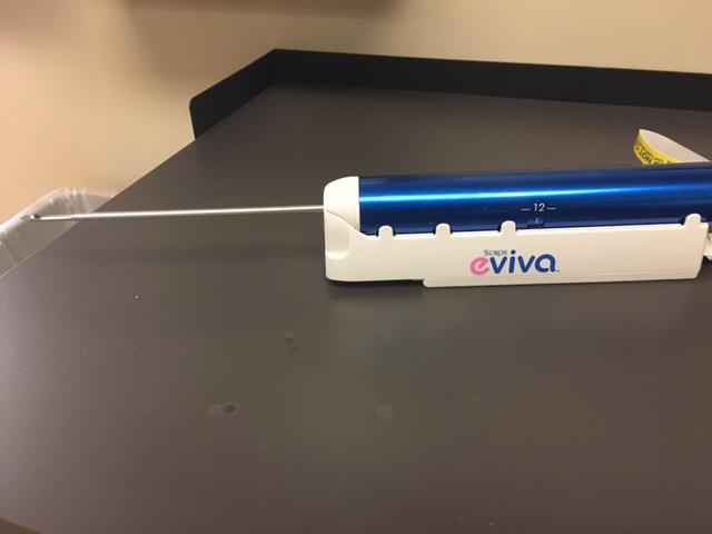
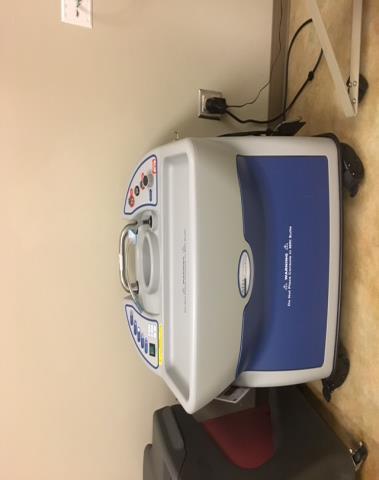
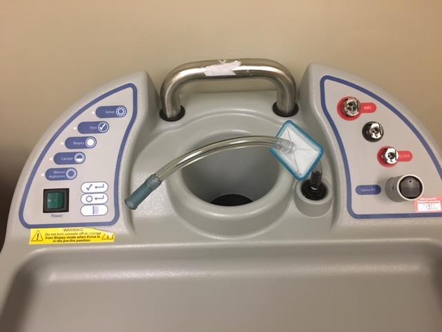
7 The TVAB system is mounted onto the mammogram equipment s C-arm and locked into place. After consent is given and a time out is performed the patient is positioned and a scout tomosynthesis view is performed to acquire the target. The target is marked and the coordinates are sent to the TVAB system. Target 7


8 The needle is then attached to the system and the needle is homed and then targeted to the appropriate coordinates. The patient s skin is cleaned and lidocaine is injected into the skin and subcutaneous tissues. Then the needle is advanced along the z-axis to the appropriate depth. A second set of tomosynthesis images is preformed to ensure accurate targeting. 8


9 Biopsy is then performed taking 6-12 cores and a clip is then placed. A tomosynthesis is acquired to ensure clip deployment and appropriate placement. Pressure is held for 10 minutes to stop bleeding and a post biopsy 2D CC and ML mammogram is performed. 9


10 NEW PRONE 2D/3D TABLE Enables biopsy of lesions only visible with tomosynthesis. Amorphous Selenium Detector (same detector technology as the upright). X-ray translucent paddles for better visualization of breast tissue surrounding biopsy window. 6.5 times larger FOV 14.3 cm X 11.7 cm. Fast one-click targeting. True 360 degree access Lateral arm (no need to take patient out of compression moving from standard approach to lateral approach). Clear paddles (easier to see landmarks). NEW PRONE 2D/3D TABLE Visualize challenging subtle masses and faint calcifications Including those only visible under tomosynthesis imaging Same proven detector technology found in Selenia Dimensions System 10
11 CASES REFERENCES Tomosynthesis Imaging: At a Translational Crossroads. Medical Physics Vol. 36, No. 6, June Digital Breast Tomomsynthesus From Concept to Clinical Care. AJR2014; 202: Advances in Digital Breast Tomosynthesis. AJR. 2017; 208: Basics of Digital Breast Tomosynthesis. Applied Radiology. March 2014: Tomosynthesis-guided vacuum-assisted breast biopsy: A feasibility study. European Radiology : Comparison of digital breast tomosynthesis and 2D digital mammography using a hybrid performance test. Physics in Medicine & Biology : Effects on short-term quality of life of vacuum-assisted breast biopsy: comparison between digital breast tomosynthesis and digital mammography. BRJ Digital Breast Tomosynthesis-guided Vacuum-assisted Breast Biopsy: Initial Experiences and Comparison with Prone Stereotactic Vacuum-assisted Biopsy. Radiology. 3/2015. Vol. 274: Number 3. Tomosynthesis-detected Architectural Distortion: Management Algorithm with Radiologic -Pathologic Correlation. Radiographics. 2016;36:
Digital breast tomosynthesis-guided biopsy: preliminary experience
 Digital breast tomosynthesis-guided biopsy: preliminary experience Poster No.: C-2207 Congress: ECR 2016 Type: Authors: Keywords: DOI: Scientific Exhibit F. Pipan, E. Cimino, E. Zanelli, A. Dal Col, R.
MultiCare Platinum & StereoLoc II Prone and Upright Stereotactic Biopsy Solutions
 B R E A S T I M A G I N G S O L U T I O N S MultiCare Platinum & StereoLoc II Prone and Upright Stereotactic Biopsy Solutions Solutions At Hologic, our unwavering commitment to women s health inspires
Introduction. FDA Approval. Breast Tomosynthesis From the Ground Up
 Breast Tomosynthesis From the Ground Up Bill Geiser, MS DABR Senior Medical Physicist wgeiser@mdanderson.org 1 Introduction Why Breast Tomosynthesis? Facility Design Requirements PACs and Storage Requirements
Breast Biopsy. Breast Tissue Markers. Cassi II. Beacon. Cassi Star Breast Tissue Marker with exceptional visualisation
 Cassi II Rotational Core Ultrasound Breast Biopsy with Stick-Freeze Technology Beacon Breast Tissue Marker with exceptional visualization Cassi Star Breast Tissue Marker with exceptional visualisation
Core Biopsy of the Breast Lesions: Review of Technical Problems and Solutions: A Pictorial Review
 Canadian Association of Radiologists Journal 62 (2011) 73e82 Thoracic and Cardiac Imaging / Imagerie cardiaque et imagerie thoracique www.carjonline.org Core Biopsy of the Breast Lesions: Review of Technical
Spiculated Lesions and Architectural Distortions Detection in Digital Breast Tomosynthesis Datasets
 Spiculated Lesions and Architectural Distortions Detection in Digital Breast Tomosynthesis Datasets Giovanni Palma 1,2, Isabelle Bloch 2, and Serge Muller 1 1 GE Healthcare, 283 rue de la minière 78530
R/F. Experiences Using SONIALVISION safire and the Utility of Tomosynthesis. 1. Introduction. 2. Basics of Tomosynthesis.
 R/F Experiences Using SONIALVISION safire and the Utility of Tomosynthesis Radiology Division, Dokkyo Medical University Koshigaya Hospital Masahiro Nakajima Mr. Masahiro Nakajima 1. Introduction The hospital
experienced. focused. evolving. just like you.
 experienced. focused. evolving. just like you. CONTROL MODULE Together, we re changing the outcome Over the past 17 years, we ve partnered with clinicians to achieve some remarkable firsts in breast cancer
i n t e r v e n t i o n a l b r e a s t b i o p s y s o l u t i o n s The Right Biopsy Solution for every patient
 i n t e r v e n t i o n a l b r e a s t b i o p s y s o l u t i o n s The Right Biopsy Solution for every patient i n t e r v e n t i o n a l b r e a s t b i o p s y s o l u t i o n s The Total Solution
Selenia Digital Breast Imaging Saving Time, Saving Lives.
 B R E A S T I M A G I N G S O L U T I O N S Selenia Digital Breast Imaging Saving Time, Saving Lives. Saving Time, Saving Lives... Selenia reveals life-saving details, improves workflow, and gives you
siemens.com/inspiration Mammomat Inspiration with PRIME Technology The reference in low-dose mammography
 siemens.com/inspiration Mammomat Inspiration with PRIME Technology The reference in low-dose mammography Mammomat Inspiration with PRIME Technology: The reference in low-dose mammography 2 Imagine a system
Seno Iris TM Speed-up diagnosis
 Speed-up diagnosis gehealthcare.com/senoiris Digital tomosynthesis is making it easier to spot breast cancer. But this data-intensive technology also pushes the limits of some viewing tools, potentially
HOLOGIC products available from ALKO Enterprises, Inc.
 HOLOGIC products available from ALKO Enterprises, Inc. Click on item to be viewed. Skeletal Health Bone Densitometry Extremity MRI Mini C-arm Interventional Breast Solutions Stereotactic Breast Biopsy
Innomed Medical Inc. development for healthcare. Ákos Horváth. Senior Project Manager Image Processing Department of X-Ray Systems
 Innomed Medical Inc. development for healthcare Ákos Horváth Senior Project Manager Image Processing Department of X-Ray Systems horvath.akos@innomed.hu Table of contents Content Innomed history Main competences
Quality ID #262: Image Confirmation of Successful Excision of Image-Localized Breast Lesion National Quality Strategy Domain: Patient Safety
 Quality ID #262: Image Confirmation of Successful Excision of Image-Localized Breast Lesion National Quality Strategy Domain: Patient Safety 2018 OPTIONS FOR INDIVIDUAL MEASURES: REGISTRY ONLY MEASURE
Temporal subtraction versus dual-energy contrastenhanced digital breast tomosynthesis: a pilot study
 Temporal subtraction versus dual-energy contrastenhanced digital breast tomosynthesis: a pilot study Ann-Katherine Carton, Jean Anne Currivan, Emily Conant, Andrew Maidment University of Pennsylvania,
Measure #262: Image Confirmation of Successful Excision of Image-Localized Breast Lesion National Quality Strategy Domain: Patient Safety
 Measure #262: Image Confirmation of Successful Excision of Image-Localized Breast Lesion National Quality Strategy Domain: Patient Safety 2016 PQRS OPTIONS FOR INDIVIDUAL MEASURES: REGISTRY ONLY DESCRIPTION:
Investor Relations Hologic
 Investor Relations Hologic Hologic Encourages Health Professionals to Get the Facts about Women s Imaging at RSNA 2010 Hologic Technologies Help Bring Imaging Excellence and Clinical Confidence to Hospitals
1. Executive Summary
 1. Executive Summary 1.1 General The fluoroscope is defined as an instrument used chiefly in industry and in the practice of medicine for observing the internal structure of objects (such as the living
RADIATION ONCOLOGY RESIDENCY PROGRAM Competency Evaluation of Resident
 Resident s Name: RADIATION ONCOLOGY RESIDENCY PROGRAM Competency Evaluation of Resident Rotation: PHYS 705: Clinical Rotation 3 Inclusive dates of rotation: Aug. 25, 2015 Feb. 25, 2016 Director or Associate
A step by step guide to stereotactic - vacuum assisted breast biopsy
 A step by step guide to stereotactic - vacuum assisted breast biopsy Poster No.: C-1857 Congress: ECR 2016 Type: Educational Exhibit Authors: A. Morales, R. Mirón Mombiela, M. Flores Fuentes, R. Garcia
Not for publication in the USA Erlangen, November 26, 2017
 Press Not for publication in the USA Erlangen, November 26, 2017 RSNA 2017 in Chicago: South Building, Hall A, Booth 1937 strengthens its CT portfolio by improving patient experience and expanding precision
Press. Innovative systems for radiology from Siemens. Healthcare Erlangen, March 3, ECR 2016 in Vienna: Hall X5, Booth 12
 Press Healthcare Erlangen, March 3, 2016 ECR 2016 in Vienna: Hall X5, Booth 12 Innovative systems for radiology from Siemens Faster MRI applications for neurology provide better diagnostic results First
Image Quality in Medical Imaging:
 Image Quality in Medical Imaging: Applications to Tomosynthesis and DOT Stefano Young March 4, 2010 Fig. 1. A: A representative transverse fat-suppressed T2-weighted fast spin-echo (SE) image shows T2a
FLORIDA HOSPITAL DIAGNOSTIC RADIOLOGY RESIDENCY PROGRAM PHYSICS GOALS AND OBJECTIVES
 FLORIDA HOSPITAL DIAGNOSTIC RADIOLOGY RESIDENCY PROGRAM PHYSICS GOALS AND OBJECTIVES Goals and objectives are based on recommendations and requirements from the AAPM, RSNA, NRC, FL DOH, and ACGME Module
In-vivo Targeting of Liver Lesions with a Navigation System based on Fiducial Needles
 In-vivo Targeting of Liver Lesions with a Navigation System based on Fiducial Needles L. Maier-Hein 1, A. Tekbas 2, A. Seitel 1, F. Pianka 2, S. A. Müller 2, S. Schawo 3, B. Radeleff 3, R. Tetzlaff 1,4,
Comprehensive Dose Management for Improved Patient Care.
 Comprehensive Dose Management for Improved Patient Care. Set the standard for dose management Toshiba is committed to continuously advancing dose reduction technologies and incorporating these into its
Workflow. Quality Assurance. Workflow. Hologic 3D Tomosynthesis Connectivity Information OBJECTIVES 3/31/2015
 Recognize the current 2D connectivity workflow Gain insight into tomosynthesis connectivity Understand the anatomy of a tomosynthesis image State the variety of tomosynthesis image sizes Indentify the
Percutaneous Spinal Injections problem presentation for HW1
 Percutaneous Spinal Injections problem presentation for HW1 Gabor Fichtinger, PhD Director of Engineering, Associate Research Professor of Computer Science and Radiology NSF-Funded Funded Engineering Research
Automated Breast Volumes. Simplified.
 www.siemens.com/ultrasound Automated Breast Volumes. Simplified. ACUSON S2000 Automated Breast Volume Scanner (ABVS) Answers for life. Automated Breast Volumes. Simplified. ACUSON S2000 Automated Breast
Deliverable 2.1: Definition of paradigms representing exemplary breast lesions cases
 Project title: Smart Optical and Ultrasound Diagnostics of Breast Cancer Grant Agreement: 731877 Call identifier: H2020-ICT-2016-1 Topic: ICT-29-2016 Photonics KET 2016 Deliverable 2.1: Definition of paradigms
Vacuum-Assisted Ultrasound excision of a breast lesion
 Vacuum-Assisted Ultrasound excision of a breast lesion Radiology Department Patient information leaflet This leaflet provides you with information about our ultrasound guided vacuum-assisted breast lesion
Pulmonary Nodule Localization - Best Method. Jay M. Lee, M.D. Chief and Associate Professor Division of Thoracic Surgery, UCLA
 Pulmonary Nodule Localization - Best Method Jay M. Lee, M.D. Chief and Associate Professor Division of Thoracic Surgery, UCLA 1 Localization of pulmonary nodules Lung cancer screening has lead to frequent
Computed Tomography: Optimization of acquisition protocols & Justification of clinical referrals. Koos Geleijns, medical physicist
 Computed Tomography: Optimization of acquisition protocols & Justification of clinical referrals Koos Geleijns, medical physicist CT delivers excellent 3D image quality CT delivers excellent 3D image quality
Integrated planning, navigation and robotic targeting for tumor ablation
 Integrated planning, navigation and robotic targeting for tumor ablation Tumor ablation Current practice Today, clinicians plan their interventional oncology procedures by viewing 2 dimensional CT slices,
First Experiences with the Ziehm Vision FD Mobile C-Arm with Flat-Panel Detector
 01 White Paper No. 02/2009 First Experiences with the Ziehm Vision FD Mobile C-Arm with Flat-Panel Detector Leiden University Medical Center (LUMC) in the Netherlands is the first hospital in the world
CT procedure with needle guidance for heptatocellular carcinoma
 CT procedure with needle guidance for heptatocellular carcinoma Philips EPIQ image fusion and navigation case study Hepatocellular carcinoma (HCC) is a common condition worldwide. There has been increasing
Clarity CT Technology
 Clarity CT Technology WHITE PAPER January 2013 Using state of the art algorithms Sapheneia Clarity CT allows physicians to lower radiation dose when acquiring CT data while maintaining image quality. The
The SAVI TM Applicator: Breast Brachytherapy Training
 The SAVI TM Applicator: Breast Brachytherapy Training SAVI Breast Brachytherapy Greater flexibility Treats the widest array of cavity & breast sizes Enhanced performance Eliminates skin spacing restrictions
40TH ANNUAL MEETING. CTA Dose Reduction: Special Considerations in Children. Jeffrey C. Hellinger, MD FACC. October 13 16, 2012 Pasadena, CA
 40TH ANNUAL MEETING October 13 16, 2012 Pasadena, CA CTA Dose Reduction: Special Considerations in Children Jeffrey C. Hellinger, MD FACC New York Cardiovascular Institute Lenox Hill Radiology and Medical
HARMONIZATION OF RADIATION SAFETY. Emerging challenges in the management of medical exposures Views from PAHO, WHO, EC, IAEA
 HARMONIZATION OF RADIATION SAFETY Emerging challenges in the management of medical exposures Views from PAHO, WHO, EC, IAEA IRPA 12. Buenos Aires, October 2008 Pablo Jiménez Regional Advisor in Radiological
Radiography Curriculum Analysis
 Program Number Program Name Date / /20 Radiography Curriculum Analysis DIRECTIONS: Determine the course(s) in which each of the following content area is covered and enter the course number(s) and/or title(s).
MQSA Physicist Qualifications Greg Sackett, M.S., CHP. Contents
 Integrated Science Support, Inc 2027 N. 36 th Street St. Joseph, MO 64506 (816) 390-9011 (800) 306-4477 MQSA Physicist Qualifications Greg Sackett, M.S., CHP Contents FDA Mammography Physicist Qualification
Quantitative biomarker for clinical decision making
 Deep imaging Quantitative biomarker for clinical decision making Joerg Aumueller English, October 2017 54% of healthcare leaders see an expanding role of in medical decision support.* * The future of healthcare
Setting The study setting was secondary care. The economic study was carried out in the USA.
 Stereotactic breast biopsy of noncalcified lesions: a cost-minimization analysis comparing 14-gauge multipass automated core biopsy to 14- and 11-gauge vacuum-assisted biopsy Soo M S, Kliewer M A, Ghate
On the Importance of Computation in Clinical Radiology
 On the Importance of Computation in Clinical Radiology Manuel Arreola, Ph.D., DABR Director of Radiological Physics Radiology Department University of Florida/Shands Healthcare My Disclaimer I ve received
Novel Kidney Imaging. Objectives. Imaging surveillance in VHL. Disclosures. From nephrology perspective 10/11/2018
 Disclosures Novel Kidney Imaging Contract with Lantheus Medical Imaging (Definity) Contrast-enhanced ultrasound Emily Chang, MD Nephrology October 4, 2018 13 th International VHL Symposium 10/11/2018 2
Press Presse Press Presse
 Press Presse Press Presse Healthcare Sector Imaging & IT Division Erlangen,, March 4, 2010 European Congress of Radiology 2010: Siemens introduces innovations for imaging and diagnostics One of the most
in Ultrasound-guided breast biopsy
 Comprehensive Solutions in Ultrasound-guided breast biopsy Outstanding Interventional Options Comprehensive Solutions in Ultrasound-guided Breast Biopsy AST AFE IMPLE Hologic s breast biopsy solutions
PHYSICS OF DIAGNOSTIC RADIOLOGY: MDPH
 PHYSICS OF DIAGNOSTIC RADIOLOGY: MDPH 614 McGill University: Medical Physics Unit GENERAL INFORMATION Physics of Diagnostic Radiology, MDPH 614 (3 credits) Mon & Wed, 11:00-12:30 / Glen Site, DS1-7001
Image based Brachytherapy- HDR applications in Partial Breast Irradiation
 Image based Brachytherapy- HDR applications in Partial Breast Irradiation Yakov Pipman, Ph.D. Long Island Jewish Medical Center Long Island Jewish Medical Center North Shore-LIJ Health System Acknowledgements
2017 ACR Computed Tomography Quality Control Manual FAQS
 Updated 11-15-2017 2017 ACR Computed Tomography Quality Control Manual FAQS Q. The updated 2017 ACR Computed Tomography Quality Control Manual has been released. (Visit www.acr.org/education/education-catalog.)
7/28/2017. Michael Speidel. University of Wisconsin - Madison
 Scanning-Beam Digital X-ray (SBDX) is a technology for real-time fluoroscopy and angiography in the cardiac cath lab Michael Speidel Characterized by high speed beam scanning with a 2D array of focal spots
SPECIFICATION DIGITAL MAMMOGRAPHY UNIT AND ASSOCIATED SERVICES
 1 Digital Mammography Unit 1. Scope SPECIFICATION DIGITAL MAMMOGRAPHY UNIT AND ASSOCIATED SERVICES This specification describes the requirements for a digital mammography equipment and associated services
Comprehensive Solutions
 Comprehensive Solutions in Ultrasound-guided Breast Biopsy ATEC Vacuum-assisted Breast Biopsy System Celero Vacuum-assisted, Spring loaded Core Breast Biopsy Device Tru-Core II Spring loaded Core Breast
Interventional CT: More Procedures, Reduced Dose, Improved Speed at Minimal Cost
 www.usa.siemens.com/healthcare Interventional CT: More Procedures, Reduced Dose, Improved Speed at Minimal Cost St. Nicholas Hospital Answers for life. Key Benefits Clinical: Improved image quality including
Clinical Applications. ImagingRite. Interventional Radiology
 Clinical Applications ImagingRite Interventional Radiology ImagingRite, a comprehensive suite of imaging tools offered with Infinix -i angiographic systems, was designed to assist clinicians in optimizing
The Role of Tomosynthesis Imaging at Our Institute and X-ray Dose Optimization
 R/F The Role of Tomosynthesis Imaging at Our Institute and X-ray Dose Optimization Tetsuta Izumi R.T. Osaka International Cancer Institute, Diagnostic and Interventional Radiology Section 1 Osaka International
The Consistent Quality of Connected Radiology
 The Consistent Quality of Connected Radiology Radiology is Evolving New compliance requirements, heightened radiation awareness and increasing technical complexity present new challenges to radiology professionals.
2/13/12. Brachytherapy: What, Where, Why, How, Kent A. Gifford, Ph.D. What is it? Where? Brachy- Treatment sites
 Brachytherapy: What, Where, Why, How, When Kent A. Gifford, Ph.D. What is it? Brachy- Greek word for near or short distance Notice no mention of internal Can involve sources placed in or on patient Can
The Latest in Radiation Dose Reduction Techniques in CT
 The Latest in Radiation Dose Reduction Techniques in CT Cynthia H. McCollough, PhD, FAAPM, FACR Professor of Radiologic Physics Director, CT Clinical Innovation Center Department of Radiology Mayo Clinic,
Trusted Performance. Smart Investment. 80 detector row Ultra Helical CT
 TM Trusted Performance. Smart Investment. 80 detector row Ultra Helical CT 2 High performance, highly economical Increased productivity and patient safety Maximum clinical capabilities Are you looking
Portable and Mobile X-ray Units
 RADIOLOGY Portable and Mobile X-ray Units Tecmed Africa offers a full range of mobile Radiology systems from a very select group of high quality suppliers. Specialised mobile systems with either manual
Ultrasound Bi-dimensional Navigation Technology for Breast application
 Ultrasound Bi-dimensional Navigation Technology for Breast application Poster No.: C-1824 Congress: ECR 2016 Type: Scientific Exhibit Authors: A. Morresi, V. Girardi, S. de Beni, M. battaglia, L. Forzoni,
Vue Radiology Client Module. Simple. Powerful. Efficient. A Single Workspace with the Ideal Tools for Accurate Reading and Insightful Reporting.
 Vue Radiology Client Module Simple. Powerful. Efficient. A Single Workspace with the Ideal Tools for Accurate Reading and Insightful Reporting. Raising the Stature of Radiology. Radiology is facing the
Leksell. Vantage Stereotactic System. Advancing stereotactic neurosurgery
 Leksell Vantage Stereotactic System Advancing stereotactic neurosurgery Innovation built on strong foundations Since the founding of our company by Lars Leksell more than 45 years ago, Elekta has delivered
Senographe. Crystal. Nova. Digital mammography transformation, simplified. gehealthcare.com
 Senographe Crystal Nova Digital mammography transformation, simplified. gehealthcare.com TM Senographe Crystal Nova Rising patient volumes. Increasing competitive pressures. Tightening cost constraints.
Making the difference with Live Image Guidance
 OncoSuite Interventional X-ray Making the difference with Live Image Guidance Next generation of Image Guided Therapy in Interventional Oncology Live Image Guidance in Interventional Oncology OncoSuite:
Fluoroscopy units come in many different physical configurations but all consist of the main components shown in this slide.
 Fluoroscopy units come in many different physical configurations but all consist of the main components shown in this slide. Fluoroscopy units used in angiographic and cardiac applications are normally
Measurement of organ dose in abdomen-pelvis CT exam as a function of ma, KV and scanner type by Monte Carlo method
 Iran. J. Radiat. Res., 2004; 1(4): 187-194 Measurement of organ dose in abdomen-pelvis CT exam as a function of ma, KV and scanner type by Monte Carlo method M.R. Ay 1, M. Shahriari 2, S. Sarkar 3, P.
Integrated on-board CBCT-US imaging system for soft tissue IGRT and real-time intra-fraction monitoring
 Integrated on-board CBCT-US imaging system for soft tissue IGRT and real-time intra-fraction monitoring John Wong Radiation Oncology and Molecular Radiation Sciences Johns Hopkins University School of
Interventional Radiology
 Angiography Systems Interventional Radiology *Pictured system is INFX-8000C Unprecedented Flexibility and Integration. Today s radiology interventions require speed, flexibility and high performance. Canon
Healthcare. Philips DoseWise Portal. Radiation exposure management software
 Healthcare Philips DoseWise Portal Radiation exposure management software The first step in building an effective radiation dose management strategy is collecting complete and accurate data so you can
Magnetic Resonance Imaging-guided Breast Biopsies: Tips and Tricks
 Canadian Association of Radiologists Journal 62 (2011) 15e21 www.carjonline.org Magnetic Resonance Imaging / Formation image de résonance magnétique Magnetic Resonance Imaging-guided Breast Biopsies: Tips
Design and Preliminary Clinical Studies of an MRI-Guided Transrectal Prostate Intervention System
 Design and Preliminary Clinical Studies of an MRI-Guided Transrectal Prostate Intervention System Axel Krieger 1, Peter Guion 2, Csaba Csoma 1, Iulian Iordachita 1, Anurag K. Singh 2,3, Aradhana Kaushal
Digital Mammography Phantom. Model 156S
 156S User Guide Cover.qxd 5/28/2004 9:43 AM Page 1 Quality Control in Mammography 156S Digital Mammography Phantom Model 156S A User s Guide GAMMEX rmi blank page sm.qxd 3/19/2003 2:24 PM Page 1 156S Users
Glandular radiation dose in tomosynthesis of the breast using tungsten targets
 JOURNAL OF APPLIED CLINICAL MEDICAL PHYSICS, VOLUME 9, NUMBER 4, FALL 2008 Glandular radiation dose in tomosynthesis of the breast using tungsten targets Ioannis Sechopoulos, a and Carl J. D Orsi Department
TITLE: Fluorescence and Diffuse Reflectance Spectroscopy for Breast Cancer Diagnosis During Core Needle Biopsy
 AD AWARD NUMBER: W81XWH-05-1-0380 TITLE: Fluorescence and Diffuse Reflectance Spectroscopy for Breast Cancer Diagnosis During Core Needle Biopsy PRINCIPAL INVESTIGATOR: Changfang Zhu CONTRACTING ORGANIZATION:
Handzettel 1. CARE Right Computed Tomography, committed to the right dose Ivo Driesser Austin, July 22 nd 2014
 CARE Right Computed Tomography, committed to the right dose Ivo Driesser Austin, July 22 nd 2014 Answers for life. Answers for life. Is 1 msv the right dose for every patient? Female, 54 Abdominal CT Male,
Radiation Dose Terms Made Simple v1
 Terms Made Simple 9670970 v1 In Air Absorbed Table In Air Absorbed Table Goal To provide basic term definitions and exhibit how radiation effects the patient, surgeon and staff. In Air Absorbed Table Why
BME101 Introduction to Biomedical Engineering Medical Imaging Özlem BİRGÜL Ankara University Department of Biomedical Engineering
 BME101 Introduction to Biomedical Engineering Medical Imaging Özlem BİRGÜL Ankara University Department of Biomedical Engineering Outline What is Medical Imaging? History of Medical Imaging X-Ray Imaging
Progress Towards an International Image Quality Monitoring Framework for Quantitative Imaging
 Progress Towards an International Image Quality Monitoring Framework for Quantitative Imaging Ricardo S. Avila rick.avila@accumetra.com October 2, 2017 Quantitative Imaging Workshop XIV Hubble Space Telescope
Breast Reconstruction Using Patients Own Tissue Based on CT Angiography and 3-D Surface Scanning
 Breast Reconstruction Using Patients Own Tissue Based on CT Angiography and 3-D Surface Scanning Jalil JALALI 1, Maximilian EDER 2, Stefan RAITH 2, Alexander VOLF 2, Fee von WALDENFELS 2, Laszlo KOVACS
GE Healthcare. Senographe Crystal. The clear and simple choice in digital mammography
 GE Healthcare Senographe Crystal The clear and simple choice in digital mammography The Senographe Crystal mammography system makes it easy to transition to full-field digital mammography. Ease of use,
CTA Throughout the Ages
 CTA Throughout the Ages Suhny Abbara, MD Associate Professor, Harvard Medical School Director of Education, Cardiac MRCT Program Director Cardiovascular Imaging Fellowship, Massachusetts General Hospital
NIST Medical Imaging Informatics Activities. Medical Imaging
 NIST Medical Imaging Informatics Activities Ram Sriram Mary Brady Alden Dima Medical Imaging Biomarker Testing Improving Change Analysis in Lung Cancer Statistically Valid and Clinically Meaningful Biomarkers
Computer-Aided Surgical Navigation Coding Guide Neurosurgery. May 1, 2009
 Computer-Aided Surgical Navigation Coding Guide Neurosurgery May 1, 2009 Please direct any questions to: Kim Brew Manager, Reimbursement and Therapy Access Medtronic Surgical Technologies (904) 279-7569
Table of Contents. Adaptive Diagnostics...7 Integrated Dose Reduction Streamlined Workflow Clinical Images... 26
 2 Table of Contents Adaptive Diagnostics...7 Integrated Dose Reduction... 17 Streamlined Workflow... 21 Clinical Images... 26 3 AQUILION TM PRIME PROVIDES CLINICAL FLEXIBILITY, ENHANCED WORKFLOW FEATURES
Intracranial stent visualization for image guided interventions and therapy
 Intracranial stent visualization for image guided interventions and therapy Daniel Ruijters, Peter van de Haar, Ruben Roijers, Niels J. Noordhoek, Jan Timmer, and Drazenko Babic interventional X-Ray, Philips
8/1/2018. Optimization Strategies for Pediatric CT Imaging. Educational Objectives. Brain CT
 Image Quality Radiation Dose Contrast Dose Sedation Optimization Strategies for Pediatric CT Imaging Samuel Brady, M.S. Ph.D. DABR samuel.brady@cchmc.org 8-1-218 @CincyKidsRad facebook.com/cincykidsrad
CT post processing and low dose scanning
 CT post processing and low dose scanning Gabor Szell GE Healthcare CT Modality Manager EE Annual Scientific and Educational Meeting Innovations in Cardiothoracic Imaging 201 13-14 May 2011, Tokuda Hospital
Radiation Exposure During Endovascular Procedures: Methods to Protect Patients, Physicians and OR Personnel
 Radiation Exposure During Endovascular Procedures: Methods to Protect Patients, Physicians and OR Personnel Melissa L. Kirkwood MD Associate Professor Division of Vascular and Endovascular Surgery UT Southwestern
Power of BRANSIST safire in Neuroendovascular Therapy
 Vascular Power of BRANSIST safire in Neuroendovascular Therapy Department of Radiology, Kinki University Hospital Suguru Ueda Mr. Suguru Ueda 1. Introduction Kinki University Hospital is located in the
GENESIS Edition Transforming CT
 GENESIS Edition Transforming CT 2 Transforming clinical confidence Transforming patient experience Transforming your workspace GENESIS Edition Transforming CT Brought to you by the leaders in area detector
Imaging/Imagine Needs for Proton Therapy: Treatment Planning. Lei Dong, Ph.D. Scripps Proton Therapy Center San Diego, CA
 Imaging/Imagine Needs for Proton Therapy: Treatment Planning Lei Dong, Ph.D. Scripps Proton Therapy Center San Diego, CA AAPM Annual Meeting Indianapolis, Aug. 07, 2013 Disclosure Software licensing agreement
RADIATION DOSE MONITOR. The DACS for Building a Low-Dose Culture
 RADIATION DOSE MONITOR The DACS for Building a Low-Dose Culture Radiation Dose Monitor / WHAT IS DACS? / DACS (Dose Archiving and Communication System) is to radiation dose reports what PACS is to DICOM
RADIATION DOSE MONITOR
 RADIATION DOSE MONITOR The DACS for Building a Low-Dose Culture Radiation Dose Monitor / WHAT IS DACS? / DACS (Dose Archiving and Communication System) is to radiation dose reports what PACS is to DICOM
Clinical trials with hypo-fractionation for breast conserving therapy (BCT): Shorter overall treatment time Effective radiation treatment
: Shorter overall treatment time Effective radiation treatment") AAMD Regional Meeting, March 21st 2015 Clinical trials with hypo-fractionation for breast conserving therapy (BCT): Shorter overall treatment time Effective radiation treatment Whelan et al 2013 & Yu et
AAMD Regional Meeting, March 21st 2015 Clinical trials with hypo-fractionation for breast conserving therapy (BCT): Shorter overall treatment time Effective radiation treatment Whelan et al 2013 & Yu et
NHS Imaging and Radiodiagnostic activity
 NHS Imaging and Radiodiagnostic activity NHS Imaging and Radiodiagnostic activity 2013/14 Release Version number: 1 First published: 6 th August 2014 Prepared by: NHS England Analytical Services (Operations)
Innovations in medical imaging:
 Innovations in medical imaging: research in academics and industry Norbert J. Pelc, Sc.D. Departments of Bioengineering and Radiology Stanford University Acknowledgements Brian Hargreaves, Stanford University