Received Date : 24-Nov-2010 Accepted Date : 31-Dec-2010 Article type : Research Article
|
|
|
- Erica Wright
- 6 years ago
- Views:
Transcription
1 Received Date : 24-Nov-2010 Accepted Date : 31-Dec-2010 Article type : Research Article Rational Design of α-helical Antimicrobial Peptides to Target Gram-negative Pathogens, Acinetobacter baumannii and Pseudomonas aeruginosa: Utilization of charge, specificity determinants, total hydrophobicity, hydrophobe type and location as design parameters to improve the therapeutic ratio Ziqing Jiang 1, Adriana I. Vasil 2, Lajos Gera 1, Michael L. Vasil 2 and Robert S. Hodges 1 1 Department of Biochemistry & Molecular Genetics and 2 Department of Microbiology, University of Colorado, School of Medicine, Anschutz Medical Campus, Aurora, CO, 80045, USA Running title: Antimicrobial Peptides to Target Gram-negative Pathogens Correspondence to: Robert S. Hodges, Department of Biochemistry and Molecular Genetics, University of Colorado, School of Medicine, Aurora, CO, 80045; Phone: (303) ; Fax: (303) ; robert.hodges@ucdenver.edu. This is an Accepted Article that has been peer-reviewed and approved for publication in the Chemical Biology & Drug Design, but has yet to undergo copy-editing and proof correction. Please cite this article as an Accepted Article ; doi: /j x
2 The rapidly growing problem of increased resistance to classical antibiotics makes the development of new classes of antimicrobial agents with lower rates of resistance urgent. Amphipathic cationic α-helical antimicrobial peptides have been proposed as a potential new class of antimicrobial agents. The goal of this study was to take a broad-spectrum 26-residue antimicrobial peptide in the all D-conformation, peptide D1(K13) with excellent biological properties and address the question of whether a rational design approach could be used to enhance the biological properties if the focus was on gram-negative pathogens only. To test this hypothesis we used 11 and 6 diverse strains of Acinetobacter baumannii and Pseudomonas aeruginosa, respectively. We optimized the number and location of positively charged residues on the polar face, the number, location and type of hydrophobe on the non-polar face and varied the number of specificity determinants in the center of the non-polar face from 1 to 2 to develop four new antimicrobial peptides. We demonstrated not only improvements in antimicrobial activity, but also dramatic reductions in hemolytic activity and unprecedented improvements in therapeutic indices. Compared to our original starting peptide D1(V13), peptide D16 had a 746-fold improvement in hemolytic activity (i.e. decrease), maintained antimicrobial activity, and improved the therapeutic indices by 1305-fold and 895-fold against A. baumannii and P. aeruginosa, respectively. The resulting therapeutic index for D16 was 3,355 and 895 for A. baumannii and P. aeruginosa, respectively. D16 is an ideal candidate for commercialization as a clinical therapeutic to treat gram-negative bacterial infections. Acinetobacter baumannii and Pseudomonas aeruginosa are two increasingly problematic hospital-associated Gram-negative pathogens due to dramatic increases in the incidence of antibiotic-resistant species (1). The mechanisms of resistance to classical antibiotics generally fall into three categories: 1) antimicrobial-inactivating enzymes, 2) reduced access to bacterial targets, or 3) mutations that change targets or cellular functions (2,3). Antimicrobial resistance among Acinetobacter species has increased substantially in the past decade (1) and were the only Gram-negative pathogens associated with consistently increasing proportions of hospital-acquired pneumonias, surgical site infection, and urinary tract infection in all hospitals of the National Nosocomial Infections Surveillance System from (1). During this period of time, the proportion of resistance to antibiotics, amikacin (aminoglycoside class), imipenem (β-lactam class), and ceftazidime (ceftazidime class) increased by approximately 4-fold, 20-fold and 3-fold, respectively (1). P. aeruginosa is a difficult organism to control with antibiotics because of the high intrinsic resistance of these organisms due to their low outer-membrane permeability coupled with secondary resistance mechanisms such as an inducible cephalosporinase or antibiotic efflux pumps (4). The proportion of resistance to imipenem and ceftazidime both increased by approximately 2-fold according to National Nosocomial Infections Surveillance System from (1). In a recent study 550 clinical isolates of A. baumannii and 250 clinical isolates of P. aeruginosa were analyzed for the prevalence of multi-drug resistance (5). 74% of A. baumannii and 34% of P. aeruginosa were multi-drug resistant. The mechanisms of resistance in A. baumannii and P. aeruginosa generally fall into 3 paths (2,3):
3 (i) reduced access to bacterial targets by either down-regulating the porin channels through which antibiotics enter the cell or by removing antibiotics through multidrug efflux pumps; (ii) antimicrobial-inactivating enzymes such as β-lactamases and aminoglycoside-modifying enzymes; or (iii) mutations that change targets or cellular functions. The indiscriminate use of broad-spectrum antibiotics in both hospital and community settings creates environments in which resistant pathogenic bacteria have a significant survival advantage (3). Although new antibiotics with new targets may be developed, the above circumstances will still inevitably lead to resistance to classical antibiotics. Effective infection control measures and development of new classes of antimicrobial agents with lower rates of resistance should be continually emphasized and are urgently required. Amphipathic cationic antimicrobial peptides (AMPs) have been proposed as a potential new class of antibiotics with the ability to kill target cells rapidly, with broad spectrum activity and effectiveness against some of the most serious antibiotic-resistant pathogens isolated in clinics. Cationic AMPs of the α-helical class have two unique features: a net positive charge of at least +2 and an amphipathic character, with a non-polar face and a polar/charged face (6). In our recent review of α-helical AMPs we found that the vast majority of peptides contain between 3 to 10 positively charged residues with a positive charge density of 1 to 3 positively charged residues for every 10 residues in the peptide (7). The largest number of amphipathic α-helical AMPs are in the range of residues in length (7). Also it is thought that the development of resistance is considerably reduced with membrane active peptides whose sole target is the cytoplasmic membrane and whose interactions with membrane components are non-specific. However, even if their sole target is the cell membrane, hemolytic activity or toxicity to mammalian cells is always a potential barrier preventing them from being used as systemic therapeutics. The goal in the development of antimicrobial peptides is to optimize hydrophobicity, to minimize eukaryotic cell toxicity and maximize antimicrobial activity, which in turn optimizes the therapeutic index. The vast majority of native AMPs are very hemolytic. To that end, the introduction of the specificity determinant design concept was developed in our laboratory as a biophysical mechanism to remove toxicity of amphipathic α-helical antimicrobial peptides (as measured by hemolytic activity against human red blood cells). We successfully introduced a specificity determinant, a positively charged lysine residue (K13) in the center of the non-polar face of our starting compound, D1(V13) (Valine at position 13 in the center of the non-polar face) (Fig. 1). This 26-residue amphipathic α-helical antimicrobial peptide without a specificity determinant had excellent antimicrobial activity, but was highly toxic to human red blood cells leading to an unacceptable low therapeutic index. For example, the geometric mean of the minimal inhibitory concentration (MIC) value against a series of Gram-negative and Gram-positive bacteria gave a value of 2.9 μm and 2.1 μm, respectively. The minimal hemolytic concentration (MHC) gave a value of 5.2 μm. The therapeutic index is the ratio of MHC/MIC, thus, 5.2/2.9=1.8 for Gram-negative bacteria and 5.2/2.1=2.5 for Gram-positive bacteria (8). We introduced a single substitution of a lysine residue at position 13 and referred to this analog as D1(K13) (Fig. 1). This valine to lysine substitution in the center of the non-polar face (denoted as specificity determinant ) achieved the following biophysical characteristics: (i) decreased the number of hydrophobic interactions from 9 to 6 compared to peptide D1(V13) (helical net representation, Fig. 1); (ii) disrupted the
4 continuous hydrophobic surface into two separated patches (Fig. 1), which in turn, resulted in the peptide having no helical structure in aqueous conditions; (iii) reduced the overall hydrophobicity and (iv) prevented peptide self-association in aqueous condition (8). This substitution also had dramatic effects on biological activity: (i) reduced toxicity by greater than 32-fold as measured by hemolytic activity against human red blood cells; (ii) enhanced antimicrobial activity by 3-fold for Gram-negative bacteria and (iii) improved the therapeutic index by 90-fold and 17-fold compared to the starting peptide D1(V13) against Gram-negative bacteria and Gram-positive bacteria, respectively (8). Generally speaking, the specificity determinant design technique allowed our antimicrobial peptides to discriminate between eukaryotic and prokaryotic cell membranes, that is, exhibit pronounced selectivity for prokaryotic cell membranes. This effect has also been recently validated by another group who re-synthesized our two key peptides, D1(V13) and D1(K13), and also demonstrated the importance of a positively charged residue ( specificity determinant ) in the non-polar face of a native 16-residue antimicrobial peptide, RTA3, derived from Streptococcus mitis (9). We have also demonstrated that the sole target of D1(K13) was the membrane and its interactions with the membrane did not involve a stereoselective interaction with a chiral enzyme, lipid or protein receptor since the all-l and all-d conformations had similar biological and biophysical properties (8). Thus, the peptide could be prepared in the all-d-conformation, which is completely resistant to proteolytic enzyme degradation, and which enhances the potential of D1(K13) as a clinical therapeutic (8,10). We have also demonstrated the role of hydrophobicity and importance of net positive charge on antimicrobial and hemolytic activity (11,12). In addition, we have shown that there is a threshold hydrophobicity at which optimal antimicrobial activity can be obtained. That is, decreasing peptide hydrophobicity on the non-polar face reduces antimicrobial activity, while increasing peptide hydrophobicity improves antimicrobial activity to a point until an optimum is reached, and further increases in hydrophobicity beyond the optimum can decrease antimicrobial activity (11). This effect is likely due to increased peptide dimerization, which prevents peptide access to the membrane in prokaryotic cells. Peptide dimers in their folded α-helical conformation would be inhibited from passing through the capsule and cell wall to reach the target membrane, unlike a less hydrophobic unstructured monomer. Thus, hydrophobicity affects the unstructured monomer to folded dimer equilibrium and antimicrobial activity. Interestingly, increasing hydrophobicity on the non-polar face of antimicrobial peptides results in stronger hemolysis of erythrocytes, which supports the view that compositional differences between prokaryotic and eukaryotic cells (capsule, cell wall and membrane lipid composition) have dramatic effects on the role hydrophobicity plays on antimicrobial and hemolytic activity. We have shown that D1(K13) due to its antimicrobial activities including antibacterial (Gram-negative and Gram-positive), antifungal and antituberculosis activities along with other desired biological and biophysical properties has potential as a broad spectrum therapeutic (8,10,11,13,14). However, the question remained; could an antimicrobial peptide with enhanced biological properties be rationally designed if the focus was on Gram-negative pathogens only, rather than broad-spectrum activity. In the current study, we chose two Gram-negative pathogens: A. baumannii (11 isolates) and P. aeruginosa (6 isolates) to evaluate antimicrobial peptide activity. We used peptide D1(K13) as the starting peptide for optimizing the number and location of positively charged
5 residues on the polar face, the number of specificity determinants on the non-polar face and overall hydrophobicity on the non-polar face including type and location of hydrophobes. We were able to develop four new antimicrobial peptides with improvements in antimicrobial activity against Gram-negative pathogens and dramatic reductions in hemolytic activity and unprecedented improvements in therapeutic indices. EXPERIMENTAL PROCEDURES Peptide Synthesis and Purification- Synthesis of the peptides was carried out by standard solid-phase peptide synthesis methodology using t-butyloxycarbonyl (t-boc) chemistry and 4-methylbenzhydrylamine resin (substitution level 0.97 mmol/g) followed by cleavage of the peptide from the resin as described previously (8,10,11). Peptide purification was performed by reversed-phase high-performance liquid chromatography (RP-HPLC) on a Zorbax 300 SB-C 8 column ( mm I.D.; 6.5 μm particle size, 300 Å pore size; Agilent Technologies, Little Falls, DE, USA) with a linear AB gradient (0.1% acetonitrile/min) at a flow rate of 2 ml/min, where eluent A was 0.2% aqueous trifluoroacetic acid (TFA), ph 2, and eluent B was 0.18% TFA in acetonitrile, where the shallow 0.1% acetonitrile/min gradient started 12% below the acetonitrile concentration required to elute the peptide on injection of analytical sample using a gradient of 1% acetonitrile/min (15). Analytical RP-HPLC and Temperature Profiling of Peptides-The purity of the peptides was verified by analytical RP-HPLC and the peptides were characterized by mass spectrometry (LC/MS). Crude and purified peptides were analyzed on an Agilent 1100 series liquid chromatograph (Little Falls, DE, USA). Runs were performed on a Zorbax 300 SB-C8 column ( mm I.D.; 5 μm particle size, 300 Å pore size) from Agilent Technologies using a linear AB gradient (1% acetonitrile/min) and a flow rate of 0.25 ml/min, where eluent A was 0.2% aqueous TFA, ph 2, and eluent B was 0.18% TFA in acetonitrile. Temperature profiling analyses were performed on the same column in 3 C increments, from 5 C to 80 C using a linear AB gradient of 0.5% acetonitrile/min, as described previously (8,10,11,16). Characterization of Helical Structure-The mean residue molar ellipticities of peptides were determined by circular dichroism (CD) spectroscopy, using a Jasco J-815 spectropolarimeter (Jasco Inc. Easton, MD, USA) at 5 C under benign (non-denaturing) conditions (50 mm NaH 2 PO 4 / Na 2 HPO 4 / 100 mm KCl, ph 7.0), hereafter referred to as benign buffer, as well as in the presence of an α-helix inducing solvent, 2,2,2-trifluoroethanol, TFE, (50 mm NaH 2 PO 4 / Na 2 HPO 4 / 100 mm KC1, ph 7.0 buffer/50% TFE). A 10-fold dilution of an approximately 500 μm stock solution of the peptide analogs was loaded into a 0.1 cm quartz cell and its ellipticity scanned from 195 to 250 nm. Peptide concentrations were determined by amino acid analysis. Determination of Peptide Amphipathicity- Amphipathicity of peptides were determined by the calculation of hydrophobic moment (17), using the software package Jemboss version (18), modified to include a hydrophobicity scale determined in our laboratory (19,20). The hydrophobicity scale used in this study is listed as followed: Trp, 33.0; Phe, 30.1; Leu, 24.6; Ile, 22.8; Met, 17.3; Tyr,
6 16.0; Val, 15.0; Pro, 10.4; Cys, 9.1; His, 4.7; Ala, 4.1; Thr, 4.1; Arg, 4.1; Gln, 1.6; Ser, 1.2; Asn, 1.0; Gly, 0.0; Glu, -0.4; Asp, -0.8 and Lys, These hydrophobicity coefficients were determined from reversed-phase chromatography at ph7 (10 mm PO 4 buffer containing 50 mm NaCl) of a model random coil peptide with a single substitution of all 20 naturally occurring amino acids (19). We proposed that this HPLC-derived scale reflects the relative difference in hydophilicity/hydrophobicity of the 20 amino acid side-chains more accurately than previously determined scales (see recent review where this scale was compared to other scales (20)). Gram-negative bacteria strains used in this Study-All the A. baumannii strains used in this study were, either obtained from the collection of Dr. Anthony A. Campagnari at the University of Buffalo and originally isolated from different patients and organs/tissues: strain 649, blood; strain 689, groin; strain 759, gluteus; strain 821, urine; strain 884, axilla; strain 899, perineum; strain 964, throat; strain 985, pleural fluid and strain 1012, sputum; or were purchased from the American Type Culture Collection (ATCC, Manassas, Virginia); strain ATCC 17978, fatal meningitis; and strain ATCC 19606, urine. P. aeruginosa strains used are as follows: strain PAO1 was isolated from a human wound in 1955 in Australia (21); strain WR5 was isolated from a burn patient at Walter Reed Army Hospital, Washington, DC, in 1976 and is a natural toxa - mutant but is virulent in experimental mouse models (22,23); strain PAK was originally isolated at Memorial University, St. John s, Newfoundland, Canada, and is widely used in the analysis of pili (24,25); strain PA14 was originally isolated as a clinical isolate in 1995 at the Massachusetts General Hospital, Boston, and is virulent in a variety of plant and animal models of infection (26); strain M2 was originally isolated in 1975 from the gastrointestinal tract of a healthy CF1 mouse, University of Cincinnati College of Medicine, and Shriners Burns Institute, Cincinnati, OH, and is virulent in a burn mouse model of P. aeruginosa infection(27); and strain CP204 was isolated from a cystic fibrosis patient in 1989 at the National Jewish Medical and Research Center, Denver, CO. All strains have been maintained at -80 C in the laboratory of Michael Vasil. Measurement of Antimicrobial Activity (MIC)-MICs were determined by a standard microtiter dilution method in Mueller Hinton (MH) medium. Briefly, cells were grown overnight at 37 C in MH broth and were diluted in the same medium. Serial dilutions of the peptides were added to the microtiter plates in a volume of 50 μl, followed by the addition of 50 μl of bacteria to give a final inoculum of colony-forming units (CFU)/mL. The plates were incubated at 37 C for 24 h, and the MICs were determined as the lowest peptide concentration that inhibited growth. Measurement of Hemolytic Activity (HC 50 )- The length of time erythrocytes are exposed to AMPs during the hemolysis assay is the least standardized parameter of the method. Reported protocols have exposures ranging from 10 minutes (28) to 24 hours (28,29). The most commonly cited times are 30 minutes (30-32) and 1 hour (33-35). Such short exposures provide valuable information about relative acute toxicity across a peptide series. However, higher exposure times are necessary to evaluate the longer-term toxicity that could result if AMPs are not fully metabolized and cleared within 1 hour in vivo. Therefore, we suggest hemolysis should be measured using a time
7 course approach extending to at least 18 hours of exposure time. Peptide samples (concentrations determined by amino acid analysis) were added to 1% human erythrocytes in phosphate-buffered saline (100 mm NaCl, 80 mm Na 2 HPO 4, 20 mm NaH 2 PO 4, ph 7.4) and the reaction mixtures were incubated at 37 C for 18 h in microtiter plates. Twofold serial dilutions of the peptide samples were carried out. This determination was made by withdrawing aliquots from the hemolysis assays and removing unlysed erythrocytes by centrifugation (800 g). Hemoglobin release was determined spectrophotometrically at 570 nm. The control for 100% hemolysis was a sample of erythrocytes treated with water. The control for no release of hemoglobin was a sample of 1% erythrocytes without any peptide added. Since erythrocytes were in an isotonic medium, no detectable release (<1% of that released upon complete hemolysis) of hemoglobin was observed from this control during the course of the assay. The hemolytic activity was determined as the peptide concentration that caused 50% hemolysis of erythrocytes after 18 h (HC 50 ). HC 50 was determined from a plot of percent lysis versus peptide concentration. When a HC 50 value could not be measured at 1000 μg/ml, an estimated value was obtained by linear extrapolation of the slope of the line between 500 and 1000 μg/ml (Fig. 6). For example, D16 showed only 10.7% lysis after 18 hours at 1000 μg/ml. Calculation of Therapeutic Index (HC 50 /MIC Ratio)-The therapeutic index is a widely accepted parameter to represent the specificity of antimicrobial peptides for prokaryotic versus eukaryotic cells. It is calculated by the ratio of HC 50 (hemolytic activity) and MIC (antmicrobial activity); thus, larger values of therapeutic index indicate greater specificity for prokaryotic cells. RESULTS In this study we designed and synthesized five new antimicrobial peptides as analogs of our starting 26-residue peptide D1(V13) and our lead broad-spectrum peptide D1(K13). The five analogs involve a minimum of 6 to a maximum of 12 substitutions in the sequence of peptide D1(V13) (Table 1). Fig. 2 and 3 show the amino acid sequences in helical net representations. The polar faces (top panels) display the polar face residues along the center of the helical net and are boxed (positively charged residues are colored blue). The non-polar faces (bottom panels) display the non-polar residues along the center of the helical net and are circled with the large hydrophobes colored green (Trp, Phe, Val and Ile) and yellow (Leu). The positively charged residue(s) in the center of the non-polar face (specificity determinant(s)) are denoted as pink triangles. The potential i to i+3/ i to i+4 electrostatic repulsions between positively charged residues are shown as black dotted lines. The i to i+3/ i to i+4 hydrophobic interactions between large hydrophobes are shown as solid black lines. These representations allow easy comparison of different analogs to explain their biological and biophysical properties describe below. Peptide Hydrophobicity - RP-HPLC of peptides is a particularly good method to characterize overall peptide hydrophobicity, and the retention times of peptides are highly sensitive to the conformational status of peptides upon interaction with the hydrophobic environment of the column matrix (8,36). The nonpolar face of an amphipathic α-helical peptide represents a preferred binding
8 domain for interaction with the hydrophobic matrix of a reversed-phase column (37). Peptide Secondary Structure-Fig. 4 shows the CD spectra of the peptides in different environments, i.e., under benign (non-denaturing) conditions (50 mm NaH 2 PO 4 /Na 2 HPO 4 / 100 mm KCl, ph 7.0; Fig. 4A) and in buffer with 50% 2,2,2-trifluoroethanol (TFE) to mimic the hydrophobic environment of the membrane (Fig. 4B). It should be noted that the all D-conformation of the peptides show CD spectra that are exact mirror images compared to their L-enantiomers, with ellipticities equivalent but of opposite sign (10). All the peptides except D22 and D1 (V13) showed negligible secondary structure in benign buffer (Fig. 4A and Table 2). D1 (V13) showed the most helical structure in benign conditions due to its uninterrupted hydrophobic surface along the non-polar face of the molecule, which stabilizes the helical structure. D22 exhibited a slight α-helical spectrum under benign conditions (Fig. 4A) compared to the spectra of the other analogs. A highly helical structure was induced by the nonpolar environment of 50% TFE, a mimic of hydrophobicity and the α-helix-inducing ability of the membrane (Fig. 4B and Table 2). All the peptide analogs in 50% TFE showed a typical α-helix spectrum with double maxima at 208 nm and 222 nm. The helicities of the peptides in benign buffer and in 50% TFE relative to that of peptide D15 (taken as 100% helix) in 50% TFE were determined (Table 2). Peptide Self-association - Peptide self-association (i.e., the ability to oligomerize / dimerize) in aqueous solution is a very important parameter for antimicrobial activity (8,10,11). We assume that monomeric random-coil antimicrobial peptides are best suited to pass through the capsule and cell wall of microorganisms prior to penetration into the cytoplasmic membrane, induction of α-helical structure and disruption of membrane structure to kill target cells (11). Thus, if the self-association ability of a peptide in aqueous media is too strong (e.g., forming stable folded dimers/oligomers through interaction of their non-polar faces) this could decrease the ability of the peptide to dissociate to monomer where the dimer cannot effectively pass through the capsule and cell wall to reach the membrane (11). The ability of the peptides in the present study to self-associate was determined by the technique of reversed-phase high-performance liquid chromatography (RP-HPLC) temperature profiling at ph 2 over the temperature range of 5 o C to 80 o C (16,38,39). The reason ph 2 is used to determine self-association of cationic AMPs is that highly positively charged peptides are frequently not eluted from reversed-phase columns at ph 7 due to non-specific binding to negatively charged silanols on the column matrix. This is not a problem at ph 2 since the silanols are protonated (i.e., neutral) and non-specific electrostatic interactions are eliminated. At ph 2, the interactions between the peptide and the reversed-phase matrix involve ideal retention behavior, i.e., only hydrophobic interactions between the preferred binding domain (nonpolar face) of the amphipathic molecule and the hydrophobic surface of the column matrix are present (37). Fig. 5A shows the retention behavior of the peptides after normalization to their retention times at 5 C. Control peptide C shows a linear decrease in retention time with increasing temperature and is representative of peptides which have no ability to self-associate during RP-HPLC. Control peptide C is a monomeric random coil peptide in both aqueous and hydrophobic media; thus, its linear decrease in peptide retention behavior with increasing temperature within the range of 5 C to 80 C represents only the general effects of temperature due to greater solute diffusivity and enhanced mass transfer between the stationary and mobile phase at higher temperatures (40). To allow for these general temperature effects, the data for
9 the control peptide was subtracted from each temperature profile as shown in Fig. 5B. Thus, the peptide self-association parameter, P A, represents the maximum change in peptide retention time relative to the random coil peptide C. Note that the higher the P A value, the greater the self-association. The P A value varies from the lowest value of 2.78 for peptide D1(K13) to the highest value of 7.40 for peptide D15 (Table 2). Peptide D1(V13) the original starting peptide has the second highest P A value and an overall hydrophobicity of min compared to D15 with a value of 93.0 min. Hemolytic activity-the hemolytic activities of the peptides against human erythrocytes were determined as a measure of peptide toxicity toward higher eukaryotic cells. The effect of peptide concentration on erythrocyte hemolysis is shown in Fig. 6. From these plots the peptide concentration that produced 50% hemolysis was determined (HC 50 ). Peptide D1(V13) was the most hemolytic with a HC 50 value of 1.8 μm compared to peptide D16 where a HC 50 value could not be determined. Comparison of peptides To best understand the structure-activity relationship in our designs, we compared small groups of peptides with their structures and corresponding activities. Peptides D1(K13) versus D11-These peptides were designed with a different net charge and charge distribution on the polar face (Fig. 2). Both peptides have identical non-polar faces: 8 large hydrophobes, 6 hydrophobic interactions and 1 specificity determinant at position 13 (K13); but different polar faces: D1(K13) has a net positive charge of +7 and D11 has a net positive charge of +10 with a cluster of four positively charged residues in the center of the polar face (K11, K14, K15 and K18) plus an extended narrow strip of positively charged residues (K3 and K7 at the N-terminal of the polar face and K22 and K26 at the C-terminal of polar face). The position of positively charged residues K1, K3, K7, K14 and K22 are identical in both peptides. K10 in peptide D1(K13) is replaced by S10 in peptide D11; T15 in peptide D1(K13) is replaced by K15 in peptide D11, H18 is replaced by K18 in peptide D11 and S26 is replaced by K26 in peptide D11 (Fig. 2). This dramatic change on the polar face increased overall peptide hydrophobicity (76.8 min for peptide D1 (K13) to 85.4 min for peptide D11), amphipathicity (4.92 for peptide D1(K13) to 5.57 for peptide D11) and association parameter (2.78 for peptide D1(K13) to 3.31 for peptide D11) (Table 2). This change on the polar face enhanced antimicrobial activity of D11 against A. baumannii (geometric mean of MIC for the 11 different strains) by 1.8-fold and P. aeruginosa (geometric mean of MIC for the 6 different strains) by 2.6-fold compared to D1(K13) (Table 3). Hemolytic activity decreased (i.e. improved) by 1.8-fold. Overall, the therapeutic index increased by 3.3-fold against A. baumannii and 4.6-fold for P. aeruginosa. Thus, D11 is a significant improvement over D1(K13). D11 has the poorest hemolytic activity among our D-analogs, which have only one specificity determinant (a single lysine residue in the center of the non-polar face, K13). These results suggest that enhancing the positive charge on the polar face from +7 to +10 improved the therapeutic index. Peptides D11 versus D22-These peptides were designed with a subtle difference in hydrophobicity (Fig. 2). Both peptides have identical polar faces: the positively charged cluster in the center and an extended narrow strip of positively charged residues as described above. Each peptide has one specificity determinant (K13) on their non-polar face, but D22 has more hydrophobic interactions (6 for peptide D11 and 8 for peptide D22 and the same number of large hydrophobes (8)
10 with V16 in peptide D11 changed to A16 in peptide D22 and A20 in peptide D11 changed to L20 in peptide D22). These changes are in the C-terminal half of the molecules creating two similar separated hydrophobic clusters in peptide D22 compared to peptide D11 (Fig. 2). These substitutions increased overall peptide hydrophobicity (85.4 min for peptide D11 to 90.7 min for peptide D22) and amphipathicity (5.57 for peptide D11 to 6.07 for peptide D22) (Table 2). A large increase in the association parameter was observed: from 3.31 for peptide D11 to 5.13 for peptide D22 (Table 2). Peptide D11 and D22 have very similar antimicrobial activity against A. baumannii (0.6 μm vs 0.8 μm, respectively) and P. aeruginosa (1.6 μm vs 2.3 μm, respectively) (Table 3). However, increasing the number of hydrophobic interactions on the non-polar face increased hemolytic activity by 3-fold (HC 50 from μm for peptide D11 to 81.3 μm for peptide D22) and thus decreased the therapeutic index greater than 4-fold for both Gram-negative pathogens (423.5 for D11 to for D22 against A. baumannii, and for D11 to 35.3 for D22 against P. aeruginosa). Thus, peptide D11 is a significant improvement over peptide D22 and D1 (K13) described above (Table 3). These results suggest that increasing hydrophobicity of D22 compared to D11 increased hemolytic activity and decreased the therapeutic index. Peptide D22 versus D14-These peptides were designed to be identical on both the polar and non-polar face, except that peptide D14 has two specificity determinants (K13/K16) while peptide D22 has only one specificity determinant (K13). Both peptides have two clusters of large hydrophobes on both N- and C- terminus of their non-polar face: W2, F5, L6, F9 and L17, L20, L21, I24. The only difference on the non-polar face is the change of A16 in peptide D22 to K16 in peptide D14. K16, the second specificity determinant on peptide D14 decreased overall hydrophobicity by 8.9 min (Table 2) while maintaining the same hydrophobic interactions. This important substitution also lowered the amphipathicity (6.07 to 5.92) and association parameter from 5.13 for peptide D22 to 3.07 for peptide D14 similar to the association parameter of peptide D11 (3.31). In our previous study we showed that a single valine to lysine substitution in the center of non-polar face (V13K) dramatically reduced toxicity and increased the therapeutic index (8). Comparing peptide D22 and D14, an extra Ala to Lys substitution generated a second specificity determinant, which maintained the same level of antimicrobial activity, but had a large improvement (i.e. decrease) in hemolytic activity (351.5 μm HC 50 value for peptide D14 compared to 81.3 μm HC 50 value for peptide D22) thereby increasing the therapeutic index by 4-fold (439.4 for peptide D14 and for peptide D22 against A. baumannii and for peptide D14 and 35.3 for peptide D22 against P. aeruginosa). As a consequence, the second specificity determinant in peptide D14 results in significant decrease (i.e. improvement) in therapeutic indices over peptide D1(K13) and D22 with D14 having very similar properties to peptide D11 (therapeutic indices of for peptide D14 and for peptide D11 against A. baumannii and for peptide D14 and for D11 against P. aeruginosa) (Table 3). In other words, if you enhance hydrophobicity (D22 vs D11) it has a disadvantage in the therapeutic index, but this hydrophobicity can be maintained as long as a second specificity determinant is introduced to counter the effect of increased hydrophobicity and the therapeutic index can be restored (D11 vs D14) (Fig. 2 and Table 3).
11 Peptide D11 versus D15 and D14 versus D16-These peptides were designed to examine the effect of different types of hydrophobes and different locations of the hydrophobes (Fig. 3). All the peptides discussed above have 5 different types of large hydrophobes in the non-polar face: tryptophan (position 2), phenylalanine (position 5 and 9), valine (position 16), isoleucine (position 24), and leucine (position 6, 17 and 21). To test the change in the type of hydrophobe, we modified peptide D11 (with one specificity determinant) and D14 (with two specificity determinants) by substituting all large hydrophobes (other than leucine) to leucine. Two new peptides D15 and D16 were generated. All the basic characteristics of D11 and D14 were maintained: net charge, number of specificity determinants, number of large hydrophobes and number of hydrophobic interactions. Only the type of large hydrophobe was changed. Trp, Phe, Val and Ile were changed to Leu to give 8 Leu residues on the nonpolar face of peptide D15 and D16 (Fig. 3). The change to all Leu residues in peptides D15 and D16 had the following effects: (i) comparing peptide D11 to D15 (Fig. 3) where both peptides have one specificity determinant (K13) the change in hydrophobicity is 7.6 min (85.4 min for peptide D11 to 93.0 min for peptide D15) and (ii) the change in association parameter is 4.09 (3.31 for peptide D11 increases to 7.40 for peptide D15) as expected due to the dramatic increase in overall hydrophobicity on the non-polar face. The similar change in hydrophobes to Leu residues in peptide D16 versus peptide D14 had the following effects. Both peptides have the same two specificity determianants K13 and K16 (Fig. 3). However, the change in hydrophobicity by changing 1 Trp, 2 Phe and 1 Ile residue to Leu residues had only a very small effect on overall hydrophobicity of 1.8 min (Table 2), which is 4-fold lower than observed for peptide D11 to D15 above. The change in association parameter was 2.1 (from 3.07 for peptide D14 to 5.17 for peptide D16), which is 2-fold lower than the observed for peptide D11 to D15 above. Thus, the change in hydrophobicity and association parameter is much greater in peptide D11 to D15 (analogs with one specificity determinant) than for peptide D14 to D16 (analogs with two specificity determinants). These results agree with the concept of specificity determinants, having two Lys residues instead of one in the center of the non-polar face decreases hydrophobicity and disrupts dimerization significantly more than one specificity determinant does even though the same 8 Leu residues exist in both D15 and D16 on the non-polar face. This change in the type of hydrophobe had an interesting effect on hemolytic activity (Table 3). Hemolytic activity decreased from μm HC 50 value for peptide D11 to μm HC 50 value for peptide D15, which was expected with the overall increase in hydrophobicity on D15 from the increased number of Leu residues (Fig. 3 and Table 2). However, the increase in hydrophobicity had the opposite effect with peptide D16 showing a 3.8-fold improvement (i.e. decrease) in hemolytic activity compared to D14 (1342 μm HC 50 value for peptide D16 versus μm HC 50 value for peptide D14). The change in type of hydrophobe to Leu residues had no significant effect on antimicrobial activity against A. baumannii (0.6 μm MIC GM value for peptide D11 to 0.5 μm MIC GM value for peptide D15); while there was a 2-fold improvement in antimicrobial activity in changing to all Leu residue in D16 compared peptide D14 (0.8 μm MIC GM value) to D16 (0.4 μm MIC GM value) against A. baumannii. In the case of P. aeruginosa, the change from peptide D11 to D15 (1.6 μm to 1.0 μm of MIC GM value, respectively) was similar to the change from peptide D14 to D16 (2.5 μm to 1.5 μm of MIC GM, respectively). The huge decrease in hemolytic activity made D16 the best among
12 these four analogs: therapeutic index against A. baumannii for D16 was 3,355, while D11, D14 and D15 were 423.5, and 339.2, respectively. Similarly, the therapeutic index against P. aeruginosa for D16 was while D11, D14 and D15 were 158.8, 140.6, 169.6, respectively. There was an 8~10 fold improvement in the therapeutic index for D16 compared to D11, D14 and D15 against A. baumannii and a 5~6 fold improvement in the therapeutic index for D16 compared to D11, D14 and D15 against P. aeruginosa. DISCUSSION We have shown that there are three important characteristics that affect the activity profile of amphipathic α-helical antimicrobial peptides: (i) the number and location of the positively charged residues on the polar face of the molecule; (ii) the number and location of the hydrophobic residues on the non-polar face including their hydrophobicity and type of hydrophobe; and (iii) the location and number of specificity determinants on the non-polar face. The net positive charge is a very important characteristic affecting the activity of AMPs. The positively charged AMPs are attracted to the negatively charged surface of the microorganism to interact with the negatively charged phospholipids on the cell membrane. Structure-activity studies showed that increasing net positive charge without changing the length of the peptide maintained or increased antimicrobial activity without increasing hemolytic activity (41-43). In our previous studies we used V13K as a lead compound to systematically decrease and increase the net positive charge on the polar face by varying the number of positively charged residues from 0 to 10 and the number of negatively charged residues from 0 to 6 in various combinations such that the net charge varied from -5 to +10. These results showed that the number of positively charged residues on the polar face and net charge are both important for antimicrobial activity and hemolytic activity (12). In a follow-up study we examined the effect of net positive charge and location of the positively charged residues on the polar face where the number of positively charged residues in the peptide varied from 5 to 10 (44). Based on these results we selected the following polar face for the present study: a cluster of four positively charged residues in the center of the polar face (K11, K14, K15 and K18) plus an extended narrow strip of positively charged residues K3 and K7 at the N-terminal end of the polar face and K22 and K26 at the C-terminal end of the polar face (Fig. 2). The dramatic change of the location of the positively charged lysine residues and the increase of net charge from +7 for peptide D1 (K13) to +10 for peptide D11 decreased hemolytic activity while increasing antimicrobial activity resulting in a 3.3 to 4.6 fold improvement in therapeutic indices against the two Gram-negative pathogens compared to peptide D1(K13) (Table 3). Since the non-polar faces on peptides D1(K13) and D11 are identical (Fig. 2), these results clearly show that the number of positively charged residues and their location on the polar face can have a large effect on hemolytic and antimicrobial activity resulting in a large improvement in the therapeutic index. The number, location and type of hydrophobic residues on the non-polar face, and their effect on overall hydrophobicity of the peptide is another important characteristic affecting the activity of AMPs. Amphipathic α-helical AMPs must have a certain minimum hydrophobicity to penetrate into the hydrophobic membrane of prokaryotic and eukaryotic cells. It is generally accepted that
13 increasing the hydrophobicity of the non-polar face of amphipathic α-helical antimicrobial peptides would increase the hemolytic activity (11,45,46). In our previous research, we investigated the role of hydrophobicity of the non-polar face and showed that there was an optimum hydrophobicity on the non-polar face required to obtain the best therapeutic index (11). Increases in hydrophobicity beyond this optimum resulted in a dramatic reduction in antimicrobial activity, which correlated with an increase of peptide self-association. High hydrophobicity on the non-polar face will cause stronger peptide dimerization/oligomerization in solution, which in turn results in the monomer-dimer/oligomer equilibrium favoring the dimer/oligomer conformation. Peptide dimers/oligomers in their folded α-helical conformation are much larger in size than an unstructured monomer and could be inhibited from passing through the capsule and cell wall of microorganisms to reach the target membranes. This would explain the decrease in antimicrobial activity beyond the optimum hydrophobicity (11). On the other hand, increasing hydrophobicity on the non-polar face is directly related to increased toxicity in eukaryotic cells, or increased hemolytic activity of human red blood cells (11) since there is no capsule or cell wall in eukaryotic cells to prevent their access to the cytoplasmic membrane. In this study D11 and D22 have identical polar faces and differ only in the location of the hydrophobes (Fig. 2). D11 and D22 both have a N-terminal hydrophobic cluster consisting of W2, F5, L6 and F9 and only differ in the C-terminal of the non-polar face (Fig. 2). D22 has a 4-residue hydrophobic cluster of L17, L20, L21 and I24, which can interact by i to i+3/i to i+4 hydrophobic interactions (4 hydrophobic interactions) whereas D11 does not have the same hydrophobic cluster. The four hydrophobes in D11 provide only two hydrophobic interactions and the overall peptide is less hydrophobic (V16 in D11 and L20 in D22). This subtle change in location of the hydrophobes results in a dramatic increase hemolytic activity for D22 compared to D11 and a resulting decrease in the therapeutic index by 4 to 5 fold (Table 4). Thus, the location of hydrophobes and overall hydrophobicity play an important role in achieving the desired activity profile for an amphipathic α-helical AMP. If we compare peptides D11 and D15, which have identical polar faces and the hydrophobes are located in the identical positions on the non-polar faces, the only difference between the two peptides is the change in type of hydrophobe. D11 has 1 Trp, 2 Phe, 1 Val and 1 Ile residue, which are changed to Leu residues in D15 (Fig. 3). This change increases the overall hydrophobicity of D15 relative to D11 and increases hemolytic activity of D15 as expected. Thus, the therapeutic index for D15 is worse than D11 against A. baumannii. On the other hand, D15 is more active than D11 against P. aeruginosa, which results in similar therapeutic indices (Table 4). Clearly, the type of hydrophobe can affect both hemolytic and antimicrobial activity and the resulting effect on the therapeutic index is dependent on the organism. In a similar manner compare peptides D14 and D16 (Fig. 3), which have identical polar faces and non-polar faces with the only difference the change of 1 Trp, 2 Phe, and 1 Ile residue to Leu residues. In this case the change of hydrophobes had little effect on overall hydrophobicity (Table 2), but had a dramatic effect on hemolytic activity (3.8 fold improvement for D16, Table 4), an improved effect on antimicrobial activity resulting in a large effect on the therapeutic index of 7.6 fold against A. baumanni and a 6.4 fold against P. aeruginosa. These results show that very similar sequence changes in D11 to D15 and D14 to D16 (Fig. 3) can have dramatically different effects. The differences between D11/D15 and D14/D16 lie in the arrangement
14 of the hydrophobes in the C-terminal of the peptides on the non-polar face and the incorporation of one specificity determinant in the D11/D15 pair and two specificity determinants in the D14/D16 pair. In support of our results that changing the type of hydrophobe can have very significant effects on the activity profile of an AMP are the results of Hawrani et al (9) who showed that a single Phe to Trp substitution on the non-polar face significantly enhanced peptide binding to neutral membranes (100% phosphatidylcholine, a mimic of eukaryotic membranes). These results suggest that removal of Trp residues from AMPs might be an additional strategy for reducing eukaryotic cell toxicity and in our case removal of aromatics in general (1 Trp and 2 Phe residues) maybe responsible for reducing eukaryotic cell toxicity for peptide D16. Interestingly, Avrahami et al (47) used de novo designed peptide analogs with the sequence KXXXKWXXKXXK (where X=Val, Ile or Leu) to show that if X is all Leu, the analog has the highest hemolytic activity and is active against most of the bacteria tested, while if X is all Val or all Ile, the analogs have lower hemolytic activity but are only active against select bacteria. Their results are directly opposite to ours where all Leu residues on the non-polar face (D16, Fig. 3) had the most desirable properties, extremely low hemolytic activity, excellent gram-negative antimicrobial activity and unprecedented therapeutic indices (Table 4). These observations suggest that our 26-residue AMPs may be dramatically different than shorter antimicrobial peptides. The specificity determinant(s) design concept was developed in our laboratory and refers to positively charged residue(s) in the center of the non-polar face of amphipathic α-helical antimicrobial peptides to create selectivity between eukaryotic and prokaryotic membranes, that is, antimicrobial activity is maintained and hemolytic activity or cell toxicity to mammalian cells is decreased or eliminated (8,10-14). Our results with specificity determinants have been recently validated by other groups (9,46). Hawrani et al (9) showed that peptide RTA3 (a 16-residue amphipathic α-helical AMP isolated from Gram-positive bacteria Streptococcus mitis) with Arg at position 5 in the center of non-polar face, lowered the hemolytic activity by 20-fold, while maintaining the same level of antimicrobial activity compared to the analog with Leu at position 5. Conlon et al (46) showed that substituting Lys to Leu at position 16 in the center of non-polar face of peptide B2RP (a 21-residue α-helical AMP isolated from mink frog Lithobates septentrionalis) increased hemolytic activity by 5-fold without changing antimicrobial activity. In the present study we designed antimicrobial peptides with identical polar and non-polar faces with the only change being the presence of one specificity determinant at position 13 (K13) or two specificity determinants at positions 13 and 16 (K13, K16), for example, comparison of D22 and D14 (Fig. 2). Peptide D22 has K13 and A16 and D14 has K13 and K16 in the center of the non-polar face (Fig. 2). The advantage of the second specificity determinant is that the hemolytic activity of D14 is decreased by 4.3 fold compared to D22 (Table 4) and the resulting therapeutic indices for D14 against A. baumannii and P. aeruginosa were 4.3 fold and 4.0 fold better, respectively. Thus, if one increases the hydrophobicity on the non-polar face (compare D11 to D22) (Fig. 2) hemolytic activity increases, however this can be overcome by inserting a second specificity determinant that decreases hemolytic activity (D14) (Table 4). With the correct combination of positively charged residues (number and location) on the polar face, the correct combination of number, location and type of hydrophobe on the non-polar face, overall hydrophobicity and the correct number of specificity determinants
15 antimicrobial peptides with the desired properties can be rationally designed. The goal of this study, was to determine if it was possible to further enhance the therapeutic indices of our lead peptide D1(K13) if we focused our studies to Gram-negative bacteria only (Acinetobacter baumannii and Pseudomonas aeruginosa) rather than attempting to develop a broad spectrum compound with activity against Gram-negative, Gram-positive bacteria, fungi and Mycobacterium tuberculosis (8,10-14). Peptides D11, D14, D15 and D16 all have significant improvements in therapeutic indices compared to our lead peptide D1(K13) with peptide D16 emerging as our most promising compound with unprecedented properties. In Fig. 7 we have compared the sequences and structure of peptides D1 (V13) and original starting peptide, D1 (K13) our lead peptide with broad-spectrum activity to our new lead antimicrobial peptide, D16 for treatment of Gram-negative infection. D16 is totally different than D1 (V13). The number of lysine residues and their location on the polar face are dramatically different. D1 (V13) has a net positive charge of +6 and D1 (K13) has a net positive charge of +7 compared to D16 with a net charge of +11. The lysine residues in D16 are organized to establish a cluster of four positively charged residues in the center of the polar face (K11, K14, K15 and K18), plus an extended narrow strip of positively charged residues (K3 and K7 at the N-terminal of the polar face and K22 and K26 at the C-terminal of polar face). On the non-polar face D1 (V13) has an uninterrupted hydrophobic face with 9 i to i+3 / i to i+4 hydrophobic interactions among large hydrophobes. This hydrophobic surface is disrupted in peptide D1(K13) with the introduction of a single specificity determinant K13. This specificity determinant reduces toxicity by 78-fold (Table 4). The effect of introducing a second specificity determinant (compare D22 and D14 (Fig. 2)) reduced the HC 50 value by 4.3 fold or a combined effect of 195-fold on the HC 50 value compared to D1 (V13) with no specificity determinant (Table 4). The non-polar face of D16 has two hydrophobic clusters of Leu residues at positions 2, 5, 6 and 9 and 17, 20, 21 and 24 to create two hydrophobic patches separated by the two specificity determinants (K13 and K16). Though D1 has the same four-residue hydrophobic cluster at the N-terminal as D16 the hydrophobes in D1 consist of Trp2, Phe5, Leu6 and Phe9 rather than 4 Leu residues in D16. D1 in the C-terminal of the non-polar face has two Leu residues in common with D16, Leu17 and Leu21 but has V16, A20 and Ile 24 compared to Leu20 and Leu24 in D16 (Fig. 7). All these changes on the polar face and non-polar face of D16 compared to D1 (V13) (Table 4) resulted in a combined effect on hemolytic activity of a 746-fold decrease (Table 4) and unprecedented improvements in the therapeutic indices of 1,305-fold and 895-fold against A. baumannii and P. aeruginosa, respectively (Table 4). In conclusion our data suggest that peptide D16 is an ideal antimicrobial peptide for further in vivo safety and efficacy studies in animal models and ultimately for its commercialization as a therapeutic agent for the treatment of human infections caused by these opportunistic gram negative pathogens.
16 ACKNOWLEDGEMENTS This research was supported by a NIH grant from the National Institute of Allergy and Infectious Diseases (NIAID) R01 AI (R.S.H.), the John Stewart Chair in Peptide Chemistry to R.S.H and the Department of Defense, Office of Naval Research through a STTR grant to BioAmps International Inc. The content is solely the responsibility of the authors and does not necessarily represent the official views of NIAID or NIH. REFERENCES 1. Gaynes, R., and Edwards, J. R. (2005) Overview of nosocomial infections caused by gram-negative bacilli. Clin Infect Dis; 41: Maragakis, L. L., and Perl, T. M. (2008) Acinetobacter baumannii: epidemiology, antimicrobial resistance, and treatment options. Clin Infect Dis; 46: Rice, L. B. (2006) Challenges in identifying new antimicrobial agents effective for treating infections with Acinetobacter baumannii and Pseudomonas aeruginosa. Clin Infect Dis; 43 Suppl 2: S Hancock, R. E. (1998) Resistance mechanisms in Pseudomonas aeruginosa and other nonfermentative gram-negative bacteria. Clin Infect Dis; 27 Suppl 1: S Garza-Gonzalez, E., Llaca-Diaz, J. M., Bosques-Padilla, F. J., and Gonzalez, G. M. (2010) Prevalence of multidrug-resistant bacteria at a tertiary-care teaching hospital in Mexico: special focus on Acinetobacter baumannii. Chemotherapy; 56: Hancock, R. E., and Lehrer, R. (1998) Cationic peptides: a new source of antibiotics. Trends Biotechnol; 16: Hodges R. S., Jiang Z., Whitehurst J., Mant C. T., Development of antimicrobial peptides as therapeutic agents In Development of Therapeutic Agents, Handbook in Pharmaceutical Sciences (Editor-in-Chief, Shayne Gad and Managing Editor, Michael Eventhal) John Wiley and Sons (in press) 8. Chen, Y., Mant, C. T., Farmer, S. W., Hancock, R. E., Vasil, M. L., and Hodges, R. S. (2005) Rational design of alpha-helical antimicrobial peptides with enhanced activities and specificity/therapeutic index. J Biol Chem; 280: Hawrani, A., Howe, R. A., Walsh, T. R., and Dempsey, C. E. (2008) Origin of low mammalian cell toxicity in a class of highly active antimicrobial amphipathic helical peptides. J Biol Chem; 283: Chen, Y., Vasil, A. I., Rehaume, L., Mant, C. T., Burns, J. L., Vasil, M. L., Hancock, R. E., and Hodges, R. S. (2006) Comparison of biophysical and biologic properties of alpha-helical enantiomeric antimicrobial peptides. Chem Biol Drug Des; 67: Chen, Y., Guarnieri, M. T., Vasil, A. I., Vasil, M. L., Mant, C. T., and Hodges, R. S. (2007) Role of peptide hydrophobicity in the mechanism of action of alpha-helical antimicrobial peptides. Antimicrob Agents Chemother; 51: Jiang, Z., Vasil, A. I., Hale, J. D., Hancock, R. E., Vasil, M. L., and Hodges, R. S. (2008) Effects of net charge and the number of positively charged residues on the biological activity of amphipathic alpha-helical cationic antimicrobial peptides. Biopolymers (Peptide Science); 90:
17 13. Jiang, Z., Kullberg, B. J., van der Lee, H., Vasil, A. I., Hale, J. D., Mant, C. T., Hancock, R. E. W., Vasil, M. L., Netea, M. G., and Hodges, R. S. (2008) Effects of Hydrophobicity on the Antifungal Activity of a-helical Antimicrobial Peptides. Chem Biol Drug Des; 72: Jiang, Z., Hggins, M. P., Whitehurst, J., Kisich, K. O., Voskuil, M. I., and Hodges, R. S. (2010) Anti-tuberculosis activity of alpha-helical antimicrobial peptides: de novo designed L- and D-enantiomers versus L- and D-LL-37. Protein Pept Lett, 2010 (in press). 15. Chen, Y., Mant, C. T., and Hodges, R. S. (2007) Preparative reversed-phase high-performance liquid chromatography collection efficiency for an antimicrobial peptide on columns of varying diameters (1mm to 9.4mm I.D.). J Chromatogr A; 1140: Lee, D. L., Mant, C. T., and Hodges, R. S. (2003) A novel method to measure self-association of small amphipathic molecules: temperature profiling in reversed-phase chromatography. J Biol Chem; 278: Eisenberg, D., Weiss, R. M., and Terwilliger, T. C. (1982) The helical hydrophobic moment: a measure of the amphiphilicity of a helix. Nature; 299: Carver, T., and Bleasby, A. (2003) The design of Jemboss: a graphical user interface to EMBOSS. Bioinformatics; 19: Kovacs, J. M., Mant, C. T., and Hodges, R. S. (2006) Determination of intrinsic hydrophilicity/hydrophobicity of amino acid side chains in peptides in the absence of nearest-neighbor or conformational effects. Biopolymers (Peptide Science); 84: Mant, C. T., Kovacs, J. M., Kim, H. M., Pollock, D. D., and Hodges, R. S. (2009) Intrinsic amino acid side-chain hydrophilicity/hydrophobicity coefficients determined by reversed-phase high-performance liquid chromatography of model peptides: comparison with other hydrophilicity/hydrophobicity scales. Biopolymers (Peptide Science); 92: Holloway, B. W. (1955) Genetic recombination in Pseudomonas aeruginosa. J Gen Microbiol; 13: Bjorn, M. J., Vasil, M. L., Sadoff, J. C., and Iglewski, B. H. (1977) Incidence of exotoxin production by Pseudomonas species. Infect Immun; 16: Pavlovskis, O. R., Pollack, M., Callahan, L. T., 3rd, and Iglewski, B. H. (1977) Passive protection by antitoxin in experimental Pseudomonas aeruginosa burn infections. Infect Immun; 18: Frost, L. S., and Paranchych, W. (1977) Composition and molecular weight of pili purified from Pseudomonas aeruginosa K. J Bacteriol; 131: Watts, T. H., Kay, C. M., and Paranchych, W. (1982) Dissociation and characterization of pilin isolated from Pseudomonas aeruginosa strains PAK and PAO. Can J Biochem; 60: Rahme, L. G., Ausubel, F. M., Cao, H., Drenkard, E., Goumnerov, B. C., Lau, G. W., Mahajan-Miklos, S., Plotnikova, J., Tan, M. W., Tsongalis, J., Walendziewicz, C. L., and Tompkins, R. G. (2000) Plants and animals share functionally common bacterial virulence factors. Proc Natl Acad Sci U S A; 97: Stieritz, D. D., and Holder, I. A. (1975) Experimental studies of the pathogenesis of infections due to Pseudomonas aeruginosa: description of a burned mouse model. J Infect Dis; 131: Chen, H. C., Brown, J. H., Morell, J. L., and Huang, C. M. (1988) Synthetic magainin analogues with improved antimicrobial activity. FEBS Lett; 236:
18 29. Mor, A., and Nicolas, P. (1994) The NH2-terminal alpha-helical domain 1-18 of dermaseptin is responsible for antimicrobial activity. J Biol Chem; 269: Blazyk, J., Wiegand, R., Klein, J., Hammer, J., Epand, R. M., Epand, R. F., Maloy, W. L., and Kari, U. P. (2001) A novel linear amphipathic beta-sheet cationic antimicrobial peptide with enhanced selectivity for bacterial lipids. J Biol Chem; 276: Chekmenev, E. Y., Vollmar, B. S., Forseth, K. T., Manion, M. N., Jones, S. M., Wagner, T. J., Endicott, R. M., Kyriss, B. P., Homem, L. M., Pate, M., He, J., Raines, J., Gor'kov, P. L., Brey, W. W., Mitchell, D. J., Auman, A. J., Ellard-Ivey, M. J., Blazyk, J., and Cotten, M. (2006) Investigating molecular recognition and biological function at interfaces using piscidins, antimicrobial peptides from fish. Biochim Biophys Acta; 1758: Gibson, B. W., Tang, D. Z., Mandrell, R., Kelly, M., and Spindel, E. R. (1991) Bombinin-like peptides with antimicrobial activity from skin secretions of the Asian toad, Bombina orientalis. J Biol Chem; 266: Conlon, J. M., Sonnevend, A., Davidson, C., Smith, D. D., and Nielsen, P. F. (2004) The ascaphins: a family of antimicrobial peptides from the skin secretions of the most primitive extant frog, Ascaphus truei. Biochem Biophys Res Commun; 320: Shin, S. Y., and Hahm, K. S. (2004) A short alpha-helical antimicrobial peptide with antibacterial selectivity. Biotechnol Lett; 26: Tencza, S. B., Douglass, J. P., Creighton, D. J., Jr., Montelaro, R. C., and Mietzner, T. A. (1997) Novel antimicrobial peptides derived from human immunodeficiency virus type 1 and other lentivirus transmembrane proteins. Antimicrob Agents Chemother; 41: Chen, Y., Mant, C. T., and Hodges, R. S. (2002) Determination of stereochemistry stability coefficients of amino acid side-chains in an amphipathic alpha-helix. J Pept Res; 59: Zhou, N. E., Mant, C. T., and Hodges, R. S. (1990) Effect of preferred binding domains on peptide retention behavior in reversed-phase chromatography: amphipathic alpha-helices. Pept Res; 3: Mant, C. T., Chen, Y., and Hodges, R. S. (2003) Temperature profiling of polypeptides in reversed-phase liquid chromatography. I. Monitoring of dimerization and unfolding of amphipathic alpha-helical peptides. J Chromatogr A; 1009: Mant, C. T., Tripet, B., and Hodges, R. S. (2003) Temperature profiling of polypeptides in reversed-phase liquid chromatography. II. Monitoring of folding and stability of two-stranded alpha-helical coiled-coils. J Chromatogr A; 1009: Dolan, J. W. (2002) Temperature selectivity in reversed-phase high performance liquid chromatography. J Chromatogr A; 965: Yang, Y. X., Feng, Y., Wang, B. Y., and Wu, Q. (2004) PCR-based site-specific mutagenesis of peptide antibiotics FALL-39 and its biologic activities. Acta Pharmacol Sin; 25: Dathe, M., Nikolenko, H., Meyer, J., Beyermann, M., and Bienert, M. (2001) Optimization of the antimicrobial activity of magainin peptides by modification of charge. FEBS Lett; 501: Ahn, H. S., Cho, W., Kang, S. H., Ko, S. S., Park, M. S., Cho, H., and Lee, K. H. (2006) Design and synthesis of novel antimicrobial peptides on the basis of alpha helical domain of Tenecin 1, an insect defensin protein, and structure-activity relationship study. Peptides; 27: Jiang, Z., Vasil, A. I., Vasil, M. L., and Hodges, R. S. (2010) Effect of Net Positive Charge and Charge
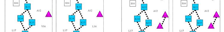
19 Distribution on the Polar Face of Amphipathic α-helical Antimicrobial Peptides on their Biological and Biophysical Properties. In: Lebl, M. (ed). In, Breaking Away: Proceedings of the 21st American Peptide Symposium (2009), Bloomington, IN, USA 45. Asthana, N., Yadav, S. P., and Ghosh, J. K. (2004) Dissection of antibacterial and toxic activity of melittin: a leucine zipper motif plays a crucial role in determining its hemolytic activity but not antibacterial activity. J Biol Chem; 279: Conlon, J. M., Ahmed, E., and Condamine, E. (2009) Antimicrobial properties of brevinin-2-related peptide and its analogs: Efficacy against multidrug-resistant Acinetobacter baumannii. Chem Biol Drug Des; 74: Avrahami, D., Oren, Z., and Shai, Y. (2001) Effect of multiple aliphatic amino acids substitutions on the structure, function, and mode of action of diastereomeric membrane active peptides. Biochemistry; 40: FIGURE LEGENDS Fig. 1. Helical net representation and space-filling model of peptide D1 (V13) and D1 (K13). In the helical net (left panel), the one-letter code is used for amino acid residues. The specificity determinant lysine residue at position 13 in the center of the nonpolar face of peptide D1 (K13) is denoted by a pink triangle. The amino acid residues on the polar face are boxed and the positively charged lysine residues are colored blue. The amino acid residues on the nonpolar face are circled and the large hydrophobes are colored yellow (Trp, Phe, Leu, Val and Ile). The i i+3 and i i+4 hydrophobic interactions between large hydrophobes along the helix are shown as black bars. In the space-filling model (right panel), hydrophobic amino acids on the nonpolar face are colored yellow; hydrophilic amino acids on the polar face are colored blue; the peptide backbone is colored white. The specificity determinant lysine residue at position 13 in the center of the nonpolar face of peptide D1 (K13) is colored pink. The models were created with the PyMOL (version 0.99) program. Fig. 2. Helical net representation of peptides D1 (K13), D11, D22 and D14. The one-letter code is used for amino acid residues. D denotes that all residues in the peptides are in the D-conformation. The specificity determinant(s) lysine residue(s) at position 13 only or position 13 and 16 in the center of the nonpolar face are denoted by a pink triangle(s). The amino acid residues on the polar face are boxed and the positively charged lysine residues are colored blue. The potential i i+3 and i i+4 electrostatic repulsions between positively charged residues along the helix are shown as dotted bars. The amino acid residues on the nonpolar face are circled; the large hydrophobes other then leucine residues (Trp, Phe, Val and Ile) are colored green and the leucine residues are colored
20 yellow. The i i+3 and i i+4 hydrophobic interactions between large hydrophobes along the helix are shown as black bars. Fig. 3. Helical net representation of peptides D11, D15, D14 and D16. The one-letter code is used for amino acid residues. D denotes that all residues in the peptides are in the D-conformation. The specificity determinant(s) lysine residues at position 13 only or position 13 and 16 in the center of the nonpolar face are denoted by a pink triangle(s). The amino acid residues on the polar face are boxed and the positively charged lysine residues are colored blue. The potential i i+3 and i i+4 electrostatic repulsions between positively charged residues along the helix are shown as dotted bars. The amino acid residues on the nonpolar face are circled; the large hydrophobes other then leucine residues (Trp, Phe, Val and Ile) are colored green and the leucine residues are colored yellow. The i i+3 and i i+4 hydrophobic interactions between large hydrophobes along the helix are shown as black bars. Fig. 4. Circular dichroism (CD) spectra. Panel A shows the CD spectra of peptides in aqueous benign buffer (100 mm KCl, 50 mm NaH 2 PO 4 /Na 2 HPO 4 at ph 7.0, 5 and panel B shows the spectra in the presence of buffer-trifluoroethanol (TFE) (1:1, v/v). Fig. 5. Peptide self-association ability as monitored by temperature profiling in reversed-phased chromatography (RP-HPLC). In panel A, the retention time of peptides are normalized to 5 through the expression (t R t t R 5 ), where t R t is the retention time at a specific temperature of an antimicrobial peptide or control peptide C, and t R 5 is the retention time at 5. In panel B, the retention behavior of the peptides was normalized to that of control peptide C through the expression (t R t t R 5 for peptides) (t R t -t R 5 for control peptide C). The maximum change in retention time from the control peptide C defines the peptide association parameter, denoted P A (Table 2). The sequences of the peptides and the random coil control peptide are shown in Table 1. Fig. 6. The hemolytic activity of peptide D1 and analogs. The concentration-response curves of peptides for percentage lysis of human red blood cells (hrbc) are shown. The peptide concentration is in microgram/ml. Peptide D16 caused only 10.7% lysis after 18 hours at 1000 μg/ml. Fig. 7. Helical net representations of peptides D1 (V13), D1 (K13) and D16. In the helical nets (left panels), the one-letter code is used for amino acid residues. The specificity determinant(s)
21 lysine residues at position 13 only or position 13 and 16 in the center of the nonpolar face are denoted by a pink triangle(s). The amino acid residues on the polar face are boxed and the positively charged lysine residues are colored blue. The potential i i+3 and i i+4 electrostatic repulsions between positively charged residues along the helix are shown as dotted bars. The amino acid residues on the nonpolar face are circled; the large hydrophobes other then leucine residues (Trp, Phe, Val and Ile) are colored green and the leucine residues are colored yellow; the alanine residues (A12, A20 and A23 in D1; A12 and A23 in D16) are colored orange. The i i+3 and i i+4 hydrophobic interactions between large hydrophobes along the helix are shown as black bars.
22 Table 1: Peptides used in this study Peptide Sequence b Substitution a Name D1 (V13) Ac-K-W-K-S-F-L-K-T-F-K-S-A-V-K-T-V-L-H-T-A-L-K-A-I-S-S-amide D1 (K13) D-(V13K) Ac-K-W-K-S-F-L-K-T-F-K-S-A-K-K-T-V-L-H-T-A-L-K-A-I-S-S-amide D11 D-(V13K, K10S, S11K, T15K, Ac-K-W-K-S-F-L-K-T-F-S-K-A-K-K-K-V-L-K-T-A-L-K-A-I-S-K-amide H18K, S26K) D22 D-(V13K, V16A, K10S, S11K, Ac-K-W-K-S-F-L-K-T-F-S-K-A-K-K-K-A-L-K-T-L-L-K-A-I-S-K-amide T15K, H18K, A20L, S26K) D14 D-(V13K, V16K, K10S, S11K, Ac-K-W-K-S-F-L-K-T-F-S-K-A-K-K-K-K-L-K-T-L-L-K-A-I-S-K-amide T15K, H18K, A20L, S26K) D15 D-(V13K, V16L, W2L, F5L, Ac-K-L-K-S-L-L-K-T-L-S-K-A-K-K-K-L-L-K-T-A-L-K-A-L-S-K-amide F9L, K10S, S11K, T15K, H18K, I24L, S26K) D16 D-(V13K, V16K, W2L, F5L, Ac-K-L-K-S-L-L-K-T-L-S-K-A-K-K-K-K-L-K-T-L-L-K-A-L-S-K-amide F9L, K10S, S11K, T15K, H18K, A20L, I24L, S26K) Control C Ac-E-L-E-K-G-G-L-E-G-E-K-G-G-K-E-L-E-K-amide a. The D- denotes that all amino acid residues in each peptide are in the D conformation, except for the control peptide, which is in the all L-conformation. b. Peptide sequences are shown using the one-letter code for amino acid residues; Ac- denotes N α -acetyl and -amide denotes C α - amide. The "specificity determinant(s)", Lys residues incorporated in the center of the nonpolar face are bolded (position 13 or positions 13 and 16).
23 Table 2: Biophysical data of D1 analogs Peptide Net Hydrophobicity Benign 50% TFE e Amphi- P Name charge a t R (min) Δt R (X-D1(V13)) b c (min) [θ] 222 %Helix d c [θ] 222 %Helix d A pathicity f D1(V13) , , D1(K13) , , D , D , , D , , D , , D , , a. t R denotes retention time in RP-HPLC at ph 2 and room temperature, and is a measure of overall peptide hydrophobicity. b. Δt R (X-D1(V13)) is the difference in retention time between the peptide analogs and peptide D1(V13), as a measure of the change in hydrophobicity. c. The mean residue molar ellipticities [θ] 222 (deg cm 2 /dmol) at wavelength 222 nm were measured at 5 o C in benign conditions (100 mm KCl, 50 mm NaH 2 PO 4 /Na 2 HPO 4, ph 7.0) or in benign buffer containing 50% trifluoroethanol (TFE) by circular dichroism spectroscopy. d. The helical content (as a percentage) of a peptide relative to the molar ellipticity value of peptide D15 in the presence of 50% TFE. e. P A denotes oligomerization/dimerization parameter of each peptide during RP-HPLC temperature profiling, which is the maximal retention time difference of (t t R -t 5 R for peptide analogs)-(t t R -t 5 R for control peptide C) within the temperature range; t t R -t 5 R is the retention time difference of a peptide at a specific temperature (t t R ) compared with that at 5 o C (t 5 R ). The sequence of control peptide C is shown in Table 1. f. Amphipathicity was determined by calculation of hydrophobic moment (17) using hydrophobicity coefficients determined by reversed-phase chromatography (19, 20) see methods for details.
24 Table 3: Antimicrobial activity of D1 analogs against Acinetobacter baumannii (A) and Pseudomonas aeruginosa strains (B) compared to peptide D1(V13) A Antimicrobial activity against Acinetobacter baumannii B Peptide MIC(μΜ) a c Name ATCC ATCC Fold GMb D1(V13) D1(K13) D D D D D Peptide Name Antimicrobial activity against Pseudomonas aeruginosa MIC(μΜ) a Fold c PAO1 PA14 PAK M2 WR5 CP204 GM b D1 (V13) D1 (K13) D D D D D a. MIC is minimal inhibitory concentration that inhibited growth of different strains in Mueller-Hinton (MH) medium at 37 o C after 24h. MIC is given based on three sets of determinations. b. GM, geometric mean of the MIC values. c. The fold improvement in antimicrobial activity (geometric mean data) compared to that of D1(V13).
25 Table 4: Summary of biological activity of D1(V13) analogs Hemolytic Antimicrobial activity Peptide activity Acinetobacter baumannii Pseudomonas aeruginosa a c c Name HC 50 Fold b MIC GM Therapeutic (μm) (μm) Index d Fold e MIC GM Therapeutic (μm) index d Fold e D1(V13) D1(K13) D D D D D f a. HC 50 is the concentration of peptide that results in 50% hemolysis after 18 hours at 37 o C. The hemolytic activities that are better than the lead peptide D1(V13) are bolded. b. The fold improvement in HC 50 compared to that of D1(V13). c. MIC is the minimum inhibitory concentration of peptide that inhibits growth of bacteria after 24 hours at 37 o C. MIC GM is the geometric mean of the MIC values from 11 different isolates of A. baumannii or 6 different isolates of P. aeruginosa. d. Therapeutic index is the ratio of the HC 50 value (μm) over the geometric mean MIC value (μm). Large values indicate greater antimicrobial specificity. The therapeutic indices with values 100 for A. baumannii and P. aeruginosa are bolded. e. The fold improvement in therapeutic index compared to that of D1(V13). f. The percent lysis for peptide D16 was only 10.7% after 18 hours. We estimated the HC 50 value based on linear extrapolation of the slope of the line between 500 and 1000 μg/ml (Fig. 6). The HC 50 could be much larger.
26
27
28
29
30
31


32
Biofilm Protocol Optimization For Pseudomonas aeruginosa. Introduction. Materials and Methods. Culture Media, Incubation Time, and Biofilm Measurement
 Biofilm Protocol Optimization For Pseudomonas aeruginosa Culture Media, Incubation Time, and Biofilm Measurement Introduction In addition to the conventional arsenal of antibiotic resistance mechanisms
Case 7 A Storage Protein From Seeds of Brassica nigra is a Serine Protease Inhibitor Last modified 29 September 2005
 Case 7 A Storage Protein From Seeds of Brassica nigra is a Serine Protease Inhibitor Last modified 9 September 005 Focus concept Purification of a novel seed storage protein allows sequence analysis and
Bivalirudin Purification:
 Bivalirudin Purification: Sorbent Screening and Overload Experiments Marc Jacob, Joshua Heng, and Tivadar Farkas Phenomenex, Inc., 411 Madrid Ave., Torrance, CA 90501 USA PO94190412_W Abstract In this
Zwitterion Chromatography ZIC
 Zwitterion Chromatography ZIC A novel technique, with unique selectivity, suitable for preparative scale separations? PhD Einar Pontén What is Zwitterion Chromatography? Our definition: Liquid chromatography
Purification: Step 1. Lecture 11 Protein and Peptide Chemistry. Cells: Break them open! Crude Extract
 Purification: Step 1 Lecture 11 Protein and Peptide Chemistry Cells: Break them open! Crude Extract Total contents of cell Margaret A. Daugherty Fall 2003 Big Problem: Crude extract is not the natural
Purification: Step 1. Protein and Peptide Chemistry. Lecture 11. Big Problem: Crude extract is not the natural environment. Cells: Break them open!
 Lecture 11 Protein and Peptide Chemistry Margaret A. Daugherty Fall 2003 Purification: Step 1 Cells: Break them open! Crude Extract Total contents of cell Big Problem: Crude extract is not the natural
11 questions for a total of 120 points
 Your Name: BYS 201, Final Exam, May 3, 2010 11 questions for a total of 120 points 1. 25 points Take a close look at these tables of amino acids. Some of them are hydrophilic, some hydrophobic, some positive
The study of protein secondary structure and stability at equilibrium ABSTRACT
 The study of protein secondary structure and stability at equilibrium Michelle Planicka Dept. of Physics, North Georgia College and State University, Dahlonega, GA REU, Dept. of Physics, University of
Zool 3200: Cell Biology Exam 3 3/6/15
 Name: Trask Zool 3200: Cell Biology Exam 3 3/6/15 Answer each of the following questions in the space provided; circle the correct answer or answers for each multiple choice question and circle either
Examining the components of your peptide sample with AccuPep QC. Lauren Lu, Ph.D. October 29, 2015, 9:00-10:00 AM EST
 Examining the components of your peptide sample with AccuPep QC Lauren Lu, Ph.D. October 29, 2015, 9:00-10:00 AM EST When do I need custom peptides? Custom peptides play an important role in many research
Introduction to Protein Purification
 Introduction to Protein Purification 1 Day 1) Introduction to Protein Purification. Input for Purification Protocol Development - Guidelines for Protein Purification Day 2) Sample Preparation before Chromatography
6/28/2016. Control of Microbial Growth. Method. Terminology. Disinfectants and Antiseptics
 Control of Microbial Growth Disinfectants and Antiseptics 1 Method Three approaches for the control of microbial growth Chemical Disinfectants and antiseptics Physical Heat Ultraviolet Irradiations Mechanical
CFSSP: Chou and Fasman Secondary Structure Prediction server
 Wide Spectrum, Vol. 1, No. 9, (2013) pp 15-19 CFSSP: Chou and Fasman Secondary Structure Prediction server T. Ashok Kumar Department of Bioinformatics, Noorul Islam College of Arts and Science, Kumaracoil
ProSEC 300S. Protein Characterization columns
 ProSEC 300S Protein Characterization columns Agilent s ProSEC 300S is a silica-based material specifically designed for the analysis of proteins by aqueous size exclusion chromatography. With a proprietary
Fundamentals of Protein Structure
 Outline Fundamentals of Protein Structure Yu (Julie) Chen and Thomas Funkhouser Princeton University CS597A, Fall 2005 Protein structure Primary Secondary Tertiary Quaternary Forces and factors Levels
Ezy MIC Strip FEATURES AND ADVANTAGES
 Imipenem with & without EDTA Ezy MIC Strips (IPM+EDTA/IPM) (Imipenem + EDTA: 1-64 mcg/ml) (Imipenem : 4-256 mcg/ml) Antimicrobial Susceptibility Testing For In Vitro Diagnostic use EM078 Not for Medicinal
Structure formation and association of biomolecules. Prof. Dr. Martin Zacharias Lehrstuhl für Molekulardynamik (T38) Technische Universität München
 Technische Universität München") Structure formation and association of biomolecules Prof. Dr. Martin Zacharias Lehrstuhl für Molekulardynamik (T38) Technische Universität München Motivation Many biomolecules are chemically synthesized
Structure formation and association of biomolecules Prof. Dr. Martin Zacharias Lehrstuhl für Molekulardynamik (T38) Technische Universität München Motivation Many biomolecules are chemically synthesized
Peptide libraries: applications, design options and considerations. Laura Geuss, PhD May 5, 2015, 2:00-3:00 pm EST
 Peptide libraries: applications, design options and considerations Laura Geuss, PhD May 5, 2015, 2:00-3:00 pm EST Overview 1 2 3 4 5 Introduction Peptide library basics Peptide library design considerations
Promix TM. Enter a New Era in Biomolecule Analysis with. Columns. Unsurpassed Selectivity and Peak Capacity for Peptides and Proteins
 Promix TM Enter a New Era in Biomolecule Analysis with Promix TM Columns Unsurpassed electivity and Peak Capacity for Peptides and Proteins Applications: Proteomics Peptide/Protein Analysis Peptide/Protein
Enabling Systems Biology Driven Proteome Wide Quantitation of Mycobacterium Tuberculosis
 Enabling Systems Biology Driven Proteome Wide Quantitation of Mycobacterium Tuberculosis SWATH Acquisition on the TripleTOF 5600+ System Samuel L. Bader, Robert L. Moritz Institute of Systems Biology,
Analysis and Purification of Polypeptides by Reversed-Phase HPLC
 Analysis and Purification of Polypeptides by Reversed-Phase HPLC Reversed-phase HPLC is a valuable tool for the analysis and purification of proteins and peptides. It is effective in separating peptide
Analysis of biomolecules by SEC and Ion-Exchange UPLC
 Analysis of biomolecules by SEC and Ion-Exchange UPLC Anders Feldthus, Waters Nordic 2011 Waters Corporation 1 Waters Commitment To develop, commercialize and market columns that when used on Waters ACQUITY
MOLEBIO LAB #3: Electrophoretic Separation of Proteins
 MOLEBIO LAB #3: Electrophoretic Separation of Proteins Introduction: Proteins occupy a central position in the structure and function of all living organisms. Some proteins serve as structural components
Biology (Microbiology): Exam #3
: Exam #3") NAME: PLEDGE: Biology 50-384 (Microbiology): Exam #3 1. You have isolated a series of mutants that have altered patterns of ß-galactosidase and lactose permease activity (i.e. they are different that the
NAME: PLEDGE: Biology 50-384 (Microbiology): Exam #3 1. You have isolated a series of mutants that have altered patterns of ß-galactosidase and lactose permease activity (i.e. they are different that the
UV Fluorescence Polarization as a Means to Investigate Protein Conformational and Mass Change
 A p p l i c a t i o n N o t e UV Fluorescence Polarization as a Means to Investigate Protein Conformational and Mass Change Using Intrinsic Tryptophan Fluorescence in Conjunction with UV-capable Polarizers
Technical Overview. Author. Abstract. Edgar Naegele Agilent Technologies, Inc. Waldbronn, Germany
 New Features of the Agilent Method Scouting Wizard for Automated Method Development of Complex Samples Analysis of Large Data Sets by Method Scouting Reports and Automated Adjustment of Flow Rates and
Types of chromatography
 Chromatography Physical separation method based on the differential migration of analytes in a mobile phase as they move along a stationary phase. Mechanisms of Separation: Partitioning Adsorption Exclusion
Section A: Prokaryotes Types and Structure 1. What is microbiology?
 Section A: Prokaryotes Types and Structure 1. What is microbiology? 2. Compare and contrast characteristics of each bacterial type: Eubacteria and Archaebacteria. Eubacteria Both Archaebacteria 3. Label
RAM Direct Injection. (Restricted Access Media) A Tool for the Separation of Small Molecules in the Presence of Large Biomolecules
 A Tool for the Separation of Small Molecules in the Presence of Large Biomolecules") RAM Direct Injection (Restricted Access Media) A Tool for the Separation of Small Molecules in the Presence of Large Biomolecules HPLC analysis of small molecules contained within a protein matrix can
RAM Direct Injection (Restricted Access Media) A Tool for the Separation of Small Molecules in the Presence of Large Biomolecules HPLC analysis of small molecules contained within a protein matrix can
1. DNA replication. (a) Why is DNA replication an essential process?
 Why is DNA replication an essential process?") ame Section 7.014 Problem Set 3 Please print out this problem set and record your answers on the printed copy. Answers to this problem set are to be turned in to the box outside 68120 by 5:00pm on Friday
ame Section 7.014 Problem Set 3 Please print out this problem set and record your answers on the printed copy. Answers to this problem set are to be turned in to the box outside 68120 by 5:00pm on Friday
PolyCAT A For Cation-Exchange
 PolyCAT A For Cation-Exchange The PolyCAT A is made though a unique process for attaching Poly(aspartic acid) covalently to silica. Proteins elute from this polypeptide coating in sharp peaks with little
ARBRE-P4EU Consensus Protein Quality Guidelines for Biophysical and Biochemical Studies Minimal information to provide
 ARBRE-P4EU Consensus Protein Quality Guidelines for Biophysical and Biochemical Studies Minimal information to provide Protein name and full primary structure, by providing a NCBI (or UniProt) accession
PLRP-S Polymeric Reversed-Phase Column for LC/MS Separation of mabs and ADC
 PLRP-S Polymeric Reversed-Phase Column for LC/MS Separation of mabs and ADC Analysis of Intact and Fragmented mabs and ADC Application Note Biotherapeutics and Biologics Author Suresh Babu C.V. Agilent
Proteins Higher Order Structures
 Proteins Higher Order Structures Dr. Mohammad Alsenaidy Department of Pharmaceutics College of Pharmacy King Saud University Office: AA 101 msenaidy@ksu.edu.sa Previously on PHT 426!! Protein Structures
7.014 Quiz II 3/18/05. Write your name on this page and your initials on all the other pages in the space provided.
 7.014 Quiz II 3/18/05 Your Name: TA's Name: Write your name on this page and your initials on all the other pages in the space provided. This exam has 10 pages including this coversheet. heck that you
Improving Resolution and Column Loading Systematically in Preparative Liquid Chromatography for Isolating a Minor Component from Peppermint Extract
 Improving Resolution and Column Loading Systematically in Preparative Liquid Chromatography for Isolating a Minor Component from Peppermint Extract Jo-Ann M. Jablonski and Rui Chen Waters Corporation,
Introduction to Proteins
 Introduction to Proteins Lecture 4 Module I: Molecular Structure & Metabolism Molecular Cell Biology Core Course (GSND5200) Matthew Neiditch - Room E450U ICPH matthew.neiditch@umdnj.edu What is a protein?
Method Development Considerations for Reversed-Phase Protein Separations
 Method Development Considerations for Reversed-Phase Protein Separations Hillary B. Hewitson, Thomas E. Wheat, Paula Hong, Kenneth J. Fountain APPLICATION BENEFITS n The BEH00 C 4 chemistry is available
Key Concept Translation converts an mrna message into a polypeptide, or protein.
 8.5 Translation VOBLRY translation codon stop codon start codon anticodon Key oncept Translation converts an mrn message into a polypeptide, or protein. MIN IDES mino acids are coded by mrn base sequences.
R R Innovation Way P/N SECKIT-7830 Newark, DE 19711, USA Tel: Fax: Website: Published in November 2013
 5-100 Innovation Way Newark, DE 19711, USA Tel:302-3661101 Fax:302-3661151 Website: www.sepax-tech.com Published in November 2013 P/N SECKIT-7830 These Phases are developed based on innovative surface
Chapter 8 DNA Recognition in Prokaryotes by Helix-Turn-Helix Motifs
 Chapter 8 DNA Recognition in Prokaryotes by Helix-Turn-Helix Motifs 1. Helix-turn-helix proteins 2. Zinc finger proteins 3. Leucine zipper proteins 4. Beta-scaffold factors 5. Others λ-repressor AND CRO
Chem 465 Biochemistry II
 Chem 465 Biochemistry II Name: 2 points Multiple choice (4 points apiece): 1. Which of the following is not true of trna molecules? A) The 3'-terminal sequence is -CCA. B) Their anticodons are complementary
36. The double bonds in naturally-occuring fatty acids are usually isomers. A. cis B. trans C. both cis and trans D. D- E. L-
 36. The double bonds in naturally-occuring fatty acids are usually isomers. A. cis B. trans C. both cis and trans D. D- E. L- 37. The essential fatty acids are A. palmitic acid B. linoleic acid C. linolenic
MATH 5610, Computational Biology
 MATH 5610, Computational Biology Lecture 2 Intro to Molecular Biology (cont) Stephen Billups University of Colorado at Denver MATH 5610, Computational Biology p.1/24 Announcements Error on syllabus Class
mab Titer Analysis with the Agilent Bio-Monolith Protein A Column
 mab Titer Analysis with the Agilent Bio-Monolith Protein A Column Application Note Biopharmaceuticals and Biosimilars Authors Emmie Dumont, Isabel Vandenheede, Pat Sandra, and Koen Sandra Research Institute
Lifetime stability of size exclusion chromatography columns for protein aggregate analysis
 APPLICATION NOTE 72362 Lifetime stability of size exclusion chromatography columns for protein aggregate analysis Authors Amy Farrell 1, Craig Jakes 1, Alexander Ley 2, Mauro De Pra 2, Frank Steiner 2,
Solutions to Quiz II
 MIT Department of Biology 7.014 Introductory Biology, Spring 2005 Solutions to 7.014 Quiz II Class Average = 79 Median = 82 Grade Range % A 90-100 27 B 75-89 37 C 59 74 25 D 41 58 7 F 0 40 2 Question 1
Investigation of Solid Phase Microextraction as an Alternative to Dried Blood Spot
 Investigation of Solid Phase Microextraction as an Alternative to Dried Blood Spot Craig Aurand and Robert Shirey Supelco, Div. of Sigma-Aldrich Bellefonte, PA 16823 USA T414068 sigma-aldrich.com/analytical
Microbial Biotechnology agustin krisna wardani
 Microbial Biotechnology agustin krisna wardani 1. The Structure of Microbes Microbes (microorganisms) are tiny organisms that are too small to be seen individually by the naked eye and must be viewed with
Disulfide Linkage Analysis of IgG1 using an Agilent 1260 Infinity Bio inert LC System with an Agilent ZORBAX RRHD Diphenyl sub 2 µm Column
 Disulfide Linkage Analysis of IgG1 using an Agilent 126 Infinity Bio inert LC System with an Agilent ZORBAX RRHD Diphenyl sub 2 µm Column Application Note Biotherapeutics & Biosimilars Author M. Sundaram
Folding simulation: self-organization of 4-helix bundle protein. yellow = helical turns
 Folding simulation: self-organization of 4-helix bundle protein yellow = helical turns Protein structure Protein: heteropolymer chain made of amino acid residues R + H 3 N - C - COO - H φ ψ Chain of amino
Protein Synthesis. Application Based Questions
 Protein Synthesis Application Based Questions MRNA Triplet Codons Note: Logic behind the single letter abbreviations can be found at: http://www.biology.arizona.edu/biochemistry/problem_sets/aa/dayhoff.html
Daily Agenda. Warm Up: Review. Translation Notes Protein Synthesis Practice. Redos
 Daily Agenda Warm Up: Review Translation Notes Protein Synthesis Practice Redos 1. What is DNA Replication? 2. Where does DNA Replication take place? 3. Replicate this strand of DNA into complimentary
Virtual bond representation
 Today s subjects: Virtual bond representation Coordination number Contact maps Sidechain packing: is it an instrumental way of selecting and consolidating a fold? ASA of proteins Interatomic distances
7.88J Protein Folding Problem Fall 2007
 MIT OpenCourseWare http://ocw.mit.edu 7.88J Protein Folding Problem Fall 2007 For information about citing these materials or our Terms of Use, visit: http://ocw.mit.edu/terms. 7.88 Lecture Notes - 8 7.24/7.88J/5.48J
PROCEDURE FOR USE NICKEL NTA Magnetic Agarose Beads (5%)
") 1 AFFINITY HIS-TAG PURIFICATION PROCEDURE FOR USE NICKEL NTA Magnetic Agarose Beads (5%) DESCRIPTION Nickel NTA Magnetic Agarose Beads are products that allow rapid and easy small-scale purification of
1 AFFINITY HIS-TAG PURIFICATION PROCEDURE FOR USE NICKEL NTA Magnetic Agarose Beads (5%) DESCRIPTION Nickel NTA Magnetic Agarose Beads are products that allow rapid and easy small-scale purification of
number Done by Corrected by Doctor Hamed Al Zoubi
 number 3 Done by Neda a Baniata Corrected by Waseem Abu Obeida Doctor Hamed Al Zoubi Note: it is important to refer to slides. Bacterial genetics *The main concepts we will talk about in this lecture:
3. Close the bottom end of the column and apply the packing device on top. Pump water through the upper adaptor to remove air.
 INSTRUCTIONS FOR USE WorkBeads Protein A Product name Pack size Article number WorkBeads Protein A Bulk Media 1.5 ml 40605001 Bulk Media 5 ml 40605002 Bulk Media 10 ml 40605003 Bulk Media 100 ml 40605004
nanodsf 2bind: Your service provider for biophysical characterization of proteins Precisely revealing protein folding and stability
 nanodsf Precisely revealing protein folding and stability 2bind: Your service provider for biophysical characterization of proteins This booklet was written and designed by 2bind 08 2015 Any reproduction
Algorithm for Matching Additional Spectra
 Improved Methods for Comprehensive Sample Analysis Using Protein Prospector Peter R. Baker 1, Katalin F. Medzihradszky 1 and Alma L. Burlingame 1 1 Mass Spectrometry Facility, Dept. of Pharmaceutical Chemistry,
How antimicrobial agents work
 Physical and Chemical Control of Microbes Physical Agents heat or radiation Chemical Agents disinfectants or antiseptics Important Terms 1. Sterilization process of killing all viable microbes 2. Bactericide
Phage therapy. Institute of Molecular Biomedicine Comenius University, Faculty of Medicine
 Phage therapy Institute of Molecular Biomedicine Comenius University, Faculty of Medicine www.imbm.sk tothova.lubomira@gmail.com Outline Introduction Taxonomy Cycles of phages Bacterial resistance Phage
Hmwk # 8 : DNA-Binding Proteins : Part II
 The purpose of this exercise is : Hmwk # 8 : DNA-Binding Proteins : Part II 1). to examine the case of a tandem head-to-tail homodimer binding to DNA 2). to view a Zn finger motif 3). to consider the case
Purification of mfp. from an Overnight Culture. Laboratory 17
 Purification of mfp from an Overnight Culture When scientists at a therapeutics company, like Amgen, have successfully identified a promising therapeutic protein, two objectives would be to locate and
NPTEL VIDEO COURSE PROTEOMICS PROF. SANJEEVA SRIVASTAVA
 LECTURE-06 PROTEIN PURIFICATION AND PEPTIDE ISOLATION USING CHROMATOGRAPHY TRANSCRIPT Welcome to the proteomics course. Today, we will talk about protein purification and peptide isolation using chromatography
CHAPTER 1. DNA: The Hereditary Molecule SECTION D. What Does DNA Do? Chapter 1 Modern Genetics for All Students S 33
 HPER 1 DN: he Hereditary Molecule SEION D What Does DN Do? hapter 1 Modern enetics for ll Students S 33 D.1 DN odes For Proteins PROEINS DO HE nitty-gritty jobs of every living cell. Proteins are the molecules
Bi 8 Lecture 7. Ellen Rothenberg 26 January Reading: Ch. 3, pp ; panel 3-1
 Bi 8 Lecture 7 PROTEIN STRUCTURE, Functional analysis, and evolution Ellen Rothenberg 26 January 2016 Reading: Ch. 3, pp. 109-134; panel 3-1 (end with free amine) aromatic, hydrophobic small, hydrophilic
Fraction Analysis of Cysteine Linked Antibody-Drug Conjugates Using Hydrophobic Interaction. chromatography. Agilent 1260 Infinity II Bio-Inert System
 Application Note Biologics & Biosimilars Fraction Analysis of Cysteine Linked Antibody-Drug Conjugates Using Hydrophobic Interaction Chromatography Agilent 126 Infinity II Bio-Inert System 7 6 5 4 5. 7.5
Microbial Growth and The Control of Microbial Growth (Chapter 6 & 7)
") Microbial Growth and The Control of Microbial Growth (Chapter 6 & 7) Lecture Materials for Amy Warenda Czura, Ph.D. Suffolk County Community College Eastern Campus Primary Source for figures and content:
Microbial Growth and The Control of Microbial Growth (Chapter 6 & 7) Lecture Materials for Amy Warenda Czura, Ph.D. Suffolk County Community College Eastern Campus Primary Source for figures and content:
Green Fluorescent Protein. Avinash Bayya Varun Maturi Nikhileswar Reddy Mukkamala Aravindh Subhramani
 Green Fluorescent Protein Avinash Bayya Varun Maturi Nikhileswar Reddy Mukkamala Aravindh Subhramani Introduction The Green fluorescent protein (GFP) was first isolated from the Jellyfish Aequorea victoria,
Improved Peptide Maps Using Core-Shell Media: Explaining Better Biopharmaceutical Applications With Kinetex C18 Columns
 Improved Peptide Maps Using Core-Shell Media: Explaining Better Biopharmaceutical Applications With Kinetex C18 Columns Michael McGinley, Jeff Layne, and Vita Knudson Phenomenex, Inc., 411 Madrid Ave.,Torrance,
Fluorous Affinity Purification of Oligonucleotides
 Fluorous Affinity Purification of Oligonucleotides A higher affinity alternative to RP cartridge purification. One-pass loading without ammonia removal. High recoveries (typically 70-100%). High selectivity
7.013 Problem Set 3 FRIDAY October 8th, 2004
 MIT Biology Department 7.012: Introductory Biology - Fall 2004 Instructors: Professor Eric Lander, Professor Robert. Weinberg, Dr. laudette ardel Name: T: 7.013 Problem Set 3 FRIDY October 8th, 2004 Problem
Thr Gly Tyr. Gly Lys Asn
 Your unique body characteristics (traits), such as hair color or blood type, are determined by the proteins your body produces. Proteins are the building blocks of life - in fact, about 45% of the human
From DNA to Protein Structure and Function
 STO-106 From DNA to Protein Structure and Function Teacher information Summary: Students model how information in the DNA base sequences is transcribed and translated to produce a protein molecule. They
Comparative Simulation Studies of Native and Single-Site Mutant Human Beta-Defensin-1 Peptides
 Comparative Simulation Studies of Native and Single-Site Mutant Human Beta-Defensin-1 Peptides Rabab A. Toubar, Artem Zhmurov, Valeri Barsegov, * and Kenneth A. Marx * Department of Chemistry, University
RNA Part I: Chemical Structure of RNA
 RA Part I: Chemical Structure of RA Structural differences between RA and DA Resistance of phosphate esters to basic hydrolysis The 2 - group of RA facilitates chemical cleavage in aqueous a by forming
The mechanism(s) of protein folding. What is meant by mechanism. Experimental approaches
 of protein folding. What is meant by mechanism. Experimental approaches") The mechanism(s) of protein folding What is meant by mechanism Computational approaches Experimental approaches Questions: What events occur and in what time sequence when a protein folds Is there a specified
The mechanism(s) of protein folding What is meant by mechanism Computational approaches Experimental approaches Questions: What events occur and in what time sequence when a protein folds Is there a specified
Adsorptive and Mechanical Mechanisms of Fluid Purification Using Charge Modified Depth Filtration Media. Dr. Robert Conway, Ph.D.
 Adsorptive and Mechanical Mechanisms of Fluid Purification Using Charge Modified Depth Filtration Media Dr. Robert Conway, Ph.D. Depth Filter Cartridge Construction Zeta Plus Media Materials of Construction
ZORBAX StableBond HPLC Columns
 ZORBAX HPLC Columns Longest column lifetime and best reproducibility for low ph separations down to ph 1 Patented stable column chemistry allows use at high temperature and low ph without degradation Six
SUMMARY OF DOCTORAL DISSARTATION
 Monika Łęgowska Katedra Biochemii Nr indeksu: 2541 SUMMARY OF DOCTORAL DISSARTATION In search of substrates and inhibitors of human cathepsin C. Design, chemical synthesis and biological studies Cathepsin
mabs and ADCs analysis by RP
 mabs and ADCs analysis by RP Shanhua Lin, Ph.D. The world leader in serving science Protein and mab Separation by HPLC Size difference? YES Size Exclusion Chromatography (SEC) MAbPac SEC-1 NO NO Charge
.CONCLUSIONS. There is currently no
 .CONCLUSIONS There is currently no - Ideal (alternative) antimicrobial preservative (AP) for vaccines - AP gold standard for vaccines - R&D pipeline for vaccine AP Lack of selective affinity tough AET
DNA Structure and Properties Basic Properties Predicting Melting Temperature. Dinesh Yadav
 DNA Structure and Properties Basic Properties Predicting Melting Temperature Dinesh Yadav Nucleic Acid Structure Question: Is this RNA or DNA? Molecules of Life, pp. 15 2 Nucleic Acid Bases Molecules of
Peptide Mapping: A Quality by Design (QbD) Approach
 Approach") Peptide Mapping: A Quality by Design (QbD) Approach Application Note Bio-Pharmaceutical Authors Sreelakshmy Menon and Suresh babu C.V. Agilent Technologies, Inc. Richard Verseput S-Matrix Corporation Abstract
Peptide Mapping: A Quality by Design (QbD) Approach Application Note Bio-Pharmaceutical Authors Sreelakshmy Menon and Suresh babu C.V. Agilent Technologies, Inc. Richard Verseput S-Matrix Corporation Abstract
HIC as a Complementary, Confirmatory Tool to SEC for the Analysis of mab Aggregates
 HIC as a Complementary, Confirmatory Tool to SEC for the Analysis of mab Aggregates Julia Baek, Shanhua Lin, Xiaodong Liu, Thermo Fisher Scientific, Sunnyvale, CA Application Note 2126 Key Words Size exclusion
Structural bioinformatics
 Structural bioinformatics Why structures? The representation of the molecules in 3D is more informative New properties of the molecules are revealed, which can not be detected by sequences Eran Eyal Plant
1. Bloomsbury BBSRC Centre for Structural Biology, Birkbeck College and University College London.
 Purification/Polishing of His-tagged proteins - Application of Centrifugal Vivapure Ion-exchange Membrane Devices to the Purification/Polishing of Histagged Background Multi-milligram quantities of highly
Supplemental Information. OprG Harnesses the Dynamics of its Extracellular. Loops to Transport Small Amino Acids across
 Structure, Volume 23 Supplemental Information OprG Harnesses the Dynamics of its Extracellular Loops to Transport Small Amino Acids across the Outer Membrane of Pseudomonas aeruginosa Iga Kucharska, Patrick
AFFINITY HIS-TAG PURIFICATION
 DESCRIPTION Nickel NTA Agarose Cartridges 5ml are used for purification of histidine-tagged proteins in native or denaturing conditions. This cartridge can be used with an automated chromatography system,
Problem Set Unit The base ratios in the DNA and RNA for an onion (Allium cepa) are given below.
 are given below.") Problem Set Unit 3 Name 1. Which molecule is found in both DNA and RNA? A. Ribose B. Uracil C. Phosphate D. Amino acid 2. Which molecules form the nucleotide marked in the diagram? A. phosphate, deoxyribose
Problem Set Unit 3 Name 1. Which molecule is found in both DNA and RNA? A. Ribose B. Uracil C. Phosphate D. Amino acid 2. Which molecules form the nucleotide marked in the diagram? A. phosphate, deoxyribose
M I C R O B I O L O G Y WITH DISEASES BY TAXONOMY, THIRD EDITION
 M I C R O B I O L O G Y WITH DISEASES BY TAXONOMY, THIRD EDITION Chapter 7 Microbial Genetics Lecture prepared by Mindy Miller-Kittrell, University of Tennessee, Knoxville The Structure and Replication
Rapid Peptide Catabolite ID using the SCIEX Routine Biotransform Solution
 Rapid Peptide Catabolite ID using the SCIEX Routine Biotransform Solution Rapidly Identify Major Catabolites with the SCIEX X500R QTOF System and MetabolitePilot TM 2.0 Software Ian Moore and Jinal Patel
Effect of Hydrogen Ion Concentration and Buffer Composition on Ligand Binding Characteristics and Polymerization of Cow s Milk Folate Binding Protein
 Bioscience Reports, Vol. 21, No. 6, December 2001 ( 2002) Effect of Hydrogen Ion Concentration and Buffer Composition on Ligand Binding Characteristics and Polymerization of Cow s Milk Folate Binding Protein
The dialkylglycine decarboxylase repressor DgdR. Functional aspects and relation to other LysR proteins
 11/29/2011 1 The dialkylglycine decarboxylase repressor DgdR. Functional aspects and relation to other LysR proteins 1. Dialkylglycine amino acids 2. In vivo and in vitro studies on the DgdR repressor
Dynamic Programming Algorithms
 Dynamic Programming Algorithms Sequence alignments, scores, and significance Lucy Skrabanek ICB, WMC February 7, 212 Sequence alignment Compare two (or more) sequences to: Find regions of conservation
Agilent AdvanceBio SEC Columns for Aggregate Analysis: Instrument Compatibility
 Agilent AdvanceBio SEC Columns for Aggregate Analysis: Instrument Compatibility Technical Overview Introduction Agilent AdvanceBio SEC columns are a new family of size exclusion chromatography (SEC) columns
Analysis of Intact and C-terminal Digested IgG1 on an Agilent Bio MAb 5 µm Column
 Analysis of Intact and C-terminal Digested IgG1 on an Agilent Bio MAb µm Column Application Note BioPharma Authors Xiaomi Xu and Phu T Duong Agilent Technologies, Inc. Abstract Nearly all proteins undergo
The Making of the Fittest: Natural Selection and Adaptation
 INTRODUCTION THE ROCK POCKET MOUSE THE BIOCHEMISTRY AND CELL SIGNALING PATHWAY OF MC1R The rock pocket mouse, Chaetodipus intermedius, is a small, nocturnal animal found in the deserts of the southwestern