Precision in Quantitative Imaging: Trial Development and Quality Assurance
|
|
|
- Edwin Miller
- 5 years ago
- Views:
Transcription
1 Precision in Quantitative Imaging: Trial Development and Quality Assurance Susanna I Lee MD, PhD Thanks to: Mitchell Schnall, Mark Rosen. Dan Sullivan, Patrick Bossuyt


2 Imaging Chain: Patient Data Raw data Image reconstruction Image analysis Image processing Data analysis Data output
3 Clinical Trials: Imaging is an Assay Disease Detection Screening Characterize Disease Diagnosis, eligibility or prognosis Anatomic distribution (e.g. tumor staging) Higher level features (e.g. heterogeneity, vascularity, etc.) Monitor Therapeutic Response Change in features with therapy Anatomic (e.g. RECIST) or functional (e.g. SUV)
4 Schema Diagnostic accuracy REFERENCE STANDARD OF DISEASE STATE PATHOLOGY CONFIRMATORY TEST FOLLOWUP ENROLLED PARTICIPANT INDEX TEST IMAGING EXAM IMAGE-GUIDED PROCEDURE MANAGEMENT STANDARD OF CARE STUDY DEFINED Biomarker REFERENCE STANDARD OF PATIENT OUTCOME OVERALL SURVIVAL (OS) PROGRESSION FREE SURVIVAL (PFS) OR DISEASE FREE SURVIVAL (DFS) TREATMENT RESPONSE
5 Cancer Biomarkers Imaging Tissue (e.g. hormone receptors, cytokeratins) PATIENT OUTCOME 1. Overall survival (OS) 2. Progression free survival (PFS) 3. Clinical response Serum (CA125, PSA, AFP, CA19-9) Genetic Genome Expression (e.g. RNA or protein)


6 What is a good biomarker? Stable technology Available widely Standardized image acquisition Reproducible Range of normal defined Balance state of the art with generalizability Sargent DJ, Rubinstein L, Schwartz L et al. Eur J Cancer 2009; 45: 290.
7 Variability: Test Retest Same patient, day, scanning protocol but separate imaging sessions Pre-treatment Post-treatment Range of test-retest Conclusion Index test variability precludes detecting pre- vs. post-treatment change. Lankester KJ, Taylor NJ, Stirling JJ et al. Br J Cancer :979.
8 Signal Requires Data Quality


9 Precision vs. Bias precise accurate not precise not accurate precise not accurate


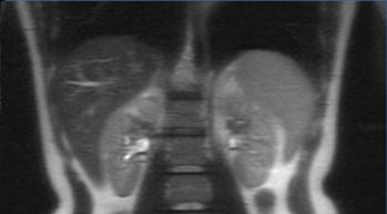
10 Multiple Sclerosis MRI Image acquisition T1 T2 Post-gadolinium T1 Image analysis Number of new or enlarging lesions Number of enhancing lesions

11 MRI endpoint in MS treatment studies 157 publications from 1995 to 2006
12 Imaging Chain: Patient Data Raw data Image reconstruction Image analysis Image processing Data analysis Data output

13 Imaging Manual Hardware and software Scanner calibration Patient preparation Scanning protocol Post-processing Image acquisition manual with a step by step description is part of any prospective study design.
14 What determines resolution? Physics of acquisition (i.e. modality) Sampling (e.g. matrix, detector size) Filtering and other contributions inherent in the reconstruction

15 Point Spread Function Patient Image
16 Image edges approximate anatomy Structure Real Edge Image Edge

17 Resolution and Sampling 160 x 160 matrix 320 x 224 matrix
18 Filtering
19 Filtering: MRI Vendor 1 Vendor 2

20 Filtering: X-ray Vendor 1 Vendor 2

21 Partial Volume Effects Completely in scan plane Partially in scan plane


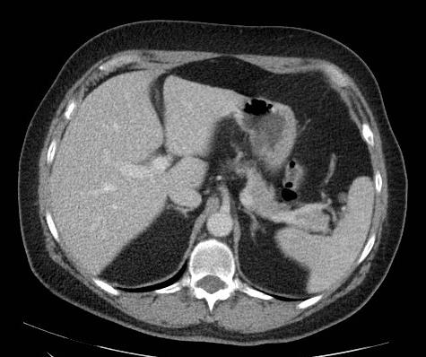
22 Partial Volume Effects: CT Completely in scan plane Partially in scan plane HU = 0 HU = 30-60

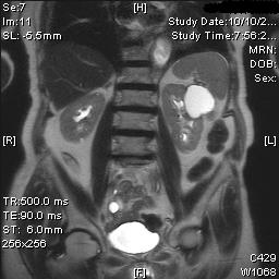
23 Partial Volume Effects: PET lesion lesion Blurred margins Lower intensity

24

25 Quantitative Imaging Biomarkers Alliance (QIBA) Started by RSNA 2007 Mission: Improve the value and practicality of quantitative imaging biomarkers by reducing variability across devices, patients and time Build imaging devices that are also measuring devices Industrialize imaging biomarkers
26 QIBA Approach 1. Identify the sources of error and variability 2. Specify potential solutions in the form of profiles 3. Test these solutions 4. Promulgate profiles to vendors and users Purpose of profiles: Advise vendors what must be implemented in their product Communicate the necessary procedures to users
27 QIBA Profile Activity Diagram Equipment Assessment Subject Preparation Image Acquisition Image Reconstruction Image Analysis Interpretation Manufacturer specification (pre-delivery) Installation specification Maintenance Quality Assurance
28 ACR Core Lab QA Site qualification Instrument performance Training Monitoring of image acquisition Scan header for protocol compliance On-line technologist qualitative review Periodic radiologist review Centralized image analysis Post-processing Reader study

29
 16-18 cm Slice Thickness 64 slices of thickness 2.5 mm Skip Correct Matrix min.")

30 Require Site Protocol Compliance Type Orientation Pulse Sequence T1 weighted GRE Sagittal Dynamic 3D Field Of View (FOV) cm Slice Thickness 64 slices of thickness 2.5 mm Skip Correct Matrix min. 256 x 192 Frequency A/P NEX 2 Phase Wrap NO Fat-Saturation YES Submitted TR Effective TE Scan Duration Flip angle 20 ms 4.5 ms Between 4.5 and 5 minutes <= 45 degrees ACRIN 6657 (I-SPY 1 trial), Nola Hylton, PI
31 Imaging Chain: Patient Data Raw data Image reconstruction Image analysis Image processing Data analysis Data output

32 Image analysis: Turning image into data User extracted features Semi automated Automated Feature 1 Feature 2 Feature 3...

33 Radiomics: Deep Learning Untrained neural network

34 Radiomics: Deep Learning Trained neural network


35 Radiomics: Automated Image Analysis Improve diagnostic accuracy


36 Radiomics: Automated Image Analysis Triage clinical workflow


37 Semi-automated: Manual Segmentation Large image datasets segmented for tumor Quantitative feature extraction Model predictive indices Integrate with genomic & clinical data for machine learning Aerts HJ et al. Nat Commun ;5:4006.

38 Reader Extracted Features Density Fluid, soft tissue, calcified Shape Round, oval, irregular Size Linear, volume Margin Sharp, blurred, spiculated Intensity high/medium/low/minimal Summary assement BIRADS level
39 ROC operating points of 108 radiologists reading same mammograms Skill TP Value judgments FP Beam, Layde, Sullivan Arch Intern Med 1996; 156:

40 Reader Variability: Size progression Increases and decreases of <10% can be a result of inherent variability. Oxnard GR et al. J Clin Oncol. 2011;29:

41 Variability Introduced by ROI Selection Slice 483 Slice 479 Slice 479 SUV=4.0 SUV=5.6 SUV=6.6
 Different ROI protocols yield different ADC values Priola AM et al. Eur Radiol.")
42 Variability Introduced by ROI Selection All ROI protocols show excellent inter-observer agreement (ICC 0.94) Different ROI protocols yield different ADC values Priola AM et al. Eur Radiol Aug 11.


43 Effect of Windowing Soft tissue window Liver window
. Kim H et al. PLoS One.")
44 Effect of Windowing Lung window Mediastinal window Significant measurement differences between window settings (p<0.001). No significant differences in measurement variability between the lung and mediastinal window settings (p>0.05). Kim H et al. PLoS One. 2016;11:e
 Definition of positive vs.")
45 Reader Study Reader blinded to reference standard Multiple readers Independent rather than consensus reads Rules for image interpretation Clinical information available to reader Image selection, windowing, order, etc. Choosing index lesions Selecting region of interest (ROI) Definition of positive vs. negative test Washout period between read sessions of paired imaging exams Digital data forms and screen to document reader study A manual defining reader rules and training cases are part of any prospective study design.


46 Overall trial framework Hypothesis and specific aims Participants Index test Reference standard Data analysis plan (statistics) Conclusions and implications Funding and compliance
47 Index Test (Imaging Exam) STARD 10 - Index test, in sufficient detail to allow replication STARD 12 - Definition of and rationale for test positivity cut-offs or result categories of the index test, distinguishing pre-specified from exploratory STARD 13 - Whether clinical information and reference standard results were available to the performers/readers of the index test STARD 25 - Any adverse events from performing the index test


48 Steps toward precision: Define image acquisition Equipment, patient preparation, protocol Balance state of the art with generalizability Define image analysis Read rules, training and testing Validate the system Test-retest, reader agreement measurements Build in procedures for ongoing QA


49
AAPM Scientific Meeting Imaging Symposium. State of the Art in Quantitative Imaging CT, PET and MRI. Which Imaging Modality is the Most Quantitative
 AAPM Scientific Meeting Imaging Symposium State of the Art in Quantitative Imaging CT, PET and MRI Michael McNitt-Gray, PhD, FAAPM; UCLA Paul Kinahan, PhD, U. Washington Ed Jackson, PhD, FAAPM, UT-MD Anderson
CQIE MRI PROCEDURES. American College of Radiology Clinical Research Center. Centers for Quantitative Imaging Excellence LEARNING MODULE
 Centers for Quantitative Imaging Excellence LEARNING MODULE CQIE MRI PROCEDURES American College of Radiology Clinical Research Center Imaging Core Laboratory v.2 Centers for Quantitative Imaging Excellence
Introduction to Quantitative Imaging as a Biomarker in Clinical Trials
 Quantitative Medical Imaging for Clinical Research and Practice Educational Session ACRIN 2009 Introduction to Quantitative Imaging as a Biomarker in Clinical Trials Katarzyna J. Macura, MD, PhD Johns
CQIE PET PROCEDURES. American College of Radiology Clinical Research Center. Centers for Quantitative Imaging Excellence LEARNING MODULE
 Centers for Quantitative Imaging Excellence LEARNING MODULE CQIE PET PROCEDURES American College of Radiology Clinical Research Center Imaging Core Laboratory v2.1 Centers for Quantitative Imaging Excellence
From Analysis Method to Quantitative Imaging Biomarker
 From Analysis Method to Quantitative Imaging Biomarker Developments in Healthcare Imaging Connecting with Industry 18 th October 2017 Sarah Lee, PhD, SMIEEE Medical Image Analysis Consultant Disclosures
NIST Medical Imaging Informatics Activities. Medical Imaging
 NIST Medical Imaging Informatics Activities Ram Sriram Mary Brady Alden Dima Medical Imaging Biomarker Testing Improving Change Analysis in Lung Cancer Statistically Valid and Clinically Meaningful Biomarkers
Images as Biomarkers potential future advances in the field as viewed by ISPY-2. Nola Hylton, PhD University of California, San Francisco
 Images as Biomarkers potential future advances in the field as viewed by ISPY-2 Nola Hylton, PhD University of California, San Francisco POTENTIAL/PROMISE: Images as Biomarkers Imaging Biomarker: a quantitative
DCE MRI Team Activity. Gudrun Zahlmann, Edward Jackson, Sandeep Gupta
 DCE MRI Team Activity Gudrun Zahlmann, Edward Jackson, Sandeep Gupta QIBA Groundwork DCE-MRI Technical Characteristics and Standards Groundwork ( precursor questions ) Diagnostic Accuracy and Reproducibility
Quantitative Imaging Biomarker DCE - MRI
 Quantitative Imaging Biomarker DCE - MRI DCE MRI: What is it about? DCE MRI: quantitative analysis of dynamic T1 contrast enhanced images Use cases: Clinical trial related UC1: pharmacodynamic investigations
Progress Towards an International Image Quality Monitoring Framework for Quantitative Imaging
 Progress Towards an International Image Quality Monitoring Framework for Quantitative Imaging Ricardo S. Avila rick.avila@accumetra.com October 2, 2017 Quantitative Imaging Workshop XIV Hubble Space Telescope
Facilitating the Use of Imaging Biomarkers in Therapeutic Clinical Trials. Michael Graham, PhD, MD President, SNM Co-chair, Clinical Trials Network
 Facilitating the Use of Imaging Biomarkers in Therapeutic Clinical Trials Michael Graham, PhD, MD President, SNM Co-chair, Clinical Trials Network Facilitating the Use of Imaging Biomarkers in Therapeutic
Jeffrey T. Yap, Ph.D. Dana-Farber Cancer Institute Brigham & Women s Hospital Harvard Medical School
 1 Case Studies of Imaging Biomarkers - Description and requirements for standardized acquisition in multicenter trials: DCE-MRI, Volumetric CT, FDG-PET/CT Jeffrey T. Yap, Ph.D. Dana-Farber Cancer Institute
UPICT template structure as modified by QIBA VolCT Technical Committee June 25, 2009
 UPICT template structure as modified by QIBA VolCT Technical Committee June 25, 2009 The QIBA VolCT Technical Committee modified and used the UPICT template for a VolCT profile on CT Lung Nodule Volume
Accuracy, Precision and Measurement Validation
 Accuracy, Precision and Measurement Validation Ángel Alberich-Bayarri, PhD 1 Biomedical Imaging Research Group GIBI230 La Fe Health Research Institute 2 QUIBIM Quantitative Imaging Biomarkers in Medicine
Evaluation of 1D, 2D and 3D nodule size estimation by radiologists for spherical and non-spherical nodules through CT thoracic phantom imaging
 Evaluation of 1D, 2D and 3D nodule size estimation by radiologists for spherical and non-spherical nodules through CT thoracic phantom imaging Nicholas Petrick * * a, Hyun J. Grace Kim b, David Clunie
QIBA Working Session
 QIBA Working Session Wednesday, November 30, 2011 November 30, 2011 QIBA Session Plenary Session Agenda 3:00 PM Year in Review: Status of the Profiles Sullivan Schwartz; Zahlmann; Kinahan QIBA visits to
2017 ACR Computed Tomography Quality Control Manual FAQS
 Updated 11-15-2017 2017 ACR Computed Tomography Quality Control Manual FAQS Q. The updated 2017 ACR Computed Tomography Quality Control Manual has been released. (Visit www.acr.org/education/education-catalog.)
REPEATABILITY, REPRODUCIBILITY AND ANALYTIC STANDARDS FOR BIOMARKER DEVELOPMENT
 REPEATABILITY, REPRODUCIBILITY AND ANALYTIC STANDARDS FOR BIOMARKER DEVELOPMENT ABBAS BANDUKWALA BIOMARKER QUALIFICATION PROGRAM ABBAS.BANDUKWALA@FDA.HHS.GOV May 17, 2018 1 Disclaimer This presentation
PET in clinical trials
 PET in clinical trials Tim Turkington, PhD Duke University How might a clinical trial differ from routine clinical PET/CT imaging? IRB Patient Preparation Radiotracer Scan Protocol Image reconstruction
Clarity CT Technology
 Clarity CT Technology WHITE PAPER January 2013 Using state of the art algorithms Sapheneia Clarity CT allows physicians to lower radiation dose when acquiring CT data while maintaining image quality. The
RADIATION ONCOLOGY RESIDENCY PROGRAM Competency Evaluation of Resident
 Resident s Name: RADIATION ONCOLOGY RESIDENCY PROGRAM Competency Evaluation of Resident Rotation: PHYS 705: Clinical Rotation 3 Inclusive dates of rotation: Aug. 25, 2015 Feb. 25, 2016 Director or Associate
BIOMARKER DISCOVERY IN LOCALLY ADVANCED CERVICAL CANCER: IQ-EMBRACE PETRA VAN HOUDT, JESPER KALLEHAUGE, UULKE VAN DER HEIDE, KARI TANDERUP MARCH 23,
 BIOMARKER DISCOVERY IN LOCALLY ADVANCED CERVICAL CANCER: IQ-EMBRACE PETRA VAN HOUDT, JESPER KALLEHAUGE, UULKE VAN DER HEIDE, KARI TANDERUP MARCH 23, 2018 Non-responder Responder DCE-MRI FOR PREDICTION
Informatics Tools for Optimized Imaging Biomarkers for Cancer Research & Discovery
 Informatics Tools for Optimized Imaging Biomarkers for Cancer Research & Discovery Bruce Rosen/Jayashree Kalpathy-Cramer/Artem Mamonov/Karl Helmer Athinoula A. Martinos Center for Biomedical Imaging, Massachusetts
Quantitative biomarker for clinical decision making
 Deep imaging Quantitative biomarker for clinical decision making Joerg Aumueller English, October 2017 54% of healthcare leaders see an expanding role of in medical decision support.* * The future of healthcare
Connect and Deliver Optima CT580 RT
 GE Healthcare Connect and Deliver Optima CT580 RT Taking aim at cancer requires more than skill and compassion it takes the right connections. Radiation oncologists must connect with a CT simulator that
Infrastructure to Integrate Imaging in Clinical Trials
 AAPM Joint Imaging/Therapy Symposium Imaging as a Biomarker Infrastructure to Integrate Imaging in Clinical Trials Financial Disclosures/COI Scientific Advisory Board, MDS Nordion Daniel C. Sullivan, M.D.
Part 3 Oral Exam Content Guide
 Initial Certification in Medical Physics Part 3 Oral Exam Content Guide The oral examination is designed to test your knowledge and fitness to practice applied medical physics in the specified specialty(ies).
Methods in Clinical Cancer Research Workshop Format for Protocol Concept Synopsis Sheet (blank) SYNOPSIS
 SYNOPSIS") B Methods in Clinical Cancer Research Workshop Format for Protocol Concept Synopsis Sheet (blank) elow is the format to follow for the concept sheet for protocols. It is based on the International Committee
B Methods in Clinical Cancer Research Workshop Format for Protocol Concept Synopsis Sheet (blank) elow is the format to follow for the concept sheet for protocols. It is based on the International Committee
QIBA Profile: Small Lung Nodule Volume Assessment and Monitoring in Low Dose CT Screening
 QIBA Profile: Small Lung Nodule Assessment in CT Screening Profile - 2017 1 2 3 4 5 6 QIBA Profile: Small Lung Nodule Volume Assessment and Monitoring in Low Dose CT Screening 7 8 Stage: Publicly Reviewed
Quality Control Concerns for New Generations of CT, MRI, & PET
 Quality Control Concerns for New Generations of CT, MRI, & PET David W. Jordan, Ph.D. Sr. Medical Physicist, UH Case Medical Center Clinical Assistant Professor, CWRU Disclosures No financial disclosures
GENESIS Edition. Transforming CT
 GENESIS Edition Transforming CT Transforming clinical confidence Transforming patient experience Transforming your workspace GENESIS Edition Transforming CT Brought to you by the leaders in area detector
8/2/2017. Key attributes of scientific excellence: rigor, innovation, and relevance. Medical Physics 3.0. Key Attributes of Scientific Excellence
 Medical Physics 3.0 in Design Key attributes of scientific excellence: rigor, innovation, and relevance Maryellen Giger, Ph.D. A. N. Pritzker Professor of Radiology / Medical Physics The University of
CT post processing and low dose scanning
 CT post processing and low dose scanning Gabor Szell GE Healthcare CT Modality Manager EE Annual Scientific and Educational Meeting Innovations in Cardiothoracic Imaging 201 13-14 May 2011, Tokuda Hospital
QIBA Profile: Small Lung Nodule Volume Assessment and Monitoring in Low Dose CT Screening
 QIBA Profile: Lung Nodule Assessment in CT Screening Profile - 2017 1 2 3 4 5 6 QIBA Profile: Small Lung Nodule Volume Assessment and Monitoring in Low Dose CT Screening 7 8 Stage: Publicly Reviewed (draft)
Advancing utility and adoption of clinical genomic diagnostics
 Advancing utility and adoption of clinical genomic diagnostics Laura J. van t Veer Director Applied Genomics, Program Leader Breast Oncology Helen Diller Family Comprehensive Cancer Center University of
USE OF PHANTOMS AND IMAGE DATASETS FOR
 USE OF PHANTOMS AND IMAGE DATASETS FOR REGULATORY DECISION MAKING Nicholas Petrick. Ph.D. Division of Imaging, Diagnostics and Software Reliability (DIDSR) Office of Science and Engineering Labs Center
COMMITTEE FOR MEDICINAL PRODUCTS FOR HUMAN USE (CHMP)
") European Medicines Agency Pre-Authorisation Evaluation of Medicines for Human Use London, 23 July 2009 Doc. Ref. EMEA/CHMP/EWP/321180/2008 COMMITTEE FOR MEDICINAL PRODUCTS FOR HUMAN USE (CHMP) APPENDIX
European Medicines Agency Pre-Authorisation Evaluation of Medicines for Human Use London, 23 July 2009 Doc. Ref. EMEA/CHMP/EWP/321180/2008 COMMITTEE FOR MEDICINAL PRODUCTS FOR HUMAN USE (CHMP) APPENDIX
RSNA/SNM/FDA. Management of Image Data Workflow. Two Topic Imaging Workshop: Standards for Imaging Endpoints in Clinical Trials
 RSNA/SNM/FDA April 13, 2010 - April 14, 2010 Two Topic Imaging Workshop: Standards for Imaging Endpoints in Clinical Trials Ted Gastineau President ICON Medical Imaging Management of Image Data Workflow
Fast enough to stop the Capable of delineating Unprecedented imaging power for the. Virtual endoscopy. The gatewa
 Fast enough to stop the Capable of delineating Unprecedented imaging power for the M U L T I S L I C E Virtual endoscopy The gatewa motion of a beating heart. anatomic structures as small as 0.25mm. earliest,
Profile: DCE MRI Quantification
 1 2 3 4 5 6 7 Profile: DCE MRI Quantification Version 1.0 May 9, 2012 Page: 1 8 9 10 11 12 13 14 15 16 17 18 19 20 21 22 23 24 25 26 27 28 29 30 31 32 33 Table of Contents I. Executive Summary... 3 II.
Imaging/Imagine Needs for Proton Therapy: Treatment Planning. Lei Dong, Ph.D. Scripps Proton Therapy Center San Diego, CA
 Imaging/Imagine Needs for Proton Therapy: Treatment Planning Lei Dong, Ph.D. Scripps Proton Therapy Center San Diego, CA AAPM Annual Meeting Indianapolis, Aug. 07, 2013 Disclosure Software licensing agreement
Trusted Performance. Smart Investment. 80 detector row Ultra Helical CT
 TM Trusted Performance. Smart Investment. 80 detector row Ultra Helical CT 2 High performance, highly economical Increased productivity and patient safety Maximum clinical capabilities Are you looking
Technical Guidance on Development of In Vitro Companion Diagnostics and Corresponding Therapeutic Products
 Administrative Notice December 26, 2013 To: Division of Pharmaceutical Affairs, Prefectural Health Department (Bureau) From: Evaluation and Licensing Division, Pharmaceutical and Food Safety Bureau Ministry
QIN: Overview of Scientific Challenges
 QIN: Overview of Scientific Challenges Goal: Robust methods for Imaging (QI) as a Biomarker for Response to Therapy RIDER: Public Resource to promote QI Methods and Standards QIN: Quantitative Imaging
Profile: DCE MRI Quantification
 1 2 3 4 5 6 7 8 Profile: DCE MRI Quantification Version 1.0 Reviewed Draft (Public Comments Addressed) July 1, 2012 Page: 1 9 10 11 12 13 14 15 16 17 18 19 20 21 22 23 24 25 26 27 28 29 30 31 32 33 34
Clinical Trial Design, Approval Process and Trial Conduct
 Clinical Trial Design, Approval Process and Trial Conduct Topic: Special Considerations in Gene Transfer: Surrogate Endpoints Michael Kalos, Ph.D. University of Pennsylvania School of Medicine May 18,
WHY QIBA: MR SPECIFICS
 Quantitative Imaging Biomarker Alliance PRINCIPAL LOGISTICAL AND FINANCIAL SUPPORT PROVIDED BY RSNA WHY QIBA: MR SPECIFICS Corporation Visit Autumn 2010 Andrew J. Buckler, MS Program Director, QIBA Our
Mirada Case Study. VentureFest, July
 Mirada Case Study VentureFest, July 2015 Synopsis of Mirada Medical Founded in 1999 as a spin-out from the University of Oxford University had 10% equity Headquartered at the OCFI, Oxford Mirada USA based
Not for publication in the USA Erlangen, November 26, 2017
 Press Not for publication in the USA Erlangen, November 26, 2017 RSNA 2017 in Chicago: South Building, Hall A, Booth 1937 strengthens its CT portfolio by improving patient experience and expanding precision
An Automated Pipeline for NGS Testing and Reporting in a Commercial Molecular Pathology Lab: The Genoptix Case
 Cartagenia Bench Lab Case Study An Automated Pipeline for NGS Testing and Reporting in a Commercial Molecular Pathology Lab: The Genoptix Case At a Glance In this case study, you will learn: How the Molecular
Optimisation of Clinical Protocols
 Optimisation of Clinical Protocols K Pathmaraj MSc, BSc, Grad Dip Comp Science Chief Technologist (PET) Dept of Molecular Imaging & Therapy Austin Health, Victoria, Australia Senior Clinical Associate
GENESIS Edition Transforming CT
 GENESIS Edition Transforming CT 2 Transforming clinical confidence Transforming patient experience Transforming your workspace GENESIS Edition Transforming CT Brought to you by the leaders in area detector
T.W.I.S.T. IDEAL Radiological Imaging Services Presented by Adrienne Coya & Sandra Marcelin
 T.W.I.S.T. IDEAL Radiological Imaging Services Presented by Adrienne Coya & Sandra Marcelin Evan O Donovan July 20, 2016 Education Program Manager, Quality Assurance Unit JCTO.WEILL.CORNELL.EDU Objectives
Buy One, Get Two for Free: Simultaneous Knee T2 Mapping and Morphological Analysis On Synthetic Images Using GRAPPATINI
 Buy One, Get Two for Free: Simultaneous Knee T2 Mapping and Morphological Analysis On Synthetic Images Using GRAPPATINI Marion Roux, Tom Hilbert, Jean-Baptise Ledoux, Fabio Becce, Tobias Kober, Patrick
Technical Assessment (TA) Summary Form (M00116)
 Summary Form (M00116)") Technical Assessment (TA) Summary Form (M00116) Please complete the following 2 table summarization of the dossier. Table 1. Summary of Evidence Validation Element For each cell below, please provide the
Technical Assessment (TA) Summary Form (M00116) Please complete the following 2 table summarization of the dossier. Table 1. Summary of Evidence Validation Element For each cell below, please provide the
HHSN C Quantitative Imaging Biomarkers Alliance (QIBA)
") HHSN268201300071C Quantitative Imaging Biomarkers Alliance (QIBA) PROGRESS REPORT: AS OF MARCH 2014 This progress report is stated in terms given in the accepted Work Plan. This progress report is organized
HHSN268201300071C Quantitative Imaging Biomarkers Alliance (QIBA) PROGRESS REPORT: AS OF MARCH 2014 This progress report is stated in terms given in the accepted Work Plan. This progress report is organized
Laboratory Accreditation Test Validation: A Brave New World for Anatomic Pathology
 Laboratory Accreditation Test Validation: A Brave New World for Anatomic Pathology Francis E. Sharkey, MD, FCAP University of Texas Health Science Center, San Antonio, TX Richard W. Brown, MD, FCAP Memorial
Introduction. FDA Approval. Breast Tomosynthesis From the Ground Up
 Breast Tomosynthesis From the Ground Up Bill Geiser, MS DABR Senior Medical Physicist wgeiser@mdanderson.org 1 Introduction Why Breast Tomosynthesis? Facility Design Requirements PACs and Storage Requirements
PET/CT imaging for response monitoring in multicenter studies: An update and future challenges
 PET/CT imaging for response monitoring in multicenter studies: An update and future challenges Paul Kinahan, PhD Director of PET/CT Physics Imaging Research Laboratory Department of Radiology University
Molecular Diagnosis Challenges & Solutions. Using Molecular Kits or Laboratory Developed Tests (Home Brew), Emphasis on Validation
, Emphasis on Validation") Using Molecular Kits or Laboratory Developed Tests (Home Brew), Emphasis on Validation Molecular Diagnosis Challenges & Solutions Behzad Poopak, DCLS PhD Tehran Medical Branch- Islamic Azad University
Using Molecular Kits or Laboratory Developed Tests (Home Brew), Emphasis on Validation Molecular Diagnosis Challenges & Solutions Behzad Poopak, DCLS PhD Tehran Medical Branch- Islamic Azad University
Integrating Biospecimen Collection into Clinical Research
 Integrating Biospecimen Collection into Clinical Research Janet E Dancey, MD, FRCPC Ontario Institute for Cancer Research Program for High Impact Clinical Trials National Cancer Institute of Canada Clinical
FFGWAS. Fast Functional Genome Wide Association AnalysiS of Surface-based Imaging Genetic Data
 FFGWAS Fast Functional Genome Wide Association AnalysiS of Surface-based Imaging Genetic Data Chao Huang Department of Biostatistics Biomedical Research Imaging Center The University of North Carolina
PRINCIPLES OF CT AND MR IMAGING Marc-André d Anjou, DMV, DACVR Faculty of Veterinary Medicine, University of Montreal Saint-Hyacinthe, Quebec, Canada
 PRINCIPLES OF CT AND MR IMAGING Marc-André d Anjou, DMV, DACVR Faculty of Veterinary Medicine, University of Montreal Saint-Hyacinthe, Quebec, Canada CT and MR imaging offer superior diagnostic possibilities
Role of PET/CT Imaging
 Quantification of 3-D PET/CT Imaging Role of PET/CT Imaging Janet R. Saffer 1,2, Joshua S. Scheuermann 1,2, Joel S. Karp 1,2, Amy Perkins 3 1. Department of Radiology, University of Pennsylvania 2. PET
FDA Regulation of Companion Diagnostics
 FDA Regulation of Companion Diagnostics Paul Radensky October 11, 2017 Disclosure + Slideset drawn from Part I of presentation made by Janice Hogan, HoganLovells, October 2016 + Updated where appropriate
Publication for the Philips MRI Community Issue 40 May 2010
 FieldStrength Publication for the Philips MRI Community Issue 40 May 2010 MRI scanner at Manipal Hospital gets a new lease on life New coils and upgrade to release 12 bring Manipal Hospital s aging Intera
Reporting Checklist for Nature Neuroscience
 Corresponding Author: Manuscript Number: Manuscript Type: Leonard Petrucelli NNA52530C Article Reporting Checklist for Nature Neuroscience # Main s: 8 # Supplementary s: 10 # Supplementary Tables: 3 #
Ingenia MR-RT. MR Systems. The comprehensive MR-sim solution to fit your planning
 Ingenia MR-RT MR Systems The comprehensive MR-sim solution to fit your planning Table of contents Experience the difference MRI makes 3 A comprehensive MR-sim solution 4 Position with precision 6 See clearly
Evidentiary Considerations for Integration of Biomarkers in Drug Development : Statistical Considerations
 Evidentiary Considerations for Integration of Biomarkers in Drug Development : Statistical Considerations August 21. 2015 Aloka Chakravarty, PhD Office of Biostatistics, OTS, CDER U.S. Food and Drug Administration
Lead the way. Molecular Imaging. GE Healthcare. imagination at work
 2010 General Electric Company All rights reserved. General Electric Company reserves the right to make changes in specifications and features shown herein, or discontinue the product described at any time
Pushing the Leading Edge in Protein Quantitation: Integrated, Precise, and Reproducible Protein Quantitation Workflow Solutions
 2017 Metabolomics Seminars Pushing the Leading Edge in Protein Quantitation: Integrated, Precise, and Reproducible Protein Quantitation Workflow Solutions The world leader in serving science 2 3 Cancer
WHY QIBA: CT SPECIFICS
 Quantitative Imaging Biomarker Alliance PRINCIPAL LOGISTICAL AND FINANCIAL SUPPORT PROVIDED BY RSNA WHY QIBA: CT SPECIFICS Corporation Visit Autumn 2010 Andrew J. Buckler, MS Program Director, QIBA Our
Creating a More Efficient Workflow for Reading High-Volume CT and MR Studies
 Creating a More Efficient Workflow for Reading High-Volume CT and MR Studies Summary Advancement in modality technologies has brought benefits to radiology, such as quicker image capture and enhanced image
Imaging Systems. Steve Rusckowski CEO Imaging Systems
 Imaging Systems Steve Rusckowski CEO Imaging Systems Agenda Market Description Innovation: Cardiovascular CT PET Time-of-Flight Operational Excellence / EBIT Improvement Care Cycle Conclusions New Imaging
Quantification in emission tomography: challenges, solutions, performance and impact
 EuroMedIm 2006 Quantification in emission tomography: challenges, solutions, performance and impact Irène Buvat U678 INSERM, Paris buvat@imed.jussieu.fr http://www.guillemet.org/irene EuroMedIm 2006 -
NCI Research Networks Model for Collaboration with Bioelectronics Round Table
 NCI Research Networks Model for Collaboration with Bioelectronics Round Table Larry Clarke, Cancer Imaging Program DCDT, NCI Detail: NIBIB Guest Scientist: NIST 1 Bioelectronics Round Table Research Triangle
Justification, Instrument Selection, and Due Diligence for Successful Implementation of MS. Objectives. Clinical Justification
 Justification, Instrument Selection, and Due Diligence for Successful Implementation of MS Robert L. Fitzgerald, PhD Professor of Pathology UCSD Objectives Create a clinical justification Create a cost
SUMMARY OF CHANGES Amendment #2, Version Date: April 25, 2006
 Amendment #2, Version Date: April 25, 2006 RTOG 0321, "Phase II Trial of Combined High Dose Rate Brachytherapy and External Beam Radiotherapy for Adenocarcinoma of the Prostate" Study Chair: I-Chow Hsu,
Making the difference with Live Image Guidance
 OncoSuite Interventional X-ray Making the difference with Live Image Guidance Next generation of Image Guided Therapy in Interventional Oncology Live Image Guidance in Interventional Oncology OncoSuite:
CT QA SOLUTIONS. Ensure Accurate Screening, Diagnosis and Monitoring ACCREDITATION CT IMAGE QUALITY QA CTDI CT CHARACTERIZATION CT PERFUSION QA
 CT QA SOLUTIONS Ensure Accurate Screening, Diagnosis and Monitoring ACCREDITATION CT IMAGE QUALITY QA CTDI CT CHARACTERIZATION CT PERFUSION QA COMPLETE CT QA With more than 40 years of experience in the
Interventional Tumor Therapy Minimally Invasive, Maximally Effective
 Cover Story Interventional Oncology Interventional Tumor Therapy Minimally Invasive, Maximally Effective There is a continuous expansion of indications for interventional therapies in oncology. Thanks
Trans NCI Initiatives Role of imaging as an enabling technology
 Oncology Co-Clinical Research Resources (Us) to Develop Best Practices for Quantitative (QI) in Mouse Models A Trans-NCI Initiative Larry Clarke, PhD, Branch Chief, CIP DCTD AAPM Meeting July th 0 Trans
What Can We Learn From Each Other?
 Imaging for Treatment Assessment in Radiation Therapy (ITART) The first biennial meeting to focus on quantitative imaging in radiation therapy. What Can We Learn From Each Other? June 21-22, 22, 2010 Washington,
Histology Pattern Recognition Software in Investigative Pathology
 Histology Pattern Recognition Software in Investigative Pathology J. Webster, DVM, PhD, DACVP Laboratory of Cancer Biology and Genetics National Cancer Institute, Bethesda, MD Pathology Visions 2011 November
Computed Tomography: Optimization of acquisition protocols & Justification of clinical referrals. Koos Geleijns, medical physicist
 Computed Tomography: Optimization of acquisition protocols & Justification of clinical referrals Koos Geleijns, medical physicist CT delivers excellent 3D image quality CT delivers excellent 3D image quality
QIBA RIC Collaboration
 QIBA RIC Collaboration Providing the Radiology Research and Development Community with the Tools toward Quantitative Imaging Methods with which to Detect, Diagnose and Treat Disease Katherine P. Andriole
Guidance for Industry
 Guidance for Industry Medical Imaging Drug and Biological Products Part 3: Design, Analysis, and Interpretation of Clinical Studies This guidance document is being distributed for comment purposes only.
N.MAFFEI, G.GUIDI, C.VECCHI, G.BALDAZZI Physics Department, University of Bologna, via Irnerio Bologna, Italy
 AN ARTIFICIAL NEURAL NETWORK TO PREDICT TIME OF REPLANNING FOR TOMOTHERAPY TREATMENTS N.MAFFEI, G.GUIDI, C.VECCHI, G.BALDAZZI Physics Department, University of Bologna, via Irnerio 40 40138 Bologna, Italy
An Automated Pipeline for NGS Testing and Reporting in a Commercial Molecular Pathology Lab: The Genoptix Case
 Bench Lab CASE STUDY An Automated Pipeline for NGS Testing and Reporting in a Commercial Molecular Pathology Lab: The Genoptix Case Authors: Matthew J. McGinniss 1, PhD FACMG, Executive Director, Molecular
03/08/2017. Advances and Challenges in Contour QA for Adaptive RT. Objective. Disclosures
 Advances and Challenges in Contour QA for Adaptive RT Kristy K Brock, PhD, DABR, FAAPM Professor, Department of Imaging Physics Director, Image Guided Cancer Therapy Program University of Texas MD Anderson
Press Presse Press Presse
 Press Presse Press Presse Healthcare Sector Imaging & IT Division Erlangen, November 30, 2009 Siemens Introduces its New Power Couple at RSNA 2009 Tim and Dot* redefine productivity in MRI up to 30 percent
GE Healthcare. Introducing Discovery MI DISCOVERY MI
 GE Healthcare Introducing Discovery MI DISCOVERY MI Introducing Discovery MI Introducing Discovery MI MEANINGFUL INSIGHTS. FROM YOUR PATIENT TO EVERY PATIENT. Meet Discovery TM MI. A PET/CT system conceptualized
Guidance for the Physics Aspects of Clinical Trials
 AAPM REPORT NO. 113 Guidance for the Physics Aspects of Clinical Trials The Report of AAPM Task Group 113 January 2018 DISCLAIMER: This publication is based on sources and information believed to be reliable,
Statistical models for supporting medical diagnosis, therapy and training
 Statistical models for supporting medical diagnosis, therapy and training Gábor Székely Computer Vision Laboratory Swiss Federal Institute of Technology Zürich 27 th January 2011 KÉPAF 2011, Szeged 1 of
Common Issues in Qualification and Validation of Analytical Procedures
 Common Issues in Qualification and Validation of Analytical Procedures Alexey Khrenov, PhD OTAT/CBER/FDA CMC Strategy Forum January 29, 2018 - Washington, DC Disclaimer These comments are an informal communication
New PET/CT from Siemens helps more patients benefit from premium technologies
 Press Healthcare Erlangen, October 9, 2015 EANM 2015, Congress Center Hamburg (CCH) New PET/CT from Siemens helps more patients benefit from premium technologies Versatile new PET/CT system addresses a
GE Healthcare. PET/CT + MR Trimodality Imaging
 Unlocking new possibilities with PET/CT + MR 1 PET/CT MR PET/CT + MR The exquisite soft-tissue-contrast of 3.0T MR. The exceptional metabolic insight of PET imaging. The precise anatomical reference of
IMAGE GUIDANCE DOSES IN RADIOTHERAPY. Scott Crowe
 IMAGE GUIDANCE DOSES IN RADIOTHERAPY Scott Crowe IMAGING DOSE ALARA requires that imaging dose is managed and optimised Imaging dose presents an increased risk for paediatric patients Increased effective
PATHOLOGICAL ASPECTS OF CLINICAL TRIALS. Prof. Dr. med. Christoph Röcken Dept. of Pathology Christian-Albrechts-University Kiel, Germany
 PATHOLOGICAL ASPECTS OF CLINICAL TRIALS Prof. Dr. med. Christoph Röcken Dept. of Pathology Christian-Albrechts-University Kiel, Germany DISCLOSURES Prof Röcken has reported no conflict of interest PATHOLOGICAL