Think outside the dot.
|
|
|
- Alexandra Shaw
- 5 years ago
- Views:
Transcription
1 Think outside the dot.


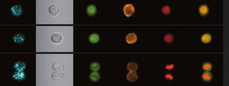
2 Insight & Analysis INNOVATIve Welcome to a Revolution in Cell Analysis: Imaging Flow Cytometry amnis The ImageStream system advances your science by combining quantitative cellular imagery with powerful population statistics With traditional cell analysis, you ve had to choose between visualizing a few cells under a microscope without quantitation or analyzing large cell populations by flow cytometry without imagery. Microscopy gives you detailed fine structure, morphology, and qualitative molecular localization. Flow cytometry gives you robust statistical information and detection of rare sub-populations. No technology has been able to give you all this at the same time and in a single experiment. Until now. The ImageStream system from Amnis gives you morphology, fluorescence localization, and population statistics for a broad range of applications. Quantitate translocation of transcription factors between cellular compartments. Examine interactions in cell conjugates. Perform high throughput FISH. Examine cells in mitosis and apoptosis. Quantitate internalization. Study the distribution and abundance of fluorescent proteins. And do it all in rare cells and highly heterogeneous samples.


3
4 New Technology distinctive An Entirely New Way to Analyze Cells amnis It s all about the numbers: 500 quantitative parameters from six simultaneous images per cell; brightfield, darkfield and multiple fluorescence images; 0.75 NA optics; sub-micron resolution; over 15,000 cells per minute. Advanced Detection Technology Delivers High Sensitivity With High Speed A unique six-channel CCD camera and a novel velocity detection system work in concert to collect 1,000 times more light than conventional technology. The technique is called Time Delay Integration. The result is high resolution imagery with fluorescence sensitivity superior to flow cytometry. Multispectral Imaging For Maximum Information Per Cell Illumination in the ImageStream system is provided by a brightfield lamp, a 488 nm laser and optional violet and red excitation lasers. Cellular imagery is split into six component colors through a unique spectral decomposition element. The result is a brightfield image, a darkfield image, and multiple fluorescence images of every cell. Simple Operation ImageStream technology is sophisticated, but operating the instrument isn t difficult. Highly automated protocols step you through calibration and set up. Focus and cell tracking are automatic. A single click of the mouse sterilizes the instrument. We ve designed the ImageStream to allow you to concentrate on your research, not your instrumentation.


5 Cells in Flow Spectral Decomposition Element Time Delay Integration Precisely-controlled fluidics position cells in the plane of focus as they flow through the system smoothly and without tumbling. A fan of dichroic mirrors splits the cell imagery into six spectral bands, one for each independent channel of the custom CCD camera. A custom six-channel CCD camera electronically tracks the motion of the cells, increasing the signal 1,000-fold. Fluorescence sensitivity exceeds standard flow cytometry. s cu r fo se to la au ir six-chan n el ccd camera h ot mirror -> b r i g h tfi e l d i l l umi n a to r v is ib le ligh t > > ir ligh t -> - -> - - -> > - A patented velocity detection system synchronizes the CCD camera readout with the motion of the cells. -> Velocity Detection A sophisticated autofocus system continually optimizes image quality. Autofocus A custom imaging objective with a numeric aperture of 0.75 and high performance optics achieve the image detail of a high quality microscope. A powerful solid state 488nm laser and optional red and violet lasers excite a wide range of dyes used commonly in microscopy and flow cytometry. Optical System Fluorescence Excitation > - au tofocu s / v elocity detector s p ectral decomp os itio n elemen t ex c la it se at rs io n > > > > > -> > > > i m a ging o bj e ctiv e Multispectral Imagery Six digital images per cell, including brightfield, darkfield, and multiple colors of fluorescence, convey tremendous quantitative information.


6


7 New Applications Leukocyte cell classification Tubulin stain in adherent cells Apoptosis Yeast budding Surface marker capping T Cell / APC conjugates Endocytic processing pathways Multicolor FISH in Suspension GFP and apoptosis Cell cycle analysis Human PBMC Immune synapse versatile One Instrument, Many Applications amnis Cell Signaling / Pathway Analysis The ImageStream system brings significant new capabilities to pathway analysis for cells in suspension. The phosphorylation states of key signaling molecules and their locations within the cell can be measured directly. Molecular association with the cell membrane, the cytoplasm, or the nucleus is easily distinguished and quantitated. Analysis of Cell Conjugates Cells communicate through cell membrane-mediated molecular interactions. The ImageStream system not only identifies cell doublets, but also quantitates molecular co-localization at the interface between the interacting cells. Fluorescence In Situ Hybridization In Suspension (FISHIS ) High throughput FISH is now possible with cells in suspension using the ImageStream system and Amnis protocols. Imagery is acquired rapidly and extended depth of field technology provides exceptionally clear visualization of multicolor chromosome spots in a range of cell types. Internalization and Intracellular Trafficking Image similarity algorithms allow you to quantitatively compare the distribution of multiple signals within single cells for co-localization, co-capping, and similar studies. Gene Expression Analysis The ImageStream system is well suited to the analysis of Green Fluorescent Protein and other fluorescent markers used in the study of gene expression. The high spatial resolution and sensitivity of the ImageStream system allows quantitation of expression levels and localization of expression to specific regions of the cell and key organelles.


8 Intracellular co-localization of multiple proteins HIV receptor mapping Multiple surface receptors Nf-κB translocation to the nucleus Phagocytosis Pseudopod formation Nuclear translocation of Raf FISH in sperm Toxoplasma gondii Gene expression in trypanosomes Analysis of TUNEL+ and TUNEL- cells Caspase and NF-κB in apoptosis Receptor Mapping and Distribution The ImageStream system not only measures the abundance of important cell surface receptors with exceptional sensitivity and resolution, but can also map their locations and co-localize them with ligands or intracellular organelles. For instance, proteins of interest may be co-localized with endosomal and lysosomal markers to follow intracellular processing and degradation. Quantitative Morphology Change in cell shape is closely correlated with function in the analysis of lymphocyte or macrophage activation, pseudopod formation, response to drugs, and many other instances. Powerful features in the IDEAS image analysis software allow you to accurately classify cells based on shape and structure. Cell Classification Characterization of peripheral blood mononuclear cell populations is a fundamental tool in hematology. The ImageStream system combines classical surface phenotyping with morphologic classification to deliver a full five-part differential analysis with room for the identification of additional sub-populations using fluorescent markers. Apoptosis Using only measurements of nuclear morphology, the ImageStream system can directly differentiate apoptotic and necrotic cells, quantify the extent of apoptosis in cell populations, and calculate sub-population frequencies. The need for surrogate markers such as Annexin V or fluorescent caspase substrates is reduced or eliminated, as are classification errors found in conventional flow cytometric apoptosis assays, reducing false positive and false negative results.
9 Analytical Power INSIGHTFUL Designed by Biologists for Biologists amnis Powerful, flexible, and extremely easy to learn, the IDEAS statistical image analysis package is integral to the ImageStream system. A Robust Feature Set, Expandable to Meet Your Needs The IDEAS feature set the heart of the image analysis package is extraordinarily robust, providing more than 500 features for every cell. IDEAS also allows you to create almost any new feature you find useful (e.g. nuclear to cytoplasmic area). Image Data and Statistical Data are Fully Integrated In IDEAS, graphs and imagery are completely integrated. Every dot on a scatter plot links directly to a cell s images click on the dot and you ll see the corresponding cell. With its virtual sorting capability, IDEAS will show you all the images of a cell population you define. An Efficient, Flexible Data Interface The IDEAS interface integrates image data, plots, and statistics. The Gallery shows you images of every cell, while the Workspace gives you graphing tools to define and analyze cell populations. The Tabular Data section allows you to view population statistics as well as individual feature values. Templates and Batch Processing Once you ve created an analysis scheme in IDEAS, you can save it as a template for batch processing future experiments or to share with your colleagues.
10 > Simple, Flexible Population Definitions Easy to use gating tools allow you to define, name and visualize cell populations quickly and intuitively. Rich Feature Set IDEAS calculates over 80 features for each image and over 500 features per cell, allowing the discrimination of subtle differences between cell populations > > Familiar Graphing Tools Quickly and easily create scatter plots and histograms to define your cell populations. Display parameters are easily adjusted > Data Linkage Every dot in a scatter plot is linked to a set of cell images. Click on the dot to see the cell, or click on a cell s images to locate it in all plots > - - > > Quantitative Data Plotting Any image analysis feature can be used in a histogram or dot plot. Extend your analysis beyond simple fluorescence intensity with localization and morphology features.

11


12 Advanced Performance FISH Spot Counting Nuclear Translocation Counting Nuclear Foci Faster Data Acquisition Standard Mode EDF Mode advanced EDF technology breaks the classical depth of field barrier amnis Image the Whole Cell in Focus EDF extended depth of field technology uses a combination of specialized optics and unique image processing algorithms to project all structures within the cell into one crisp plane of focus. Enables New Applications Many applications, such as FISH, depend critically on the resolution and accurate counting of spots within the cell. With the exceptional focus depth of EDF, high throughput analysis of FISH has become a reality. Improved Precision and Discrimination In addition to increasing depth of field, EDF improves resolution, thereby enhancing the discrimination of cellular features and improving precision in the quantitative analysis of cell imagery over a wide array of applications. Reduced Data Acquisition Time In addition to keeping the whole cell in focus, the EDF option allows the ImageStream to be run with a larger core diameter, thereby increasing throughput by up to three-fold. The EDF Extended Depth of Field Option The EDF option for the ImageStream system includes all required modifications to the instrument and software, installation, testing, documentation and user training. The EDF option can be included with a new ImageStream system or installed as a field upgrade.
13 ImageStream Specifications Advanced Engineering Creates Exceptional Performance Performance Imaging rate: up to 300 cells/second Sample throughput: ~5 min/sample Detection limit: <50 f luorescent molecules Numeric aperture: 0.75 Pixel size: 0.5 x 0.5 microns Field of view: 45 microns wide Sample volume: microliters Data Analysis Automated crosstalk compensation post-acquisition Unlimited user-defined image features Over 500 standard image features per cell Instrument Operation Automated sample load, empty, flush, and purge Automated focus and core position tracking Automated sterilization Automated calibration and quality control Automated laser alignment Requirements VAC, Hz 100 Mbps ethernet, minimum No external air or water required 36 w x 24 h x 24 d 350 lbs Illumination Sources SourcE Wavelength MAX Power Brightfield Lamp Standard nm Blue Laser Standard 488 nm 200 mw Violet Laser Optional 405 nm 350 mw Red Laser Optional 658 nm 80 mw The ImageStream system includes a solid-state 488 nm laser (200 mw) as the standard excitation source. The laser options include a red 658 nm laser (80 mw) and a high power violet 405 nm laser (350 mw). Each of these laser options is available factory installed or they may be purchased for installation on an existing ImageStream system. The dyes listed here represent just some of those that may be used on the ImageStream system configured with 405 nm, 488 nm and 658 nm lasers. The ImageStream is a Class 1 laser product. Detection Channels channel 1 channel 2 channel 3 channel 4 channel 5 channel nm nm nm nm nm nm Darkfield DAPI FITC PE 7-AAD Cy5, Cy5.5 Hoechst Alexa Fluor 488 Cy3 PE-Alexa Fluor 610 CyChrome Hoechst Alexa Fluor 500 Alexa Fluor 546 Propidium Iodide Alexa Fluor 647 Alexa Fluor 405 Alexa Fluor 514 Alexa Fluor 555 PE-Texas Red Alexa Fluor 660 Alexa Fluor 430 Syto 11, 13, 16 YFP Qdot 605 Alexa Fluor 680 Cascade Blue mitotracker Green OFP Qdot 625 Alexa Fluor 700 Pacific Blue Spectrum Green Qdot 565 ECd draq5 live/dead Violet Lucifer Yellow Qdot 585 Brightfield PerCP Vybrant DyeCycle Blue Cascade Yellow POPO-3 Qdot 705 CFP Qdot 525 PO-PRO-3 APC Brightfield Qdot 545 DsRed mitotracker Deep Red Brightfield Brightfield Brightfield 2008 Amnis Corporation All trademarks are acknowledged.
14 Amnis Corporation 2505 Third Avenue Suite 210 Seattle, WA USA Phone Fax U.S. Toll-Free
See what you ve been missing
 See what you ve been missing Seeing is Believing, but ImageStream is Proof If you rely on flow cytometry or microscopy, you need the power of the new ImageStream X A breakthrough intersection of technologies
Amnis ImageStream : Technical Reports & Applications
 Amnis ImageStream : Technical Reports & Applications ImageStream : Flow Cytometry and Microscopy in a Single Platform The ImageStream achieves true multispectral Imaging in Flow by combining microscopy
11/19/2013. Janine Zankl FACS Core Facility 13. November Cellular Parameters. Cellular Parameters. Monocytes. Granulocytes.
 DEPARTEMENT BIOZENTRUM Janine Zankl FACS Core Facility 13. November 2013 Cellular Parameters Granulocytes Monocytes Basophils Neutrophils Lymphocytes Eosinophils Cellular Parameters 1 What Is Flow Cytometry?
INTRODUCTION TO FLOW CYTOMETRY
 DEPARTEMENT BIOZENTRUM INTRODUCTION TO FLOW CYTOMETRY F ACS C ore F acility Janine Zankl FACS Core Facility 3. Dezember 2015, 4pm Cellular Parameters Granulocytes Monocytes Basophils Lymphocytes Neutrophils
Flexible, Intuitive and Affordable
 Data Sheet guava easycyte Flow Cytometry Systems Flexible, Intuitive and Affordable The guava flow cytometry systems are easy to use and deliver complete and comprehensive cell analysis right on your benchtop.
NovoCyte Flow Cytometer
 NovoCyte Flow Cytometer The Flow Cytometer for Everyone 2 Experience the NovoCyte Advantage Focus on advancing your research. Let the flow cytometer do the rest. NovoCyte Flow Cytometer High Performance
Boundary-breaking acoustic focusing cytometry
 Boundary-breaking acoustic focusing cytometry Introducing the Attune NxT Acoustic Focusing Cytometer a high-performance system that s flexible enough for any lab One of the main projects in my laboratory
A legacy of innovation and discovery
 A legacy of innovation and discovery CellInsight CX7 LZR High Content Analysis Platform Quantifiably brilliant data Since the introduction of Thermo Scientific ArrayScan High Content Analysis (HCA) Readers
Introduction to Flow Cytometry. -- BD FACSCanto II TM. Daisy Kuo Application Specialist BDBiosciences
 Introduction to Flow Cytometry -- BD FACSCanto II TM Daisy Kuo Application Specialist E-mail: daisy_kuo@bd.com BDBiosciences Outline Basic Concept of Flow Cytometry FACSCanto II System Introduction Application
Review of techniques in imaging and cytometry. Peter Meeus Onze Lieve Vrouw Ziekenhuis, Aalst 7 may 2009, SCK/CEN Mol
 Review of techniques in imaging and cytometry Peter Meeus Onze Lieve Vrouw Ziekenhuis, Aalst 7 may 2009, SCK/CEN Mol Cytometry: The counting and measuring of cells,... Mosby's Medical Dictionary, 8th edition.
Principles of flow cytometry: overview of flow cytometry and its uses for cell analysis and sorting. Shoreline Community College BIOL 288
 Principles of flow cytometry: overview of flow cytometry and its uses for cell analysis and sorting Shoreline Community College BIOL 288 Flow Cytometry What is Flow Cytometry? Measurement of cells or particles
Flow Cytometry - The Essentials
 Flow Cytometry - The Essentials Pocket Guide to Flow Cytometry: 1. Know your Cytometer 2. Understanding Fluorescence and Fluorophores 3. Gating Process 4. Controls 5. Optimization 6. Panel Building 7.
Widefield Microscopy Bleed-Through
 In widefield microscopy the excitation wavelengths which illuminate the sample, and the emission wavelengths which reach the CCD camera are selected throughout a filter cube. A filter cube consists of
Your Research, Revolutionized
 Your Research, Revolutionized Drive Your Research Forward Your research needs are evolving and with the CytoFLEX flow cytometer you ll see just how far your data can take you. CytoFLEX has the advanced
Everything counts. But nothing counts like the
 Everything counts But nothing counts like the Countess II FL Automated Cell Counter The new Countess II FL Automated Cell Counter is a benchtop cell assay platform equipped with state-of-the-art optics
Your Research, Revolutionized
 Your Research, Revolutionized Drive Your Research Forward Your research needs are evolving and with the CytoFLEX flow cytometer you ll see just how far your data can take you. CytoFLEX has the advanced
Selected Topics in Electrical Engineering: Flow Cytometry Data Analysis
 Selected Topics in Electrical Engineering: Flow Cytometry Data Analysis Bilge Karaçalı, PhD Department of Electrical and Electronics Engineering Izmir Institute of Technology Outline Experimental design
Sapphire. Biomolecular Imager THE NEXT GENERATION OF LASER-BASED IMAGING
 Sapphire Biomolecular Imager THE NEXT GENERATION OF LASER-BASED IMAGING Breakthrough image capture and analysis The Sapphire Biomolecular Imager is a next generation laser scanning system that provides
NEWTON 7.0 BIOLUMINESCENCE & FLUORESCENCE IMAGING IN VIVO - IN VITRO IMAGING
 NEWTON 7.0 BIOLUMINESCENCE & FLUORESCENCE IMAGING IN VIVO - IN VITRO IMAGING SMART IMAGING SYSTEM The NEWTON 7.0 system combines high sensitivity with advanced animal-handling features and userfriendly
FLOW CYTOMETRY. CyAn ADP. Analyzer
 FLOW CYTOMETRY CyAn ADP Analyzer Experience the Power of the CyAn ADP and its optimal performance The Power of Detection The Power of Speed The Power of Ease The CyAn ADP Analyzer is the next step in Advanced
Incorporating New, Bright Fluorochromes into Multicolor Panel Design
 Incorporating New, Bright Fluorochromes into Multicolor Panel Design Maria C. Jaimes, MD Senior Staff Scientist BD Biosciences 23-14684-00 Overview Multicolor flow: successful application prerequisites
Each question may have MULTIPLE correct answers. Select all that are correct.
 Knowledge Assessment Flow Cytometry Workshop, Part 1 April 20, 2015 Each question may have MULTIPLE correct answers. Select all that are correct. 1. Tandem dyes are a. highly stable fluorophores after
AMREP FLOW CYTOMETRY CORE FACILITY. Flow Cytometry Data Analysis Workshop Friday 24 th October
 AMREP FLOW CYTOMETRY CORE FACILITY Flow Cytometry Data Analysis Workshop Friday 24 th October Polychromatic Flow Cytometry Data Analysis: Immunophenotyping Lymphocyte sub-populations and targeting their
Introduction to Flow Cytometry. -- BD FACSCanto II TM. Daisy Kuo Assistant Product Manager BDBiosciences
 Introduction to Flow Cytometry -- BD FACSCanto II TM Daisy Kuo Assistant Product Manager E-mail: daisy_kuo@bd.com BDBiosciences Outline Basic Concept of Flow Cytometry FACSCanto II System Introduction
Cellometer Vision CBA
 Features of the Vision CBA Image Cytometry System All-in-One System Basic cell counting, primary cell viability, and cellbased assays. See for Yourself Why the Top Ten Pharmaceutical Companies Trust Cellometer
Muse Cell Analyzer. Experience simple, affordable flow cytometry.
 Muse Cell Analyzer Experience simple, affordable flow cytometry. Simple, affordable flow cytometry. Now at your side. Sophisticated cell analysis doesn t have to be exclusive, complicated or costly. With
PowERFUL. INTUITIvE. customizable. ThE NEw STAR of. BENchToP FLow cytometry
 PowERFUL. INTUITIvE. customizable. ThE NEw STAR of BENchToP FLow cytometry ABoUT AcEA Established in 2002, ACEA Biosciences, Inc. develops cutting-edge cell analysis platforms for life science research.
CyFlow Space Your flexible flow cytometer
 CyFlow Space Your flexible flow cytometer www.sysmex-partec.com CyFlow Space its flexibility gives you the space you need for your work Analysing cells and particles, be it from blood, plasma, tissue,
Review of techniques in flow cytometry. Peter Meeus Onze-Lieve-Vrouwziekenhuis, Aalst 5 may 2011, SCK/CEN Mol
 Review of techniques in flow cytometry Peter Meeus Onze-Lieve-Vrouwziekenhuis, Aalst 5 may 2011, SCK/CEN Mol Cytometry definitions The counting and measuring of cells,... Mosby's Medical Dictionary, 8th
CyFlow Space Your flexible flow cytometer
 CyFlow Space Your flexible flow cytometer www.sysmex-partec.com CyFlow Space its flexibility gives you the space you need for your work Analysing cells and particles, be it from blood, plasma, tissue,
Vybrant DyeCycle Violet stain
 Vybrant DyeCycle Violet Stain Catalog no. V35003 Table 1. Contents and storage information. Material Amount Concentration Storage Stability Vybrant DyeCycle Violet stain 200 μl 5 mm solution in deionized
Cell Proliferation and Death
 Cell Proliferation and Death Derek Davies, Cancer Research UK http://www.london-research-institute.org.uk/technologies/120 Proliferation A cell Apoptosis Cell death Proliferation signals Senescence DNA
Fluorescence Light Microscopy for Cell Biology
 Fluorescence Light Microscopy for Cell Biology Why use light microscopy? Traditional questions that light microscopy has addressed: Structure within a cell Locations of specific molecules within a cell
NEWTON 7.0 BIOLUMINESCENCE & FLUORESCENCE IMAGING IN VIVO - IN VITRO IMAGING
 NEWTON 7.0 BIOLUMINESCENCE & FLUORESCENCE IMAGING IN VIVO - IN VITRO IMAGING The NEWTON s protocol driven image acquisition is as quick as it is intuitive: adjust your exposure, save, print or quantify.
Attune NxT Acoustic Focusing Cytometer The next generation in acoustic cytometry
 Attune NxT Acoustic Focusing Cytometer The next generation in acoustic cytometry Maybelline Giam Field Application Scientist The world leader in serving science Attune NxT Flow Cytometer Attune NxT Acoustic
Best practices in panel design to optimize the isolation of cells of interest
 Sort Best practices in panel design to optimize the isolation of cells of interest For Research Use Only. Not for use in diagnostic or therapeutic procedures. Alexa Fluor is a registered trademark of Life
Cancer inflammation research applications and products
 Cancer inflammation research applications and products Flow cytometry Immunoassays Cell imaging Instrumentation Invitrogen Attune NxT Flow Cytometer Antibodies RNA flow Conjugated antibodies for flow cytometry
The Basics of Flow Cytometry
 The Basics of Flow Cytometry F ACS C ore F acility Janine Bögli, Biozentrum, 29. January 2018 The functions of the FACS Core Facility Centralization of equipment and expertise Train users Sorter operation
CRC Flow Cytometry Core Facility Knut och Alice Wallenbergs Stiftelse
 CRC Flow Cytometry Core Facility CRC Flow Cytometry Core Facility Knut och Alice Wallenbergs Stiftelse CRC Flow Cytometry Core facility Instruments: 1. FACSCalibur: analyser, 6 parameters, 4 colors (BD),
Sapphire. Biomolecular Imager THE NEXT GENERATION OF LASER-BASED IMAGING
 Sapphire Biomolecular Imager THE NEXT GENERATION OF LASER-BASED IMAGING Breakthrough image capture and analysis The Sapphire Biomolecular Imager is a next generation laser scanning system that provides
PERFORMANCE MADE EASY REAL-TIME PCR
 PERFORMANCE MADE EASY REAL-TIME PCR The MyGo Pro real-time PCR instrument provides unmatched performance in a convenient format. Novel Full Spectrum Optics deliver 120 optical channels of fluorescence
CyFlow Cube series Appealing from every angle
 CyFlow Cube series Appealing from every angle www.sysmex-partec.com CyFlow Cube 6 and Cube 8: compact, economic flow cytometers with a great performance Panta rhei a flexible solution for demands in flow
Confocal Microscopy Analyzes Cells
 Choosing Filters for Fluorescence A Laurin Publication Photonic Solutions for Biotechnology and Medicine November 2002 Confocal Microscopy Analyzes Cells Reprinted from the November 2002 issue of Biophotonics
Conquer cell counting
 Conquer cell counting Cell Countess II Automated Cell Counters Fast Accurate Affordable Countess II Automated Cell Counters Advanced technology at an affordable price Accurate counts in as little as 10
High-throughput automation with the Attune NxT Autosampler: consistent results across all wells and across plates
 APPLICATION NOTE Attune NxT Flow Cytometer with Autosampler High-throughput automation with the Attune NxT Autosampler: consistent results across all wells and across plates Introduction The emerging field
What you need to know before designing a panel
 Design What you need to know before designing a panel For Research Use Only. Not for use in diagnostic or therapeutic procedures. Alexa Fluor is a registered trademark of Life Technologies Corporation.
CyFlow Space. Your Flexible Flow Cytometer.
 CyFlow Space Your Flexible Flow Cytometer www.sysmex-partec.com Ultimate Flexibility Modular System The CyFlow Space flow cytometer is a modular system with ultimate flexibility: from a basic configuration
Principles of Multicolor Panel Design BD. BD, the BD Logo and all other trademarks are property of Becton, Dickinson and Company.
 1 Principles of Multicolor Panel Design 2 Common Multicolor Applications Intracellular cytokine staining Regulatory T cells (Tregs) Protein phosphorylation (BD Phosflow) Leukemia and lymphoma phenotyping
Phagocytosis Assay Kit (IgG PE)
") Phagocytosis Assay Kit (IgG PE) Item No. 600540 www.caymanchem.com Customer Service 800.364.9897 Technical Support 888.526.5351 1180 E. Ellsworth Rd Ann Arbor, MI USA TABLE OF CONTENTS GENERAL INFORMATION
Phagocytosis Assay Kit (IgG PE) Item No. 600540 www.caymanchem.com Customer Service 800.364.9897 Technical Support 888.526.5351 1180 E. Ellsworth Rd Ann Arbor, MI USA TABLE OF CONTENTS GENERAL INFORMATION
Flowcytometry Dirk Pacholsky
 Flowcytometry Dirk Pacholsky Flowcytometry: Overview 1 2 Overview Flow Cyto Metry Fluid Cell Measurement measuring cell properties of cells in suspension Most Flow Cytometer have Spatially separated Lasers
RaftNote. Imaging and Sorting Living Cells Stained with Vital Dyes on Cell Microsystems CytoSort Array and Automated CellRaft AIR System
 RaftNote Applications and protocols for use with CellRaft Technology Imaging and Sorting Living Cells Stained with Vital Dyes on Cell Microsystems CytoSort Array and Automated CellRaft AIR System Jacquelyn
Index. Index 425. Bacteria flow cytometry, general considerations, 33, 34 Gram-negative versus Grampositive
 Index 425 Index Acridine orange, see DNA/RNA simultaneous analysis Aerosol management system (AMS), biohazardous material sorting, 421 AMS, see Aerosol management system Annexin V, see Apoptosis, flow
Multiplex Fluorescence Assays for Adherence Cells without Trypsinization
 Multiplex Fluorescence Assays for Adherence Cells without Trypsinization The combination of a bright field and three fluorescent channels allows the Celigo to perform many multiplexed assays. A gating
High Throughput Suspension Cell and Bead Analysis. Intellicyt ique Screener PLUS
 High Throughput Suspension Cell and Bead Analysis Intellicyt ique Screener PLUS High Throughput Suspension Cell and Bead Analysis Intellicyt ique Screener PLUS the fastest path to actionable results Whether
BD LSRFortessa. Performance without peer, choice without compromise
 BD LSRFortessa Performance without peer, choice without compromise Performance without peer, choice without compromise The BD LSRFortessa cell analyzer offers the ultimate in choice for flow cytometry,
Anatomy of a flow cytometer
 Anatomy of a flow cytometer Fluidics Optics Electronics Cells in suspension flow in single-file through an illuminated volume where they scatter light and emit fluorescence that is collected, filtered,
CyFlow Cube 6. Immunology Microbiology Industrial Applications Agrosciences Aquaculture. Flow Cytometry: Simply elegant. Elegantly simple.
 CyFlow Cube 6 Immunology Microbiology Industrial Applications Agrosciences Aquaculture Flow Cytometry: Simply elegant. Elegantly simple. 01 Applications for Indus trial ApplicationS for Health care Agro
Multiplexed 3D FRET imaging in deep tissue of live embryos Ming Zhao, Xiaoyang Wan, Yu Li, Weibin Zhou and Leilei Peng
 Scientific Reports Multiplexed 3D FRET imaging in deep tissue of live embryos Ming Zhao, Xiaoyang Wan, Yu Li, Weibin Zhou and Leilei Peng 1 Supplementary figures and notes Supplementary Figure S1 Volumetric
NovoCyte Quanteon F. Flow Cytometer. When Exceptional Performance Meets Simplicity
 NovoCyte Quanteon F Flow Cytometer When Exceptional Performance Meets Simplicity A Quantum Leap In Benchtop Flow Cytometry The NovoCyte Quanteon The NovoCyte Quanteon flow cytometer builds on its successful
Apoptosis detection. Apoptosis assays for the Attune Acoustic Focusing Cytometer. APOPTOSIS DETECTION Attune Acoustic Focusing Cytometer
 POPTOSIS ETECTION ttune coustic Focusing Cytometer poptosis detection poptosis assays for the ttune coustic Focusing Cytometer poptosis is a carefully regulated process of cell death that occurs as a normal
NEWTON 7.0 BIOLUMINESCENCE & FLUORESCENCE IMAGING IN VIVO - IN VITRO IMAGING
 NEWTON 7.0 BIOLUMINESCENCE & FLUORESCENCE IMAGING IN VIVO - IN VITRO IMAGING The NEWTON s protocol driven image acquisition is as quick as it is intuitive: adjust your exposure, save, print or quantify.
Principles of Immunophenotyping
 Principles of Immunophenotyping % of Cell types? Immune activation? Changes based on health state? Moving from a heterogeneous population of blood cells to identifying the presence and proportion of different
BD FACSMelody. Cell Sorter. The simple solution for consistent, quality results
 BD FACSMelody Cell Sorter The simple solution for consistent, quality results The right instrument is needed for the best results Simplicity, affordability and quality are key Cell sorting is fast becoming
Thermo Scientific ArrayScan XTI High Content Analysis Reader. revolutionizing cell biology with the power of high content
 Thermo Scientific ArrayScan XTI High Content Analysis Reader revolutionizing cell biology with the power of high content learn more about your cells using high content technology Thermo Scientific High
Understanding Flow Cytometry
 Understanding Flow Cytometry The Basic Concepts Maree Bagnara Products Sales Specialist/Account Manager Flow Cytometry PN775136 1 Successful Flow Cytometry data is driven by.. Understanding the Biology
Introduction to. BD FACSAria TM Cell Sorter. Flow = Fluid Cyto = Cell Metry = Measurement
 What is Flow Cytometry? Introduction to BD FACSAria TM Cell Sorter Flow = Fluid Cyto = Cell Metry = Measurement BD Biosciences Application Specialist 產品應用專員 Daisy Kuo 郭正佼 A variety of measurements are
Navios EX FLOW CYTOMETER POWERFUL, DEPENDABLE CLINICAL FLOW CYTOMETRY
 Navios EX FLOW CYTOMETER POWERFUL, DEPENDABLE CLINICAL FLOW CYTOMETRY BECAUSE EVERY EVENT MATTERS The Navios EX flow cytometer offers a solution for advanced cytometry applications optimized for the clinical
Cellometer. Vision CBA. Image Cytometry System for 20µl Cell-Based Assays
 Cellometer Vision CBA Image Cytometry System for µl Cell-Based Apoptosis Cell Cycle and Others Features of the Vision CBA Image Cytometry System All-in-One System Basic cell counting, primary cell viability,
Expanding Multicolor Options in Flow Cytometry with Novel Brilliant Violet TM Fluorophores. Carsten Wiethe Scientific Application Manager BioLegend
 Expanding Multicolor Options in Flow Cytometry with Novel Brilliant Violet TM Fluorophores Carsten Wiethe Scientific Application Manager BioLegend Seminar Outline BrilliantViolet TM (BV)fluorophores Instrument
Automated Imaging and Dual-Mask Analysis of γh2ax Foci to Determine DNA Damage on an Individual Cell Basis
 A p p l i c a t i o n N o t e Automated Imaging and Dual-Mask Analysis of γh2ax Foci to Determine DNA Damage on an Individual Cell Basis Brad Larson, BioTek Instruments, Inc., Winooski, VT USA Asha Sinha
J. Philip McCoy, Jr., Ph.D., H.C.L.D. J. Philip McCoy, Jr., PhD
 J. Philip McCoy, Jr., Ph.D., H.C.L.D. Email: mccoyjp@mail.nih.gov 1 Introduction 2 How It All Works A very brief description of how a flow cytometer works. Please read it it will help you design your experiments.
Application Note. Introduction. EMD Millipore is a division of Merck KGaA, Darmstadt, Germany
 pplication Note Data Sheet Features of New InCyte Software Facilitate Sophisticated Flow Cytometry nalysis Introduction dvances in flow cytometry applications and new instrument technologies such as plate-based
Selecting Reagents for Multicolor Flow Cytometry
 HotLines Platinum Edition f a l l 0 0 6 Selecting Reagents for Multicolor Flow Cytometry By Holden Maecker and Joe Trotter The availability of flow cytometers capable of detecting 6, 8, and more colors
determine optimum instrument settings for their own instruments and establish their own daily values.
 PC7 (770/488) SETUP KIT 6607121 PN 4299504-C FLOW CYTOMETER ALIGNMENT VERIFICATION FLUOROSPHERES FLOW CYTOMETER DETECTOR STANDARDIZATION FLUOROSPHERES INTENDED USE For Research Use Only. Not for use in
Flow cytometry within reach.
 Affordable, but more than capable. The Accuri C6 Flow Cytometer is flow cytometry at its best. Its affordable price makes it accessible beyond compare. The C6 has a foot print small enough to fit most
Spherotech, Inc. 1. SPHERO TM Technical Note STN-8 Rev C
 SPHERO TM Technical Note STN- Rev C. 0070 CALIBRATION AND PERFORMANCE TRACKING OF FLOW CYTOMETERS USING SPHERO TM CALIBRATION PARTICLES Introduction The SPHERO TM Calibration Particles are versatile, stable,
GUAVA EASYCYTE SYSTEMS
 GUAVA EASYCYTE SYSTEMS Expanding the potential of flow cytometry The life science business of Merck KGaA, Darmstadt, Germany operates as MilliporeSigma in the U.S. and Canada. Guava easycyte Systems Expanding
Optical Observation - Hyperspectral Characterization of Nano-scale Materials In-situ
 Optical Observation - Hyperspectral Characterization of Nano-scale Materials In-situ Research at the nanoscale is more effective, when research teams can quickly and easily observe and characterize a wide
Everything counts. But nothing counts like Countess II automated cell counters. Fast Accurate Affordable
 Everything counts But nothing counts like Countess II automated cell counters Fast Accurate Affordable Countess II automated cell counters Advanced technology at an affordable price Precise, accurate counts
Innovations To Meet Your Needs
 Innovations To Meet Your Needs Cooled CCD Camera 1340 x 1037 pixel resolution for greatest image quality 12-bit precision provides 3 orders of linear dynamic range Windows and Power Macintosh Software
Navios EX FLOW CYTOMETER POWERFUL, DEPENDABLE FLOW CYTOMETRY
 Navios EX FLOW CYTOMETER POWERFUL, DEPENDABLE FLOW CYTOMETRY BECAUSE EVERY EVENT MATTERS The Navios EX flow cytometer offers a solution for advanced cytometry applications with optimized workflows for
Flow Cytometry For New PhDs
 Flow Cytometry For New PhDs 2012 Simon Monard SCRM and CIR smonard@staffmail.ed.ac.uk Derek Davies CRUK derek.davies@cancer.org.uk Monday 20th and Tues 21st Feb 2012 Program Day 1: Day 2: Basics of flow
BD FACSCanto II. A proven research platform for maximum reliability and the highest quality results
 BD FACSCanto II A proven research platform for maximum reliability and the highest quality results A proven platform for maximum reliability and the highest quality results Built on more than 25 years
Cell Processing Workstation. In Situ Laser-Mediated Cell Purification and Processing
 LEAP Cell Processing Workstation Cell Processing Workstation In Situ Laser-Mediated Cell Purification and Processing Expanding the Capabilities of Cell Processing Consistent, High-Yield, High-Purity Processing
Visualisation, Sizing and Counting of Fluorescent and Fluorescently-Labelled Nanoparticles
 Visualisation, Sizing and Counting of Fluorescent and Fluorescently-Labelled Nanoparticles Introduction Fluorescent molecules have long been used to specifically label particular structures and features
MF-ChemiBIS. Today s most comprehensive solution for your bio-imaging needs and applications. Documenting Nature
 MF-ChemiBIS Today s most comprehensive solution for your bio-imaging needs and applications Documenting Nature MF-ChemiBIS Excellence in bio-imaging The DNR Advantage As pioneers in bio-imaging technologies
Challenges for Flow Cytometry in Regulated Bioanalysis
 Challenges for Flow Cytometry in Regulated Bioanalysis Minesh Patel Merck Millipore Discovery & Development Solutions. Oxford, UK. Overview Flow cytometry principles Current uses and regulatory environments
iline F Capturing the Most From Your Suspension Cell Culture Process. In Real-Time
 iline F Capturing the Most From Your Suspension Cell Culture Process. In Real-Time www.iline-f.com iline F Continuous suspension cell culture monitoring in a bioreactor The iline F microscope will change
Violet Chromatin Condensation/Dead Cell Apoptosis Kit with Vybrant DyeCycle Violet and SYTOX AADvanced for Flow Cytometry
 Violet Chromatin Condensation/Dead Cell poptosis Kit with Vybrant DyeCycle Violet and SYTOX Dvanced for Flow Cytometry Catalog no. 35135 Table 1. Contents and storage information. Material mount Concentration
Flow Cytometry. Flow Cytometry Basics Guide
 Flow Cytometry Flow Cytometry Basics Guide Table of Contents Chapter 1 Chapter 2 Chapter 3 Chapter 4 Chapter 5 Principles of the Flow Cytometer Fluidics System.... 3 Optics and Detection.... 4 Signal and
Accelerating the Pace of Understanding
 Vectra 3 P R O D U C T N O T E Quantitative Pathology Imaging and Analysis Key Benefits Part of PerkinElmer's Phenoptics workflow solution for Cancer Immunology Research Detect and measure multiple expressed
Flow Cytometry Support Reagents
 Excite and inspire Flow Cytometry Support Reagents Introduction Miltenyi Biotec is a leading supplier of flow cytometry products, offering one of the broadest ranges of antibodies, kits, assays, and support
Vybrant DyeCycle Green and Orange Stains
 Vybrant DyeCycle Green and Orange Stains Catalog nos. V35004, V35005 Table 1. Contents and storage information. Material Amount Concentration Storage* Stability Vybrant DyeCycle Green stain Vybrant DyeCycle
The CQ1 Confocal Quantitative Image Cytometer and its Application to Biological Measurement
 The CQ1 Confocal Quantitative Image Cytometer and its Application to Biological Measurement Hirofumi Sakashita *1 Koji Ohashi *1 Kazuo Ozawa *2 ohei Tsubouchi *1 The CQ1 confocal quantitative image cytometer,
Designing and executing a successful flow cytometry experiment
 Designing and executing a successful flow cytometry experiment 1x1 0 5 Orange Fluorescent Protein 10000 1000 100 10 0 1000 2000 3000 4000 Forward Scatter This presentation is based on true events, however,
High-dimensional flow-cytometric analysis of human B-cell populations
 High-dimensional flow-cytometric analysis of human B-cell populations The BD FACSCelesta cell analyzer and FlowJo software together enable deep analysis of B-cell biology Features High-resolution analysis
Attune TM Acoustic Focusing Cytometer Training. Manik Punj Attune Training
 Attune TM Acoustic Focusing Cytometer Training Manik Punj Attune Training Attune Training Agenda Section 1 An Introduction to Flow Cytometry Section 2 An Introduction to Acoustic Focusing Hydrodynamic
A previous ICCS Module entitled Instrument optimization - Adjusting PMT voltages and compensation 1 should be read as a prerequisite to this module.
 Sponsored and reviewed by ICCS Quality and Standards Committee Title: Compensation Tips for Beckman Coulter 10-Color Navios Platform Written by: Salima Janmohamed-Anastasakis Ph.D., Applications Scientist,
Technical Bulletin. Multiple Methods for Detecting Apoptosis on the BD Accuri C6 Flow Cytometer. Introduction
 March 212 Multiple Methods for Detecting Apoptosis on the BD Accuri C6 Flow Cytometer Contents 1 Introduction 2 Annexin V 4 JC-1 5 Caspase-3 6 APO-BrdU and APO-Direct Introduction Apoptosis (programmed
DEPArray Technology. Sorting and Recovery of Rare Cells
 DEPArray Technology Sorting and Recovery of Rare Cells Delivering pure, single, viable cells The DEPArray system from Silicon Biosystems is the only automated instrument that can identify, quantify, and