Comprehensive guide to imaging reagents. abcam.com/cell-imaging-resources
|
|
|
- Evan Davidson
- 6 years ago
- Views:
Transcription


1 Comprehensive guide to imaging reagents abcam.com/cell-imaging-resources
2 Front cover images Anti-CaMKII antibody [EP1829Y] (Alexa Fluor 647) (ab196165) 2. Anti-GM130 antibody [EP892Y] - cis-golgi Marker (Alexa Fluor 488) (ab195302) 3. Anti-Survivin antibody [EPR2675] (Alexa Fluor 488) (ab194237) 4. Anti-p27 KIP 1 antibody [Y236] (Alexa Fluor 488) (ab194233) 5. Anti-CaMKII antibody [EP1829Y] (Alexa Fluor 647) (ab196165) 6. Anti-HDAC2 antibody [Y461] (Alexa Fluor 647) (ab196518) 7. Anti-CDX2 antibody [EPR2764Y] (Alexa Fluor 647) (ab195008) 8. Anti-CDX2 antibody [EPR2764Y] (Alexa Fluor 488) (ab195007) 9. Anti-Hsp47 antibody [EPR4217] (Alexa Fluor 488) (ab192841) Alexa Fluor is a registered trademark of Life Technologies. Alexa Fluor dye conjugates contain(s) technology licensed to Abcam by Life Technologies. 2 Comprehensive guide to imaging reagents
3 Contents 4 Your total resource for imaging 5 Our commitment to quality and validation 6 Key insights for successful immunofluorescence experiments 7 Direct versus indirect staining 9 Cell imaging protocols and webinars 10 Directly conjugated antibodies 13 Alexa Fluor conjugated secondary antibodies 16 Organelle markers and dyes 19 Ion indicators and false neurotransmitters for cell imaging 21 Ancillary imaging reagents 22 Conjugation kits and custom conjugation services Comprehensive guide to imaging reagents 3
4 Your total resource for cell imaging Imaging protocols and webinars Conjugation kits and services Ancillary imaging reagents Directly conjugated antibodies Organelle markers and dyes Alexa Fluor conjugated secondaries Ion indicators and false neurotransmitters I use Abcam antibodies because they are such good products. My experience with their products demonstrates to me that they perform very well in my experiments. Dr Scott Altmann, HDL Apomics 4 Comprehensive guide to imaging reagents
5 Our commitment to quality and validation Whether it s antibodies, kits, proteins or small molecule activators and inhibitors, our products are continuously validated to provide consistent performance and relevant data. Manufacturing excellence Over 15 years of developing and manufacturing products in-house guarantees consistency and quality. Validated in multiple applications and species A team of dedicated research scientists feed current scientific knowledge into our product validation process. Partnering with experts We maintain close connections with academic and industry experts to perform specialized testing and to provide insight on our validation processes. Stability testing Our lab performs stability testing to provide you with accurate storage temperature recommendations for maximal performance. Abpromise If for any reason the product does not work as stated on the datasheet, we will provide a replacement or refund up to 12 months from the date the product was delivered. Numbers you can trust 39,000+ Validated imaging reagents 28,000+ Reagents with a reference, image or Abreview 25,000+ Protein targets with validated antibodies Comprehensive guide to imaging reagents 5
6 Key tips for successful immunofluorescence experiments 1. Proper data interpretation is extremely important in any imaging experiment. Autofluorescence, off-target binding of the primary antibody and non-specific binding of secondary antibodies can be mis-interpreted as genuine staining. It is therefore important to ensure the correct controls are used. No primary or secondary antibodies to rule out the possibility of autofluorescence Secondary alone to dismiss the possibility of the secondary antibody binding nonspecifically to the endogenous cell proteins Often, immunofluorescence images consist of only a few cells so it is important to make sure the image is typical and representative of the population. 2. It is important not to let your cells dry out at any point during staining as this can introduce artefacts. Drying of the samples during incubation steps can be prevented by using a humidifying chamber. 3. Fixation may or may not mask the epitope of an antibody (this is particularly relevant for monoclonal antibodies). It is therefore worth adding the antibody to cells that have and have not been fixed to identify if fixation is or is not required. 4. The cell membrane must be permeabilized to allow antibodies access to the intracellular components. For each antibody the optimal permeabilization step will be different and therefore a variety of methods should be investigated, for instance trying different percentages of Triton X-100 ( %). 5. For multi-target staining, it is important to make sure your secondary antibodies are pre-adsorbed against the host species of your primary and secondary antibodies. Pre-adsorbed secondary antibodies minimize cross-species reactivity and non-specific binding with endogenous proteins within the sample. 6. Non-specific staining may be reduced by a. Blocking with serum from the host species of your secondary antibody. b. Using less antibody and/or decreasing the incubation times (overnight at 4 C or 1 hour at room temperature). c. Quenching residual aldehydes following formaldehyde fixation using 0.1M glycine. 7. After adding the fluorescently labeled antibodies to your cells, make sure all subsequent incubation steps are performed in the dark to prevent quenching of the fluorescent dye/proteins. 6 Comprehensive guide to imaging reagents
7 Direct vs indirect immunofluorescence Immunofluorescence (IF) or cell imaging techniques rely on the use of chemically conjugated antibodies to label a specific target antigen with a fluorescent dye (known as fluorophores or fluorochromes) such as fluorescein isothiocyanate (FITC). The fluorophore allows visualization of the target distribution in the sample under a fluorescent microscope (eg epifluorescence and confocal microscopes). We distinguish between two immunocytochemistry(icc)/if methods depending on whether the fluorophore is conjugated to the primary or the secondary antibody: Direct ICC/IF: uses a single primary antibody directed against the target of interest. The antibody is directly conjugated to a fluorophore. Indirect ICC/IF: uses two antibodies. The primary antibody is unconjugated and a fluorophore-conjugated secondary antibody directed against the primary antibody is used for detection. Direct Antigen Primary antibody Secondary antibody cell Fluorophore Indirect cell cell Comprehensive guide to imaging reagents 7
8 Indirect and direct methods have their advantages and disadvantage as shown in the table below. Time Cost Complexity Flexibility Sensitivity Species cross-reactivity Background Direct Protocols for direct IF are usually shorter as they only require one labeling step. Conjugated primary antibodies are usually more expensive than their unconjugated counterparts. Less steps in the protocol simplify direct methods. Commercially available pre-conjugated primary antibodies limit your flexibility. The signal obtained in direct methods may seem weak when compared to indirect methods as signal amplification provided by the use of secondary antibodies does not occur. Species cross-reactivity is minimized in direct methods as the fluorophore is already conjugated to the primary antibody. Non-specific binding is reduced through the use of conjugated primary antibodies. Indirect The fact that you have to use a conjugated secondary antibody to detect the primary antibody results in additional steps. Secondary antibodies are relatively inexpensive compared to primary antibodies. Further cost savings may be realized by the possibility of using the same conjugated secondary antibody to detect different primary antibodies. Added complexity in indirect methods may result from having to select the appropriate secondary antibody. This is particularly relevant in multiplex experiments where several secondary antibodies, each targeting a different species and conjugated to different dyes, are needed. The possibility of using different conjugated secondary antibodies adds greater flexibility. Several secondary antibodies will bind to the primary antibody resulting in an amplified signal. Secondary antibodies may cross-react with species other than the target. The use of pre-adsorbed secondary antibodies can prevent cross-reactivity. Samples with endogenous immunoglobulins may exhibit a high background with indirect methods. 8 Comprehensive guide to imaging reagents
9 Cell imaging protocols and webinars Our website offers you a wide range of cell imaging protocols and webinars. Highlighted below are some of our most popular ones. Protocols Immunocytochemistry and immunofluorescence protocol Our detailed guide on ICC/IF is a must-have resource to ensure you get optimal results in your cell imaging experiments. Whether you are doing single staining or multiple staining, the guide provides a step-by-step protocol that includes hints and tips for: Fixation Antigen retrieval (optional) Permeabilization Blocking and incubation Multicolor staining (optional): Simultaneous incubation Sequential incubation Counter staining Mounting You can find the protocol here: Find more protocols at: Webinars Our webinar series can be viewed on demand, and provides detailed examples to help you with your cell imaging experiments. Topics include: Introduction to ICC An overview of the key principles of the technique Single and multiple labeling in ICC Strategies to overcome common pitfalls in multiple labeling experiments Principles and practices of confocal microscopy Outlines basic and advanced principles of the technique Optimizing ICC Hands-on tips to improve your experiments Find more webinars at: Comprehensive guide to imaging reagents 9
10 Directly conjugated antibodies Rapid advances in fluorescence microscopy and flow cytometry have led to better and brighter imaging reagents. Whether you are performing single cell imaging or multicolor flow cytometry, our dye-labeled antibodies are the ideal immunoreagents to light up your experiment. Feature Powered by RabMAb technology Free dye removal step Optimized fluorophore:protein (F:P) labeling Suitable for multicolor analysis Thorough validation Benefit Higher affinity and specificity than traditional monoclonals Higher signal to noise ratios Superior sensitivity for each dye conjugate Eliminates cross-reactivity seen with twostep labeling Batch tested in cellular imaging applications Validated for cell imaging applications We validate each batch of antibody conjugate in key imaging applications to ensure they perform in your hands, every time. ICC/IF high-resolution cell and tissue section images taken using a confocal microscope are provided on the datasheet. Nuclear and cytoskeletal counterstains are overlaid to increase the contrast of subcellular localization. Flow cytometry histogram is provided on the datasheet showing positive events as indicated by a peak shift. Two negative controls an isotype control, and an unlabeled control are overlaid to confirm positive signal. The choice is yours We manufacture antibodies labeled with nine distinct Alexa Fluor dyes, covering a wide range of UV, visible, and near-ir wavelengths, and reducing the chances of spectral overlap. Our Alexa Fluor series Alexa 405Fluor 488 series offered 555 by 568 Abcam Discover more 10 Comprehensive guide to imaging reagents
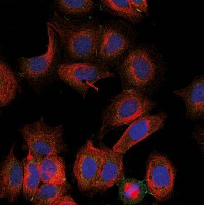
 Blue staining Nuclear counterstain with DAPI Instrument Leica-Microsystems TCS SP8 confocal microscope Sample 109 Methanol fixed HeLa cells Red Peak Counts Primary antibody ab194724")
11 Product highlights Sample PFA-fixed T47D cells Green staining Primary antibody ab rabbit monoclonal to cytokeratin (Alexa Fluor 488) Red staining Ab mouse monoclonal to alpha tubulin (Alexa Fluor 594) Blue staining Nuclear counterstain with DAPI Instrument Leica-Microsystems TCS SP8 confocal microscope Sample 109 Methanol fixed HeLa cells Red Peak Counts Primary antibody ab rabbit monoclonal to Ki67 (Alexa Fluor 647) Blue Peak Unlabeled methanol fixed HeLa cell control Black Peak Ki67 - Alexa Fluor 647 (675/30 BP) Rabbit monoclonal (Alexa Fluor 647) isotype control Instrument Beckman Coulter FC500 MPL Comprehensive guide to imaging reagents 11
12 Using multiple conjugates in flow panels? Our multicolor flow selector lets you search and compare conjugated antibodies for up to three targets of interest. Build your next multicolor flow experiment at abcam.com/flow-cytometry 12 Comprehensive guide to imaging reagents
13 Alexa Fluor conjugated secondary antibodies Feature Brightest dyes Greater photostability Broad ph tolerance Good water solubility Benefit Alexa Fluor dyes outperform other spectrally-similar dyes providing greater sensitivity Allows for longer periods of image capture Intensity of Alexa Fluor dyes remains high over a wide ph range Prevents precipitation and aggregation of Alexa Fluor conjugated antibodies We have implemented a robust manufacturing process that guarantees the quality of our Alexa Fluor conjugated secondary antibodies: Conjugation the number of dye molecules per antibody (F:P ratio) are optimized to provide superior sensitivity Purification the highest possible purity grade is achieved by removing free dye after conjugation Validation each individual antibody is validated in ICC/IF to ensure bright staining and low background The spectral location in the red made this perfect for a double label with the other fluorophore in the blue. It has stood up well to storage in the refrigerator and gives a good signal after six months. Researcher from Station Biologique de Roscoff, France Comprehensive guide to imaging reagents 13
14 Alexa Fluor Anti-mouse IgG H&L Anti-rabbit IgG H&L Anti-rat IgG H&L Anti-goat IgG H&L Antichicken IgG H&L ab ab ab ab ab ab ab ab ab ab ab ab ab ab ab ab ab ab ab ab ab ab ab ab ab Explore our broad offering at Ideal for multicolor staining We currently offer secondary antibodies conjugated to nine different Alexa Fluor dyes covering the whole spectrum from the UV to the near infra-red regions with minimal spectral overlap: Raised in different species including donkey, goat, and rabbit Targeting several species and their isotypes such as rabbit, mouse, rat, goat, and chicken A large range of pre-adsorbed antibodies ensures low species cross-reactivity Many fragments as well as whole antibodies Emission spectra Alexa Fluor conjugated secondary antibodies Alexa Fluor 405 Fluorescene emission Alexa Fluor 488 Alexa Fluor 555 Alexa Fluor 568 Alexa Fluor 594 Alexa Fluor 647 Alexa Fluor 680 Alexa Fluor Wavelength (nm) Alexa Fluor Comprehensive guide to imaging reagents
15 If you are looking for common combinations in double and triple immunostaining experiments we recommend the following combinations: Immunostaining Antibody combination Double Alexa Fluor 488 and Alexa Fluor 647 Triple Alexa Fluor 405, Alexa Fluor 555 and Alexa Fluor 647 Alexa Fluor Absorption max (nm) Emission max (nm) Emission color* Matching dyes Extinction coefficient Quantum yield Cascade Blue Cy2, FITC (fluorescein) Cy3, TRITC (Rhodamine) Rhodamine Red 35,000-73, , , Texas Red 92, APC, Cy5 270, Cy5, IR , Cy7 290, IR ,000 - * Typical emission color seen through a conventional fluorescence microscope with appropriate filters ** Human vision is insensitive to light beyond ~650 nm; it is not possible to view near-ir fluorescent dyes For more information on how to handle and use Alexa Fluor conjugated secondary antibodies, please visit our FAQs page at Comprehensive guide to imaging reagents 15
16 Organelle markers and dyes The subcellular localization of a protein is often tied to its function, so it is important to determine where the protein of interest is located. High resolution imaging allows researchers to track the location and movement of proteins within the cellular environment. To ensure a proper interpretation of these experiments, it is necessary to confirm whether the protein is actually located in the subcellular environment you expect. Tracking your protein of interest There are two different approaches that can be used to confirm the subcellular localization of a protein: organelle-specific antibodies and organelle stains. Organelle stains can be used as counterstains to help identify the location of specific proteins and targets of interest within the cell, while antibodies against proteins associated with a specific organelle can lead to a better understanding of cellular function. Organelle marker antibodies Feature Over 60 targets for multiple organelles and structures The brightest dyes available Multiple host species and clonalities Benefit Easily find the right commercial antibody for your experiment Greater sensitivity with Alexa Fluor dyes Easily study multiple targets with RabMAb reagents CytoPainter organelle dyes Feature Use in live and fixed cells Suitable for proliferating and nonproliferating cells High photostability Benefit Easy to implement in your current staining protocols Can be used in most cellular or tissue samples Minimal photobleaching to allow long exposures 16 Comprehensive guide to imaging reagents
17 When might a CytoPainter dye work best for you? Working with live cells the current range of antibodies available in the market can only be used on fixed cells, therefore they cannot be used when studying a time-lapsed event in live cells. Most organelle dyes will stain subcellular compartments in live cells as well as being retained within said compartments after a fixation step. This versatile characteristic means that organelle dyes can be used for co-staining experiments with antibodies. Studying multiple proteins the number of proteins that you can study using antibodies is limited by the number of species in which fluorescence-linked antibodies are available. Organelle dyes bypass this limitation as they are chemical compounds, making them an excellent alternative to antibodies when your experiment requires multiple targets. Morphology/distribution studies when studying disease models and mutated cells, it is possible that the protein of interest will not be localized to the expected subcellular compartment, or the morphology of the organelle may be compromised. Organelle stains will stain the subcellular structures as long as they are intact, even if its morphology has been changed. DRAQ dyes for nuclear staining DRAQ5 and DRAQ7 are far-red fluorescent dyes that are used for nuclear staining. Key features of DRAQ5 : Can be used in both live/non-fixed and fixed cells in flow cytometry, live cell imaging, and cell-based assays Rapid uptake into live cells No compensation needed with common FITC/GFP + PE combinations in flow cytometry No photobleaching Key features of DRAQ7 : Only stains nuclei in fixed/permeabilized cells and does not enter intact live cells No compensation required with common FITC/GFP + PE combinations in flow cytometry Low photobleaching Comprehensive guide to imaging reagents 17
 Red staining Microtubule marker ab195889 mouse monoclonal to alpha tubulin (Alexa Fluor 594) Blue")
 1:500 Blue staining Nucleus labeled with Hoechst nuclear staining 18 Comprehensive guide to")
18 Product highlights Sample Methanol fixed Hek293 cells Green staining Plasma membrane marker ab mouse monoclonal to alpha 1 Sodium potassium ATPase (Alexa Fluor 488) Red staining Microtubule marker ab mouse monoclonal to alpha tubulin (Alexa Fluor 594) Blue staining Nucleus labeled with DAPI Sample Whole Hydractinia fixed in 4% PFA Red staining Actin filaments stained with CytoPainter F-actin Staining Kit Red Fluorescence (ab112127) 1:500 Blue staining Nucleus labeled with Hoechst nuclear staining 18 Comprehensive guide to imaging reagents
19 Ion indicators and false neurotransmitters for cell imaging Imaging and monitoring intracellular ion changes is vital for our understanding of signaling and functional pathways in cellular systems. These are central to many fundamental processes such as muscle contraction and synaptic nerve signal transmission. Measuring the ionic concentrations with both spatial and temporal resolution has become critical in research ranging from drug discovery to neuronal function studies. We provide a wide range of ion indicators to track calcium and other ion concentrations with intense fluorescent signals over a range of different wavelengths. Ca2+ indicators Indicator Excitation (nm) Emission (nm) Kd (nm) Notable features Available with enhanced characteristics Fura-2 340/ Low affinity Leakage resistant Near membrane Available with enhanced characteristics Indo / Fluo Rhod Low affinity Leakage resistant Near membrane Brighter than Fluo-4 due to improved cell loading Ideal for use in cells and tissues that have high levels of auto fluorescence. Rhod-2 AM is cationic, which facilitates uptake into mitochondria. Find your ion, ph and membrane potential indicator today at abcam.com/indicators Comprehensive guide to imaging reagents 19


20 Product highlights Neuronal mapping with novel ph-responsive fluorescent false neurotransmitters Discovery of fluorescent false neurotransmitters The fluorescent false neurotransmitters FFN102, FFN202, and FFN511 have been designed to loosely mimic the overall topology and physical properties of monoamine neurotransmitters and have been engineered to have fluorescence properties. Images show a group of neuronal cells stained with 50 μm FFN102 (sum projection of a confocal stack). FFN102 localizes to structures on the cell soma (S) as well as neurites (arrows). Z indicates the area zoomed in for an additional z-stack. FFN102 allows measurement of activity at dopamine synapses and enables the study of synaptic plasticity by allowing optical imaging of dopaminergic presynaptic terminals. Applications suitable for FFN102, FFN202 and FFN511: Measure localization and activity of dopaminergic presynaptic terminals Measure ph of secretory vesicles Visualize dopamine release from individual presynaptic terminals Advantages of using fluorescent false neurotransmitters: Optically study various aspects of synaptic transmission Compatible with GFP tags including Alexa Fluor 488 Sufficiently bright, photostable and suitable for two-photo fluorescence microscopy Suitable for standard fluorescent microscopy To find out more please visit abcam.com/ffn 20 Comprehensive guide to imaging reagents
21 Ancillary imaging reagents We offer a wide range of high-quality ancillary reagents to support your imaging experiments, including mounting media and sera for blocking. Mounting media Fluoroshield is an aqueous mounting medium with a unique formulation that prevents rapid photobleaching of commonly used fluorescent labels during your experiment. In addition, the fluorescence of the sample is retained during prolonged storage at 4 C in the dark. Selected references Lambert KG et al. Contingency-based emotional resilience: effort-based reward training and flexible coping lead to adaptive responses to uncertainty in male rats. Front Behav Neurosci8:124 (2014). Chung CY et al. Progressive Proximal-to-Distal Reduction in Expression of the Tight Junction Complex in Colonic Epithelium of Virally-Suppressed HIV+ Individuals. PLoS Pathog10:e (2014). Hernandez-Delgadillo R et al. Bismuth oxide aqueous colloidal nanoparticles inhibit Candida albicans growth and biofilm formation. Int J Nanomedicine 8: (2013). CyGEL is a novel thermoreversible hydrogel that enables easy immobilization of live non-adherent cells and organisms, and their subsequent recovery after microscopy. CyGEL is liquid when cold and a gel at ~21 C. It is optically clear with low autofluorescence. For imaging experiments longer than 2 hours, we recommend CyGEL Sustain, which is specially formulated to allow the addition of RPMI and similar culture media, enabling cells to survive for hours. Sera for blocking Most immunostaining protocols include a blocking step to reduce non-specific binding of the antibody. We offer high-quality sera from a number of different species, including goat, donkey, guinea pig, mouse and rabbit. Simply match the serum species to the species that the secondary antibody was raised in. Selected references Guo Y et al. PEG-Like Nanoprobes: Multimodal, Pharmacokinetically and Optically Tunable Nanomaterials. PLoS One 9:e95406 (2014). Najm FJ et al. Transcription factor-mediated reprogramming of fibroblasts to expandable, myelinogenic oligodendrocyte progenitor cells. Nat Biotechnol 31: (2013). Witasp A et al. Elevated circulating levels and tissue expression of pentraxin 3 in uremia: a reflection of endothelial dysfunction. PLoS One 8:e63493 (2013). Dzinic SH et al. Identification of an intrinsic determinant critical for maspin subcellular localization and function. PLoS One 8:e74502 (2013). Comprehensive guide to imaging reagents 21
22 Conjugation kits and custom conjugation services Can t find a commercially available antibody in the conjugate or buffer format you need? Try one of our fast and reliable conjugation kits to accelerate your research. If you are working on a project requiring a larger supply, our custom reformulation and conjugation services have successfully delivered many projects. If you are struggling to find your antibody of interest labeled with the right dye, you can easily conjugate the label to the antibody yourself using one of our antibody conjugation kits. Our conjugation kits are quick and easy to use: Less than one minute hands-on time Conjugated antibody ready in under 20 minutes when using our fast conjugation kits (3 hours when using the standard kits) One-step labelling method, no separation steps required Label small amounts of antibody (as little as 10 µg) We offer a wide range of fluorescent and enzymatic labels. abcam.com/kits/antibody-conjugation-kits Selected references Guo Y et al. PEG-Like Nanoprobes: Multimodal, Pharmacokinetically and Optically Tunable Nanomaterials. PLoS One 9:e95406 (2014). Najm FJ et al. Transcription factor-mediated reprogramming of fibroblasts to expandable, myelinogenic oligodendrocyte progenitor cells. Nat Biotechnol 31: (2013). Witasp A et al. Elevated circulating levels and tissue expression of pentraxin 3 in uremia: a reflection of endothelial dysfunction. PLoS One 8:e63493 (2013). Dzinic SH et al. Identification of an intrinsic determinant critical for maspin subcellular localization and function. PLoS One 8:e74502 (2013). Custom conjugation services Abcam delivers many custom conjugates to our customers each month. We encourage you to inquire about your specific conjugation or reformulation needs. We can conjugate milligrams of material to Alexa Fluor dyes and other common labels, to provide you with a consistent supply for your research We can purify and reformulate products to suit your particular applications Learn more at abcam.com/custom-conjugation 22 Comprehensive guide to imaging reagents
23
24 Alexa Fluor is a registered trademark of Life Technologies. Alexa Fluor dye conjugates contain(s) technology licensed to Abcam by Life Technologies. abcam.com
THE BASICS OF IMMUNOHISTOCHEMISTRY
 THE BASICS OF IMMUNOHISTOCHEMISTRY Introduction Immunohistochemistry (IHC) identifies specific tissue components by means of a specific antigen/antibody reaction tagged with a visible label. IHC makes
Immunohistochemistry guide
 Immunohistochemistry guide overview immunohistochemistry Overview Immunohistochemistry is a laboratory technique utilized for the visual detection of antigens in tissue. When working with cells this technique
CF Dyes Next Generation Fluorescent Dyes Secondary antibody
 CF Dyes Next Generation Fluorescent Dyes Secondary antibody OZYME 10 AVENUE AMPÈRE - CS 30268-78053 ST QUENTIN EN YVELINES CEDEX Tél. : 01 34 60 24 24 - Fax : 01 34 60 92 12 - www.ozyme.fr/info CF Dyes
Quality Matters: Bethyl DyLight Antibody Conjugates
 Quality Matters: Bethyl DyLight Antibody Conjugates Criteria for Fluorescent Probe Selection The use of fluorescent probes has become a mainstream technique to detect speci ic molecular targets in both
A guide to selecting control, diluent and blocking reagents
 Specializing in Secondary Antibodies and Conjugates A guide to selecting control, diluent and blocking reagents Optimize your experimental protocols with Jackson ImmunoResearch Secondary antibodies and
A guide to selecting control, diluent and blocking reagents
 Specializing in Secondary Antibodies and Conjugates A guide to selecting control, diluent and blocking reagents Optimize your experimental protocols with Jackson ImmunoResearch Secondary antibodies and
Contents. 11 The Use of Epitope Tags in Histochemistry References... 98
 Contents 1 Antibodies for Immunohistochemistry... 1 1.1 Structure of Antibodies... 2 1.2 Polyclonal Antibodies... 4 1.3 Mouse Monoclonal Antibodies... 4 1.4 Rabbit Monoclonal Antibodies... 5 1.5 Protein
ab CytoPainter ER Staining Kit Red Fluorescence
 ab139482 CytoPainter ER Staining Kit Red Fluorescence Instructions for Use Designed to detect Human endoplasmic reticulum by microscopy. This product is for research use only and is not intended for diagnostic
Product Information. Before you begin. Component A 1 vial of 30 ul vial of 300 ul each Glycerol. Tris
 Glowing Products for Science Mix-n-Stain Antibody Labeling Kits Size: 1 labeling per kit Storage: -20 o C Stability: Stable for at least 1 year from date of receipt when stored as recommended. Components:
Flow Cytometry - The Essentials
 Flow Cytometry - The Essentials Pocket Guide to Flow Cytometry: 1. Know your Cytometer 2. Understanding Fluorescence and Fluorophores 3. Gating Process 4. Controls 5. Optimization 6. Panel Building 7.
Anti-MOUSE IgG (H&L) (GOAT) Antibody DyLight 488 Conjugated (Min X Bv Ch Gt GP Ham Hs Hu Rb Rt & Sh Serum Proteins)
 (GOAT) Antibody DyLight 488 Conjugated (Min X Bv Ch Gt GP Ham Hs Hu Rb Rt & Sh Serum Proteins)") Anti-MOUSE IgG (H&L) (GOAT) Antibody DyLight 488 Conjugated (Min X Bv Ch Gt GP Ham Hs Hu Rb Rt & Sh Serum Proteins) - 610-141-121 Code: 610-141-121 Size: 100 µg Product Description: Anti-MOUSE IgG (H&L)
Anti-MOUSE IgG (H&L) (GOAT) Antibody DyLight 488 Conjugated (Min X Bv Ch Gt GP Ham Hs Hu Rb Rt & Sh Serum Proteins) - 610-141-121 Code: 610-141-121 Size: 100 µg Product Description: Anti-MOUSE IgG (H&L)
For labelling sub-cellular organelles in tissue sections, cell cultures and cell free experiments using our proprietary Red fluorescence probe
 ab112127 CytoPainter F-actin Staining Kit - Red Fluorescence Instructions for Use For labelling sub-cellular organelles in tissue sections, cell cultures and cell free experiments using our proprietary
Zenon Goat IgG Labeling Kits
 Product Information Revised: 19 June 2007 Quick Facts Storage upon receipt: 2 6 C Protect from light Abs/Em: See Table 1 Unlabeled IgG antibody Zenon labeling reagent (labeled Fab fragment) Introduction
ab CytoPainter ER Staining Kit Red Fluorescence
 ab139482 CytoPainter ER Staining Kit Red Fluorescence Instructions for Use Designed to detect Human endoplasmic reticulum by microscopy. This product is for research use only and is not intended for diagnostic
Widefield Microscopy Bleed-Through
 In widefield microscopy the excitation wavelengths which illuminate the sample, and the emission wavelengths which reach the CCD camera are selected throughout a filter cube. A filter cube consists of
ApoTrack Cytochrome c Apoptosis ICC Antibody Kit: 2 color immunocytochemistry of cytochrome c and mitochondria.
 PROTOCOL ApoTrack Cytochrome c Apoptosis ICC Antibody Kit 1850 Millrace Drive, Suite 3A Eugene, Oregon 97403 MSA07 Rev.1 DESCRIPTION ApoTrack Cytochrome c Apoptosis ICC Antibody Kit: 2 color immunocytochemistry
The best and brightest
 Labeling and Detection The best and brightest Alexa Fluor 488 dye Labeling and Detection A superior alternative to FITC Brighter conjugate fluorescence Unequalled photostability Perfect spectral match
Five steps for publication-quality cell imaging the first time
 Five steps for publication-quality cell imaging the first time Follow this proven guide to capture the best possible fixed-cell images Introduction We are all driven by great scientific innovation and
ApoTrack Cytochrome c Apoptosis ICC Antibody Kit
 ab110417 ApoTrack Cytochrome c Apoptosis ICC Antibody Kit Instructions for Use For the Immunocytochemistry analysis of cytochrome c and a mitochondrial marker (Complex Vα) in apoptotic cells and non-apoptotic
CytoPainter Golgi Staining Kit Green Fluorescence
 ab139483 CytoPainter Golgi Staining Kit Green Fluorescence Instructions for Use Designed for the detection of Golgi bodies by microscopy This product is for research use only and is not intended for diagnostic
ApoTrack Cytochrome c Apoptosis ICC Antibody
 ab110417 ApoTrack Cytochrome c Apoptosis ICC Antibody Instructions for Use For the Immunocytochemistry analysis of cytochrome c and a mitochondrial marker (Complex Vα) in apoptotic cells and nonapoptotic
2-step or indirect immunofluorescence 1. Substrate on which cells are plated: plastic vs. glass; coating vs. non
 Variables in standard immunostaining protocol 2-step or indirect immunofluorescence 1. Substrate on which cells are plated: plastic vs. glass; coating vs. non 2. Plating density: sparse vs. confluent 3.
Dino-Lite knowledge & education. Fluorescence Microscopes
 Dino-Lite knowledge & education Fluorescence Microscopes Dino-Lite Fluorescence models Smallest fluorescence microscope in the world Revolution to biomedical and educational applications Flexible Easy
QImaging Camera Application Notes Multicolor Immunofluorescence Imaging
 QImaging Camera Application Notes Multicolor Immunofluorescence Imaging In order to image localization of intracellular proteins with high specificity, it is frequently necessary to multiplex antibody
Understanding Flow Cytometry
 Understanding Flow Cytometry The Basic Concepts Maree Bagnara Products Sales Specialist/Account Manager Flow Cytometry PN775136 1 Successful Flow Cytometry data is driven by.. Understanding the Biology
More on fluorescence
 More on fluorescence Last class Fluorescence Absorption emission Jablonski diagrams This class More on fluorescence Common fluorophores Jablonski diagrams to spectra Properties of fluorophores Excitation
FluoProbes dyes. FluoProbes dyes directly conjugated to secondary antibodies
 FluoProbes dyes Our large range of FluoProbes dyes is characterised with a very high fluorescence intensity and an excellent photostability. They are available alone (activated), conjugated to antibodies
Azure Biosystems Western Blotting Workflow
 Azure Biosystems Western Blotting Workflow PROBE PLAN SEPARATE ANALYZE VISUALIZE PLAN Plan your experiment and choose your detection method Chemiluminescent Western Blotting The most common method for
ab CFSE Fluorescent Cell Labeling Kit
 ab113853 CFSE Fluorescent Cell Labeling Kit Instructions for Use For the durable fluorescent labeling of live cells for fluorescent microscopy and flow cytometry, population growth studies and within sample
Secondary Detection Probes & Kits
 Secondary Detection Probes & Kits Secondary Antibody Conjugates Biotin/Streptavidin Conjugates Enzyme Labeled Conjugates Our Mission AAT Bioquest is committed to constantly meet or exceed its customer
Single cell molecular profiling using Quantum Dots. Technical Journal Club Rahel Gerosa
 Single cell molecular profiling using Quantum Dots Technical Journal Club 01.10.2013 Rahel Gerosa Molecular Profiling Powerful technique to study complex molecular networks underlying physiological and
ab CFSE Fluorescent Cell Labeling Kit
 ab113853 CFSE Fluorescent Cell Labeling Kit Instructions for Use For the durable fluorescent labeling of live cells for fluorescent microscopy and flow cytometry, population growth studies and within sample
BIO 315 Lab Exam I. Section #: Name:
 Section #: Name: Also provide this information on the computer grid sheet given to you. (Section # in special code box) BIO 315 Lab Exam I 1. In labeling the parts of a standard compound light microscope
Qdot nanocrystal. wide range of biological investigations, Qdot nanocrystals are powerful complements
 Feature nanocrystal conjugates for flow cytometry Take the easy route to multicolor flow cytometry. With applications across a wide range of biological investigations, nanocrystals are powerful complements
Goat Anti Rabbit IgG Antibodies
 Goat Anti Rabbit IgG Antibodies Table 1. Contents and storage information. Material Amount Concentration Storage Upon Receipt Stability Whole antibodies 0.5 ml F(ab ) 2 fragments 250 µl 2 mg/ml in 0.1
Selected Topics in Electrical Engineering: Flow Cytometry Data Analysis
 Selected Topics in Electrical Engineering: Flow Cytometry Data Analysis Bilge Karaçalı, PhD Department of Electrical and Electronics Engineering Izmir Institute of Technology Outline Experimental design
BIO 315 Lab Exam I. Section #: Name:
 Section #: Name: Also provide this information on the computer grid sheet given to you. (Section # in special code box) BIO 315 Lab Exam I 1. In labeling the parts of a standard compound light microscope
ab Mouse and Rabbit Specific HRP/AEC IHC Detection Kit - Micropolymer
 Version 4 Last updated 21 June 2018 ab236467 Mouse and Rabbit Specific HRP/AEC IHC Detection Kit - Micropolymer For the detection of a specific antibody bound to an antigen in tissue sections. This product
Fluorescence Microscopy. Terms and concepts to know: 10/11/2011. Visible spectrum (of light) and energy
 and energy") Fluorescence Microscopy Louisiana Tech University Ruston, Louisiana Microscopy Workshop Dr. Mark DeCoster Associate Professor Biomedical Engineering 1 Terms and concepts to know: Signal to Noise Excitation
Fluorescence Microscopy Louisiana Tech University Ruston, Louisiana Microscopy Workshop Dr. Mark DeCoster Associate Professor Biomedical Engineering 1 Terms and concepts to know: Signal to Noise Excitation
What to look for in a fluorophore. What to do with a fluorophore. Types of fluorochromes
 What to do with a fluorophore Intracellular localization (ER, Golgi, PM, nuclear, lysosome, MT, actin,...) Dynamic processes (protein synthesis, trafficking, turnover, DNA replication, cytoskeletal remodeling,
Neural Stem Cell Characterization Kit
 Neural Stem Cell Characterization Kit Catalog No. SCR019 FOR RESEARCH USE ONLY Not for use in diagnostic procedures USA & Canada Phone: +1(800) 437-7500 Fax: +1 (951) 676-9209 Europe +44 (0) 23 8026 2233
ab64254 Liquid Fast-Red Substrate Kit (75X)
") Version 1 Last updated 6 June 2018 ab64254 Liquid Fast-Red Substrate Kit (75X) For the immunohistochemical staining. This product is for research use only and is not intended for diagnostic use. Table
Version 1 Last updated 6 June 2018 ab64254 Liquid Fast-Red Substrate Kit (75X) For the immunohistochemical staining. This product is for research use only and is not intended for diagnostic use. Table
Overview of Immunohistochemistry
 Overview of Immunohistochemistry Immunohistochemistry (IHC) combines anatomical, immunological and biochemical techniques to identify discrete tissue components by the interaction of target antigens with
Understanding secondary antibodies
 Understanding secondary antibodies IgG Fragment antigen binding antibodies and isotypes D2 D2 F(ab ) 2 after pepsin cleavage www.abcam.com/secondary_antibody Antibody structure and F(ab) antibodies The
Imaging of Cells using fluorescents dyes. By: Josué A. Benjamín Rivera September 27, 2018
 Imaging of Cells using fluorescents dyes By: Josué A. Benjamín Rivera September 27, 2018 1 History Sir William Henry Perkin BRITISH CHEMIST In 1856, at the age of 18, William Henry Perkin set out with
IHC staining protocol. Paraffin, frozen and free-floating sections
 IHC staining protocol Paraffin, frozen and free-floating sections IHC staining protocol Contents Paraffin and frozen sections Immunostaining free-floating sections Signal amplification Paraffin and frozen
ab EXPOSE Mouse and Rabbit Specific HRP/DAB Detection IHC Kit
 Version 7 Last updated 17 January 2018 ab80436 - EXPOSE Mouse and Rabbit Specific HRP/DAB Detection IHC Kit For the detection of a specific antibody bound to an antigen in tissue sections. This product
More choices, better detection.
 3 Protein Detection More choices, better detection. Perform Western blotting experiments faster and easier with our new Thermo Scientific Pierce G2 Fast Blotter and accessories, and push your microscopy
ab CytoPainter Phalloidin-iFluor 488
 Version 3 Last updated 23 May 2017 ab176753 CytoPainter Phalloidin-iFluor 488 Reagent For staining actin filaments (F-actin) in formaldehyde-fixed cells and tissues. This product is for research use only
EdU Click FC ROTI kit for Flow Cytometry
 USER MANUAL EdU Click FC EdU Click FC Introduction and product description: The detection of cell proliferation is of utmost importance for assessing cell health, determining genotoxicity or evaluating
Fluorescence Light Microscopy for Cell Biology
 Fluorescence Light Microscopy for Cell Biology Why use light microscopy? Traditional questions that light microscopy has addressed: Structure within a cell Locations of specific molecules within a cell
EdU Flow Cytometry Kit. User Manual
 User Manual Ordering information: (for detailed kit content see Table 2) EdU Flow Cytometry Kits for 50 assays: Product number EdU Used fluorescent dye BCK-FC488-50 10 mg 6-FAM Azide BCK-FC555-50 10 mg
Advanced Therapeutic Antibody Discovery with Multiplexed Screening
 Advanced Therapeutic Antibody Discovery with Multiplexed Screening White Paper Scientists need powerful tools that can deliver results to fully understand the ability of candidate antibodies to interrupt
ab EXPOSE Rabbit Specific HRP/DAB Detection IHC Kit
 Version 3 Last updated 3 November 2017 ab80437 - EXPOSE Rabbit Specific HRP/DAB Detection IHC Kit For the detection of a specific antibody bound to an antigen in tissue sections. This product is for research
Immunohistochemistry. How does it look like? When do we need IHC? When do we need IHC? In clinic: In research:
 Introduction How does it look like? Immunohistochemistry Smooth muscle actin Parvalbumin Distrophyn Sandrine Bichet Head of Molecular Histology Platform Signal versus background 06.03.2012 IHC basics Introduction
Principles of flow cytometry: overview of flow cytometry and its uses for cell analysis and sorting. Shoreline Community College BIOL 288
 Principles of flow cytometry: overview of flow cytometry and its uses for cell analysis and sorting Shoreline Community College BIOL 288 Flow Cytometry What is Flow Cytometry? Measurement of cells or particles
Product Datasheet. PIEZO1 Antibody NBP Unit Size: 0.1 ml
 Product Datasheet PIEZO1 Antibody NBP1-78537 Unit Size: 0.1 ml Store at 4C short term. Aliquot and store at -20C long term. Avoid freeze-thaw cycles. Publications: 3 Protocols, Publications, Related Products,
Human Dopaminergic Neuron Immunocytochemistry Kit
 USER GUIDE Human Dopaminergic Neuron Immunocytochemistry Kit Catalog no. A29515 Pub. No. MAN0014301 (MP25515) Rev. B.0 Table 1 Contents and storage Kit component Part no. Conc. Amount Storage 1 Usage notes
Imaging Quantum Dots using FUJIFILM LAS 4000
 Imaging Quantum Dots using FUJIFILM LAS 4000 Application Note John Pizzonia, Ph.D. 9-28-07 Quantum dots (also known as nanocrystals) are a special class of materials known as semiconductors, which are
USER GUIDE. Introduction. Catalog No. C10423, C10723
 USER GUIDE CellEvent Caspase-3/7 Green Detection Reagent Catalog No. C10423, C10723 Pub. No. MAN0003556 Rev. B.0 Table 1. Contents and storage Material C10423 Amount C10723 Concentration Storage* CellEvent
LiveReceptor AMPAR <Endogenous AMPAR Labeling Reagent>
 9-7 Hongo 2-Chome, Bunkyo-Ku Tokyo 113-0033, Japan LiveReceptor AMPAR Product Background Neurotransmitter receptors including glutamate receptors and GABA receptors
PSC 4-Marker Immunocytochemistry Kit PSC (OCT4, SSEA4) Immunocytochemistry Kit PSC (SOX2, TRA-1-60) Immunocytochemistry Kit
 Immunocytochemistry Kit PSC (SOX2, TRA-1-60) Immunocytochemistry Kit") PSC 4-Marker Immunocytochemistry Kit PSC (OCT4, SSEA4) Immunocytochemistry Kit PSC (SOX2, TRA-1-60) Immunocytochemistry Kit Catalog no. A24881, A25526, A25525 Table 1 Contents and storage Kit component
PSC 4-Marker Immunocytochemistry Kit PSC (OCT4, SSEA4) Immunocytochemistry Kit PSC (SOX2, TRA-1-60) Immunocytochemistry Kit Catalog no. A24881, A25526, A25525 Table 1 Contents and storage Kit component
USER GUIDE. Introduction. Catalog No. C10730, C10731
 USER GUIDE CellEvent Caspase-3/7 Red Detection Reagent Catalog No. C10730, C10731 Pub. No. MAN0016090 Rev. B.0 Table 1. Contents and storage Material C10730 Amount C10731 Concentration Storage* CellEvent
SOX2 antibody - Embryonic Stem Cell Marker (ab15830) datasheet
 datasheet") -Embryonic Stem Cell Marker (ab15830) 1/8 ページ d Products: Stem Cells >> Germline Stem Cells >> Embryonic Germ Cells SOX2 antibody - Embryonic Stem Cell Marker (ab15830) datasheet Product Name Product type
-Embryonic Stem Cell Marker (ab15830) 1/8 ページ d Products: Stem Cells >> Germline Stem Cells >> Embryonic Germ Cells SOX2 antibody - Embryonic Stem Cell Marker (ab15830) datasheet Product Name Product type
Product Datasheet. STRO-1 Antibody (STRO-1) NBP Unit Size: 0.1 ml
 NBP Unit Size: 0.1 ml") Product Datasheet STRO-1 Antibody (STRO-1) NBP1-48356 Unit Size: 0.1 ml Store at 4C short term. Aliquot and store at -20C long term. Avoid freeze-thaw cycles. Publications: 7 Protocols, Publications, Related
Product Datasheet STRO-1 Antibody (STRO-1) NBP1-48356 Unit Size: 0.1 ml Store at 4C short term. Aliquot and store at -20C long term. Avoid freeze-thaw cycles. Publications: 7 Protocols, Publications, Related
AMREP FLOW CYTOMETRY CORE FACILITY. Flow Cytometry Data Analysis Workshop Friday 24 th October
 AMREP FLOW CYTOMETRY CORE FACILITY Flow Cytometry Data Analysis Workshop Friday 24 th October Polychromatic Flow Cytometry Data Analysis: Immunophenotyping Lymphocyte sub-populations and targeting their
Assay Name: HPC proliferation measurement using Ki-67 cellular marker
 Assay Name: HPC proliferation measurement using Ki-67 cellular marker Assay ID: Celigo_02_0014 Table of Contents Experiment: HPC proliferation measurement using Ki-67 cellular marker... 2 Celigo Setup...2
IR-Blot Secondary antibodies Rev01
 0 About us Cyanagen is a biotech company located in Bologna, dedicated to research, development and production of reagents for molecular diagnostic since 2003 and one of the leading companies in the field
Immunohistochemistry: Basics and Methods
 Immunohistochemistry: Basics and Methods Bearbeitet von Igor B Buchwalow, Werner Böcker 1st Edition. 2010. Buch. x, 153 S. Hardcover ISBN 978 3 642 04608 7 Format (B x L): 15,5 x 23,5 cm Gewicht: 445 g
ab TripleStain IHC Kit: R&R&M on human tissue (DAB, AP/Red & Green/HRP)
") ab183288 TripleStain IHC Kit: R&R&M on human tissue (DAB, AP/Red & Green/HRP) Instructions for Use For the detection of Rabbit and Mouse Primary antibodies on Human Tissue. This product is for research
ab183288 TripleStain IHC Kit: R&R&M on human tissue (DAB, AP/Red & Green/HRP) Instructions for Use For the detection of Rabbit and Mouse Primary antibodies on Human Tissue. This product is for research
IR-Blot Secondary antibodies Rev00
 0 About us Cyanagen is a biotech company located in Bologna, dedicated to research, development and production of reagents for molecular diagnostic since 2003 and one of the leading companies in the field
ab CytoPainter Phalloidin-iFluor 594
 Version 4 Last updated 23 May 2017 ab176757 CytoPainter Phalloidin-iFluor 594 Reagent For staining actin filaments (F-actin) in formaldehyde-fixed cells and tissues. This product is for research use only
ab Mouse and Rabbit AP/Fast-Red (ABC) Detection IHC Kit
 Detection IHC Kit") ab128967 - Mouse and Rabbit AP/Fast-Red (ABC) Detection IHC Kit Instructions for Use For the detection of a specific antibody bound to an antigen in tissue sections. This product is for research use only
ab128967 - Mouse and Rabbit AP/Fast-Red (ABC) Detection IHC Kit Instructions for Use For the detection of a specific antibody bound to an antigen in tissue sections. This product is for research use only
Compensation: Fundamental Principles
 Flow Cytometry Seminar Series 2017 : Fundamental Principles Spillover correction in multicolor flow cytometry 28.02.2017 http://www.cytometry.uzh.ch Contents Fluorescence and its detection Absorption and
Detection of protein expression
 Detection of protein expression by immunocytochemistry Dennis Brown, Ph. D. Program in Membrane Biology/Renal Unit MGH East Brown@receptor.mgh.harvard.edu http://membranebiology.mgh.harvard.edu Level of
How to perform-control immunostaining experiment - microscopist subjective point of view. Pawel Pasierbek
 How to perform-control immunostaining experiment - microscopist subjective point of view. Pawel Pasierbek Immunolabeling and fluorescent detection became such a standard procedure in the biomedical research
How to run Alpha assay: How to setup an Alpha assay Make your own assay!
 How to run Alpha assay: How to setup an Alpha assay Make your own assay! 1 2009 PerkinElmer AlphaLISA kits - recommendations before starting the assay Samples: Phenol red and hemoglobin: choose AlphaLISA
Axol Guide to Performing Immunocytochemistry (ICC) Application Protocol Version 2.0
 Application Protocol Version 2.0") Axol Guide to Performing Immunocytochemistry (ICC) Application Protocol Version 2.0 Table of Contents General ICC Protocol 3 Synaptic Marker ICC Protocol 5 Recommended Markers 6 Technical Support 7 2 General
Axol Guide to Performing Immunocytochemistry (ICC) Application Protocol Version 2.0 Table of Contents General ICC Protocol 3 Synaptic Marker ICC Protocol 5 Recommended Markers 6 Technical Support 7 2 General
ab CytoPainter Mitochondrial Staining Kit NIR Fluorescence
 ab176747 CytoPainter Mitochondrial Staining Kit NIR Fluorescence Instructions for Use For staining Mitochondria in live cells with our proprietary NIR probe. This product is for research use only and is
Ab-DeliverIN TM - Antibody Delivery Reagent Results
 Ab-DeliverIN TM - Antibody Delivery Reagent Results OZ Biosciences is delighted to announce the launching of the innovative Ab-DeliverIN TM - antibody delivery reagent. Ab-DeliverIN TM is a lipid based
Cancer inflammation research applications and products
 Cancer inflammation research applications and products Flow cytometry Immunoassays Cell imaging Instrumentation Invitrogen Attune NxT Flow Cytometer Antibodies RNA flow Conjugated antibodies for flow cytometry
phab Amine and Thiol Reactive Dyes for Antibody Internalization Studies Nidhi Nath, Ph.D. Group Leader, Protein Analysis Promega Corporation
 phab Amine and Thiol Reactive Dyes for Antibody Internalization Studies Nidhi Nath, Ph.D. Group Leader, Protein Analysis 1 Outline 1. phab Dyes 2. Protocols for conjugating phab Dyes to antibodies 3. Applications:
SANTA CRUZ BIOTECHNOLOGY, INC.
 TECHNICAL SERVICE GUIDE: Western Blotting 2. What size bands were expected and what size bands were detected? 3. Was the blot blank or was a dark background or non-specific bands seen? 4. Did this same
Multiplex Fluorescent Western Blot Starter Kit for the Bio- Rad ChemiDoc MP
 Page 1 of 7 INSTRUCTIONS: Z-310 Multiplex Fluorescent Western Blot Starter Kit for the Bio- Rad ChemiDoc MP Rockland Immunochemicals and Bio-Rad Laboratories have jointly developed an easy to use multiplex
Immunofluorescence Confocal Microscopy of 3D Cultures Grown on Alvetex
 Immunofluorescence Confocal Microscopy of 3D Cultures Grown on Alvetex 1.0. Introduction Immunofluorescence uses the recognition of cellular targets by fluorescent dyes or antigen-specific antibodies coupled
ab DoubleStain IHC Kit: R&Rt on Human/Mouse Tissue (Green/HRP & AP/Red)
") ab183285 DoubleStain IHC Kit: R&Rt on Human/Mouse Tissue (Green/HRP & AP/Red) Instructions for Use For the detection of Rat and Rabbit Primary antibodies on Human/Mouse Tissue. This product is for research
ab183285 DoubleStain IHC Kit: R&Rt on Human/Mouse Tissue (Green/HRP & AP/Red) Instructions for Use For the detection of Rat and Rabbit Primary antibodies on Human/Mouse Tissue. This product is for research
Glycoprotein Assay Kit. Green Fluorescence)
") Version 1 Last updated 15 June 2018 ab235629 O-GalNAc Modified Glycoprotein Assay Kit (FACS/Microscopy, Green Fluorescence) For the measurement of O-GalNAc-glycosylated proteins in suspension or adherent
Version 1 Last updated 15 June 2018 ab235629 O-GalNAc Modified Glycoprotein Assay Kit (FACS/Microscopy, Green Fluorescence) For the measurement of O-GalNAc-glycosylated proteins in suspension or adherent
FLUORESCENT PEPTIDES. Outstanding Performance and Wide Application Range
 FLUORESCENT PEPTIDES Peptides and amino acids labeled with and Tide Quencher TM We offer peptides and amino acids tagged with fluorescent dyes. They meet highest demands in fluorescence intensity and photo-stability,
11/19/2013. Janine Zankl FACS Core Facility 13. November Cellular Parameters. Cellular Parameters. Monocytes. Granulocytes.
 DEPARTEMENT BIOZENTRUM Janine Zankl FACS Core Facility 13. November 2013 Cellular Parameters Granulocytes Monocytes Basophils Neutrophils Lymphocytes Eosinophils Cellular Parameters 1 What Is Flow Cytometry?
Immunostaining Protocols
 Immunostaining Protocols Lula L. Hilenski, Ph.D. Director Microscopy in Medicine Core Emory University Variables in standard immunostaining protocol 2-step or indirect immunofluorescence 1. Substrate on
Identification of red and white blood cells from whole blood samples using the Agilent 2100 bioanalyzer. Application Note
 Identification of red and white blood cells from whole blood samples using the Agilent 2100 bioanalyzer Application Note Sylvie Veriac Valérie Perrone Madeleine Avon Abstract Agilent Equipment: 2100 bioanalyzer
Contents. SCHOOL of FLUORESCENCE. For more information, go to lifetechnologies.com/imagingbasics
 MPSF educator packet This packet contains illustrations and figures from the Molecular Probes School of Fluorescence website. They illustrate concepts from the basic physical properties that underlie fluorescence
Image-iT FX Kits with Alexa Fluor Secondary Detection Conjugates
 Image-iT FX Kits with Alexa Fluor Secondary Detection Conjugates Table 1. Contents and Storage Information. Material Amount Concentration Storage Stability Alexa Fluor IgG conjugates Alexa Fluor streptavidin
Immunohistochemistry: Basics and Methods
 Immunohistochemistry: Basics and Methods Igor B. Buchwalow l Werner Böcker Immunohistochemistry: Basics and Methods Prof. Dr. Igor B. Buchwalow Prof. Dr. Werner Böcker Gerhard-Domagk-Institut für Pathologie
In-Cell Western Kits I and II
 Odyssey and Aerius Infrared Imaging Systems In-Cell Western Assay Kits I and II Published November, 2006. The most recent version of this protocol is posted at http://biosupport.licor.com/protocols.jsp
Assay Name: Antibody-Dependent Drug Uptake Assay
 Assay Name: Antibody-Dependent Drug Uptake Assay Assay ID: Celigo_02_0019 Table of Contents Experiment: Antibody-Dependent Drug Uptake Assay... 2 Celigo Setup...2 Assay Protocol and Plate Setup...3 Results...5
ab TripleStain IHC Kit: M&M&R on Human tissue (DAB, AP/Red & HRP/Green)
") ab183286 TripleStain IHC Kit: M&M&R on Human tissue (DAB, AP/Red & HRP/Green) Instructions for Use For the detection of Rabbit and Mouse Primary antibodies on Human Tissue. This product is for research
ab183286 TripleStain IHC Kit: M&M&R on Human tissue (DAB, AP/Red & HRP/Green) Instructions for Use For the detection of Rabbit and Mouse Primary antibodies on Human Tissue. This product is for research
ab Human on human IHC kit (AP/Permanent
 Version 1 Last updated 13 September 2016 ab214753 Human on human IHC kit (AP/Permanent Red) For staining human primary antibodies on human tissues without background staining This product is for research
ab Human on human IHC kit (HRP/DAB)
") Version 1 Last updated 13 September 2016 ab214749 Human on human IHC kit (HRP/DAB) For staining human primary antibodies on human tissues without background staining This product is for research use only
Version 1 Last updated 13 September 2016 ab214749 Human on human IHC kit (HRP/DAB) For staining human primary antibodies on human tissues without background staining This product is for research use only
Definitions. What functions does a flow cytometer be able to do? The build of the Flow cytometry and sorting. Flow cytometry
 The build of the Flow cytometry and sorting Flow cytometry Seminar Mónika Tóth 11-12-13.04.2011 Definitions Flow cytometry Process or measurement method can measure discrete properties physical, chemical,