Xenotransplantation of Human Umbilical Cord Perivascular Cells in a Femoral Defect. Rano Matta
|
|
|
- Ginger Brown
- 5 years ago
- Views:
Transcription
1 Xenotransplantation of Human Umbilical Cord Perivascular Cells in a Femoral Defect by Rano Matta A thesis submitted in conformity with the requirements for the degree of Master of Applied Science Institute of Biomaterials and Biomedical Engineering University of Toronto Copyright c 2009 by Rano Matta
2 Abstract Xenotransplantation of Human Umbilical Cord Perivascular Cells in a Femoral Defect Rano Matta Master of Applied Science Institute of Biomaterials and Biomedical Engineering University of Toronto 2009 This work examines the osteogenic potential and immune-privileged properties of human umbilical cord perivascular cells (HUCPVCs) in normal Wistar rats and athymic rnu/rnu rats for up to 60 days. HUCPVCs demonstrated a mesenchymal stromal cell phenotype, assayed through flow cytometry, and RT-PCR analysis detected their expression of osteogenic genes. A bone tissue engineering construct was developed through centrifugal seeding of HUCPVCs onto calcium phosphate-coated PLGA scaffolds. These cell-scaffold constructs were transplanted into bilateral femoral defects. HUCPVCs did not induce any systemic biological response in normal rats; however, they did not engraft and impaired bone healing up to 60 days. When transplanted into athymic rats, HUCPVCs were detected up to 30 days in the femoral defects, improved bone regeneration at 15 and 30 days, as measured by micro computed tomography, and expressed osteogeneic proteins. These findings demonstrate that HUCPVCs are suitable for bone tissue engineering studies in larger animals. ii
3 Acknowledgements I wish to express my sincerest gratitude to Dr. John E. Davies, AKA JED, for giving me the opportunity to become a Bonehead. With your unique teaching style and discerning scientific perspective, the lesson s I have learned in this short time have been invaluable, and time I have spent in the Bone interface lab has been very memorable and enjoyable. JED s enthusiasm to teach, his humility, his patience with me, and his commitment to each and every student s success, are examples I will carry with me throughout my career. JED you re one of a kind! I am grateful to Dr. Vanessa Mendes, Susan Carter, and Dr. Yuko Ujiie for their technical assistance with animal surgeries and help surrounding many aspects of my work. I am also grateful to Vanessa for her friendship and for filling in as my lab mom. I am also grateful to Dr. Michel Araujo for teaching me to use the MicroCT, for his patience with my never-ending requests, and for wearing his sunglasses indoors. Thanks to Patralika for her help with the histology and especially for her kind words and reassurance during those long hours at the microtome. I am grateful to Limin Guan for always providing Osteoscaf on short notice. I wish to also express my appreciation to Elaine Cheng for her help with flow cytometry and for her friendship. A sincere thanks to Alejandro Gomez for his assistance with flow cytometry, PCR, and all things HUCPVC, and most importantly for the sushi lunches, heated debates, and memorable times in the lab. Also, a sincere thanks to Catalina Estrada for her help with PCR and for cheering me up with her infectious sense of humour. Thanks to all the members of the Bone interface group that I have worked with: Rahul, Dave Lickorish, Nazlee, Hamideh, Reynaldo, Ferryal, Val, Lorraine, Dave Grant, Elena, and James; your friendship and support will not be forgotten. To my boys Akram, Amir, Jack, John, Mike, Rami, Sam, Sherif, and Tamer, thanks for your support, friendship, and the copious amounts of chicken wings throughout this process. To Donna, the past two years would not have been the same without you, thank iii
4 you for your care, for putting up with me, and for brightening up my days. Most importantly, I wish to thank my parents and my brother for their unbelievable love and support. You have enabled me to reach higher each day, and your encouragement is why I continue to work and succeed. This one s for you! iv
5 Contents 1 Introduction Osteogenesis and Fracture Healing Early blood interactions in the fracture site Coagulation, Provisional Matrix Formation, and Inflammation Granulation Tissue and Angiogenesis Osteogenesis Clinical problem and motivation Regenerative medicine and tissue engineering for bone repair Bone biomaterials Cell-based therapies Multipotent mesenchymal stromal cells Bone marrow stromal cells as a therapeutic cell source Bone marrow stromal cells for bone tissue engineering Immunological properties of mesenchymal progenitor cells Human umbilical cord perivascular cells Human umbilical cord perivascular cells as an osteogenic cell source Rationale Hypothesis Objectives v
6 2 Materials and Methods Cell isolation and culture Cell phenotyping and gene expression Flow cytometry RT-PCR Tissue engineering of a cell-scaffold construct Preparation of a three-phase resorbable scaffold Cell seeding Assessment of cellular attachement and viability Visualizing cell attachement on cell-scaffold constructs In vivo transplantation Cell-scaffold construct preparation Animal subjects Femoral implantation Blood sampling Morphometric analysis of bone healing RT-PCR tracking of HUCPVCs implanted in rat femora Histology and Immunohistochemistry Statistics Results Cell Immunophenotyping and gene expression Flow Cytometry RT-PCR Scaffold seeding Animal Studies Xenotransplantation in Wistar rats Xenotransplantation in athymic rats vi
7 4 Discussion Phenotyping of human umbilical cord perivascular cells Human umbilical cord perivascular cells have a mesenchymal stromal cell phenotype Human umbilical cord perivascular cells express several osteogenic genes Development of a tissue engineered construct suitable for orthotopic implantation Centrifugal seeding enables efficient seeding of calcium phosphate coated scaffolds Transplantation of human umbilical cord perivascular cells in immunocompetent rats Xenotransplantation of HUCPVCs does not elicit a systemic immune response HUCPVCs cause a local immune response and do not engraft in immunocompetent rats Transplantation of human umbilical cord perivascular cells in athymic rats HUCPVCs enhance bone regeneration in athymic rats HUCPVCs engraft and contribute to bone regeneration Conclusions 104 Bibliography 106 vii
8 List of Tables 2.1 Primary antibodies for flow cytometry analysis Distribution of primary antibodies per tube of cells for flow cytometry analysis Primers for amplification of osteogenic markers used in RT-PCR for analysis of RNA isolates from HUCPVCs and excised human bone Distribution of animals and time points for evaluation p-values in blood count between the experimental group and control rats that received only the CP-PLGA scaffold without HUCPVCs viii
9 List of Figures 1.1 Timeline for normal fracture healing events Schematic of the centrifugation seeding method Photographs of the sequence of surgical steps Mineral volume measurements in the femoral defect using Micro computed tomography Flow cytometry data of surface markers comparing HUCPVCs with hbm- SCs Representative flow cytometry histograms RT-PCR data comparing cdna from HUCPVCs grown in normal growth medium to cdna from human bone with regard to osteogenic markers Scanning electron microscopy images of HUCPVCs cultured on scaffold for five days after centrifugal seeding Optimization of scaffold seeding Mean blood count parameter time-course values with standard deviations Post-operative weights of animals receiving HUCPVCs+scaffold, and control animals receiving only scaffold Healing of femoral defect in Wistar rats transplanted with HUCPVCs Progression of fibrous tissue formation and scaffold resorption in a femoral defect in Wistar rats transplanted with HUCPVCs ix
10 3.10 Idenification of fibrous tissue in a femoral defect in Wistar rats transplanted with HUCPVCs at 10 days Unusual trabecular outgrowth observed in experimental and control femurs after 30 and 60 days Human specific staining of HUCPVCs in femoral defects in Wistar rats Cellular infiltration in HUCPVC-treated defects in Wistar rats CD68 staining in HUCPVC-treated defects in Wistar rats Healing of femoral defect in athymic rats transplanted with HUCPVCs MicroCT analysis of bone healing in athymic rats transplanted with HUCPVCs HUCPVCs in culture stained with DiD and DiR Resin sections observed by phase contrast microscope under DiR optical filter HUCPVCs survive in an athymic rat after seven days HUCPVCs survive in an athymic rat after 10 days HUCPVCs contribute to new bone in an athymic rat after 15 days HUCPVCs persist in new bone until 30 days and express osteogenic proteins. 77 x
11 Chapter 1 Introduction 1.1 Osteogenesis and Fracture Healing Bone is a dynamic tissue that is constantly being remodeled through the processes of osteogenesis and resorption 1. Osteogenesis, the laying down of new bone matrix, is carried out by osteoblasts, the cell responsible for bone formation. The synthetic activity of osteoblasts is counteracted by osteoclasts, multi-nuclear cells responsible for bone resorption. Fracture healing is a complex process in which damaged bone restores its original architecture through a cascade of molecular and cellular events 2. The process of fracture healing is unique, in that it does not form a poorly organized replacement matrix, otherwise known as scar tissue, but rather regenerates the original matrix and retains its mechanical properties 3. Normal fracture and bone repair follows a known sequence of events described below: haematoma formation, inflammation, angiogenesis, osteogenesis, and bone remodeling 4 (Figure 1.1). 1


12 Chapter 1. Introduction 2 Figure 1.1: Timeline for normal fracture healing events Early blood interactions in the fracture site In the event of a fracture, blood from ruptured and destroyed tissue invades the fracture site 4. This is followed by haematoma formation and platelet degranulation, releasing mostly TGFβ and PDGF 5;6, which are important in regulating migration, proliferation, and differentiation of committed mesenchymal progenitor cells 7. At the same time, the complement system is activated releasing inflammatory products 8. Inflammatory cells invade the site and release IL-1 and IL-6, which may be important in regulating the early events of the fracture healing process Coagulation, Provisional Matrix Formation, and Inflammation Platelet activation also results in the formation of a clot. The process of clot formation by platelets occurs in concert with the coagulation pathway. Activated platelets catalyze the production of thrombin, which then stabilizes the growing thrombus. This is done through the production of fibrin from fibrinogen. The end result is a three dimensional provisional matrix composed of fibrin 9. Many signaling molecules are released in the clot, primarily by platelets. A chemotactic gradient of cytokines, growth factors and chemoattractants causes migration of inflammatory cells (neutrophils and monocytes), as well as mesenchymal cells and osteoprogenitors into the defect site. Initially neutrophils invade the defect site, lasting for the first hours, however monocytes, upon differentiating into macrophages, and fibroblasts eventually overtake them 9. Inflammatory products
13 Chapter 1. Introduction 3 released by the neutrophils and macrophages act as mediators of the inflammatory response (IL-1, IL-6, IL-8, tumor necrosis factor-α, and macrophage colony stimulating factor) Granulation Tissue and Angiogenesis The proliferation of monocytes and macrophages, and later fibroblasts and vascular endothelial cells at the defect site leads to the formation of granulation tissue to replace the clot. Granulation tissue is seen as early as three days after fracture 4. Angiogenesis accounts for almost 60% of the granulation tissue and is observed in the wound site as a result of endothelial cells migrating from existing venules and forming capillary tubes 11. The other major activity within granulation tissue is synthesis of a connective tissue matrix by fibroblasts, consisting of collagen and proteoglycans. As wound healing progresses, the clot becomes rich in collagen, specifically collagen III 9. Some fibroblasts differentiate to myofibroblasts and they are responsible for wound contraction during this stage Osteogenesis Following wound contraction in a fracture, when the fracture is not adequately stabilized, there is often formation of a callous and cartilage, which is then calcified, forming bone 2. It is important to note that most fractures show some degree of motion 12. This results in healing which includes both endochondral and intramembranous ossification, also known as primary and secondary fracture healing, respectively. Though, when there is significant stability in the fracture to prevent cartilage formation, a primary callus is formed and bone formation occurs by intramembranous ossification on both faces of the fracture site 2.
14 Chapter 1. Introduction 4 Woven Bone formation During the formation of granulation tissue, pericytes from the newly formed vessels 13 and osteoprogenitor cells from the marrow and the periosteum infiltrate the fracture site 1. Migration of mesenchymal and osteoprogenitor cells is mediated by platelet-derived signals 14. Migrating osteogenic cells attach to cell adhesion molecules such as bone sialoprotein (BSP), osteocalcin (OC), and osteopontin (OP) within the provisional matrix 3. Upon attachement, they begin to express bone morphogenetic proteins (BMPs) 15. BMPs induce the differentiation of osteoprogenitors to mature secretory osteoblasts; BMPs 2, 6 and 9 are primarily involved in the differentiation of pluripotent mesenchymal stem cells into osteoprogenitor cells, and BMPs 2, 4, 7 and 9 further differentiate them to become osteoblasts 4. Immature secretory osteoblasts first produce an afibrillar matrix on the host bone. This matrix, typically µm thick, is electron dense and consists of noncollagenous proteins, such as bone sialoprotein and osteopontin and proteoglycans from the plasma 16. These proteins provide nucleation sites for calcium mineralization, resulting in the formation of a non-collagenous, calcified layer called the cement line. After this, fully differentiated osteoblasts deposit a collagenous matrix, consisting of primarily type I collagen onto the afibrillar zone 1. Osteoblasts continue laying down collagen, which becomes mineralized, eventually becoming enveloped in their own matrix and forming osteocyte lacunae 17. This primary mineral matrix has an architecture known as woven bone 1. Woven bone is rapidly forming and allows for some mechanical stability in the fracture site until it is further remodeled into more mature lamellar bone Clinical problem and motivation A bone defect may not heal because of local factors (infection, mechanical instability, inadequate vascularity, poor bone contact, magnitude of injury) and systemic conditions (malnutrition, smoking, diabetes, metabolic bone disorders) 18. When the normal endoge-
15 Chapter 1. Introduction 5 nous mechanisms are not able to restore the lost bone, such as in non-union fractures, removal of benign bone tumors, or large-scale traumatic bone injury, surgical intervention is necessary. Current therapeutic approaches to repair large bone defects can be divided into two groups: (1)without graft transplant (metallic implants, Ilizarov technique, and distraction osteogenesis), and (2) with graft transplant (autologous, allogenic, or xenogenic bone grafts, or the use of different graft substitutes). The use of metallic implants for rigid internal fixation, using either plates or intramedullary rods, is useful in restoring shape, mechanical function, and load bearing in the bone. However, use of these implants does not allow complete bone repair and remodeling since the metal plates and rods are not resorbable and cannot be remodeled by the body. As well, one must consider the implant surface and geometry, and the anatomical site into which the implant is placed, to ensure adequate osseointegration and mechanical support for the implant 1. One method that does not make use of internal fixation, The Iliazarov technique, consists of an osteotomy followed by bone distraction and avoids complications associated with poor implant osseointegration and graft integration. However, it is often a difficult procedure both for the surgeon and the patient 19. Alternatively, there are bone grafts and bone graft substitutes, which provide an osteoconductive matrix in which endogenous bone can grow, and often osteoinductive growth factors. Autograft There are approximately 2.2 million cases of skeletal defects world wide per year that require bone graft 20. Autograft, cancellous bone harvested from one anatomical site in the patient, often the iliac crest, and implanted in the defect, is the current gold standard for bone graft procedures. It provides osteogenic cells and bone marrow, osteoinductive proteins and factors, as well as an osteoconductive collagen matrix. However, autograft requires a second operation at the tissue harvest site, and this can create complications
16 Chapter 1. Introduction 6 including increased postoperative pain, bleeding, haematoma, infection and donor-site morbidity 21;22. Moreover, there is a limited supply of bone that can be harvested, specifically in the pediatric and geriatric patient population, and this makes it ineffective for treating large volume defects. While producing the most clinically predictable results, reports show that autograft still has a failure rate of 13-30% 23. Allograft In reconstruction of large cortical and cancellous bone defects, a unique biological alternative to autograft is allograft, which is taken from a cadaver. While commonly used, allografts present the potential for disease transmission from donor to recipient, or immune responses to the mismatched antigens present in donor bone Moreover, allograft bone has been shown to decrease in strength over time, with failure rates after massive allograft reconstructions reported around 50% after 10 years in vivo 27. Given the shortcomings with current graft and non-graft procedures, researchers have turned their attention to other therapeutic strategies. Tissue engineering strategies, combining porous scaffold with adult human stem cells and growth factors as the osteogenic agents are a less intrusive alternative that can supplement current bone repair techniques and address the need for large quantities of viable bone. 1.2 Regenerative medicine and tissue engineering for bone repair When a multi-cellular organism sustains injury it restores homeostasis by one of two mechanisms. The first is the formation of a biological matrix to re-establish both physical and physiological continuity of the damaged area. The second is the process of regeneration, whereby developmental pathways are reactivated to restore the architecture and function of the original tissue. Regenerative medicine then seeks devise new therapies in
17 Chapter 1. Introduction 7 cases where the body s own ability to restore the tissue is insufficient. Strategies developed to accomplish this fall into three general categories: (1) cell and growth-factor based therapy, (2) the use of biomaterials alone, or (3) the use of functional tissue engineered constructs from cells and biomaterials. Tissue engineering is an interdisciplinary field that merges medicine, materials science and engineering in the design of functional artificial constructs for the purposes of maintenance, regeneration, or replacement of malfunctioning tissues 28. To direct tissue repair, cells are seeded onto a resorbable scaffold, which provides them with mechanical and spatial cues that can direct their growth and differentiation in vitro, and allow delivery to the transplant site. Transplanted cells directly replace damaged endogenous cells or direct tissue replacement by the endogenous machinery through the release of soluble factors. This approach can theoretically be used to repair any tissue. However, to successfully regenerate the desired tissue one must consider each element of a tissue engineered construct Bone biomaterials To differentiate between cellular therapy and tissue engineering 29, the use of a biocompatible scaffold is essential to provide a three-dimensional framework in which cells can migrate, organize, and differentiate. For bone tissue engineering, scaffolds are designed to enhance specific attributes 30 : (1) biocompatibility - the ability to surpass an immunogenic and chronic inflammatory response, (2) osteoconductivity - a porous structure and high surface roughness which enables the infiltration of cells and neovasculature and the attachement, survival, and proliferation of osteogenic cells, (3) osteointegration - the formation of a direct structural and functional connection between the mineralized tissue and the surface of the scaffold, and (4) biodegradability - damaged tissue can be initially replaced with the scaffold, but is then able to regenerate and completely replace the synthetic matrix 31.
18 Chapter 1. Introduction 8 Porosity is a decisive characteristic for scaffolds used as bone substitutes, as it has been shown to influence bone deposition 32. Porosity must be controlled to balance the ability to allow cellular infiltration and mechanical integrity. Pore shape and size must be suitable for osteoblast infiltration and complete interconnectivity is necessary to ensure adequate nutrient exchange and cell viability throughout the scaffold. Natural biomaterials Bone biomaterials are either naturally or synthetically derived. Natural bone substitutes include demineralized bone matrix (DBM) and collagen. Allogenic DBM has been used for several decades in humans for the treatment of non-unions and large defects, and is typically used as a bone graft extender or enhancer rather than a substitute 32;33. DBM is produced by decalcification of cortical bone, typically by hydrochloric acid, leaving a trabecular collagenous backbone that can serve as an osteoinductive and osteoconductive scaffold. The decalcification also decreases DBM s antigenic stimulation and may enhance the release of bone morphogenic protein and other bone growth factors 33. Collagen is another naturally-derived bone biomaterial also typically used as a graft extender or carrier material for growth factors and stem cells 34;35. It is processed from purified collagen material (normally from animal sources) into aqueous solutions or suspensions at adequate ph. It can then be manufactured into porous collagen scaffolds or sponges, either through freeze-drying or exogenous cross-linking using a variety of chemical agents. As with any polymer, the degree of cross linking of collagenous materials directly affects its chemical, physical, and mechanical properties 23. Collagen sponges are currently the most effective delivery vehicle for recombinant bone morphogenetic proteins in bone tissue engineering applications 23.
19 Chapter 1. Introduction 9 Synthetic biomaterials Synthetic materials used for bone substitutes generally fall under three categories: ceramics, degradable polymers, or polymer-ceramic composites. Synthetic bone substitutes must be biocompatible and capable of osseointegration. They should also possess similar strength and elastic modulus to the bone being replaced, in order to prevent stress shielding and maintain adequate toughness for cyclic loading. 20. Ceramics are made from inorganic, non-metallic minerals that can possess a crystalline structure. Calcium phosphates, calcium sulfates, calcium carbonates, and bioactive glass are the most commonly used ceramics for bone substitutes 20;32;36. Ceramic materials are favoured as bone substitutes for their high compressive strength. While ceramics do not exist naturally, and do not have intrinsic osteoconductive or osteoinductive capabilities, they have been used successfully for bone reconstruction both alone and when combined with stem cells and growth factors 32;37;38. The commonly used tricalcium phosphate (TCP) has a stoichiometry similar to precursor bone, whereas hydroxyapatite (HA) is the mineral component of bone 20. These ceramics work by preventing soft tissue formation and promoting the formation of an osteoid directly onto their surfaces. This osteoid then mineralises and the resulting new bone undergoes remodeling. While both TCP and HA are biocompatible, porous TCP is resorbable and is eventually replaced by the endogenous bone, while HA will not degrade and does not integrate well with host bone 37. The large difference in mechanical strength of the HA implants as compared to the surrounding lamellar structure can result in stress shielding in the healing bone and fracture upon cyclic loading. Thus, HA is often combined with TCP or other materials for improved resorption and functionality 36;39;40. There is an increasing focus on the use of degradable polymers and polymer-ceramic composites for bone tissue engineering, owing to their biocompatibility and simple manufacturing 31; Many different synthetic polymer matrices have been used for bone tissue engineering including polycaprolactones 41;44, polylactide, polyglycolide 45, and as-
20 Chapter 1. Introduction 10 sociated copolymers (polylactic-co-glycolic acid) 31;42. Although polymeric biomaterials are often biocompatible, some materials may trigger acute and chronic inflammatory responses 46. Different polymer matrices have different mechanical properties, degradation times, and degradation mechanisms. For example, polylactic-co-glycolic acid will undergo bulk degradation and can elicit a chronic inflammatory response in vivo 31. Polymer-ceramic composites can overcome some of the disadvantages of both materials, while maintaining their advantageous properties - namely, the array of different mechanical properties and degradation times of polymers and the mechanical strength and biocompatibility of ceramics. One example of a polymer-ceramic composite used for bone tissue engineering is a calcium phosphate (CP) coated CP-polylactic-co-glycolic acid (PLGA) composite scaffold developed by Lickorish et al. 31. This scaffold draws upon the ability of PLGA to be manufactured with a highly porous architecture, similar to trabecular bone, and combines this with embedded calcium phosphate particles, which enhance the stiffness of the polymeric matrix. The third phase, a biomimetic CP film, covers the external surface of the scaffold and minimizes the fibrous tissue encapsulation that occurs at the host/material interface Cell-based therapies Cell-based therapies, as they apply to bone tissue engineering, focus on the development of engineered cells and tissues for the purposes of restoring function through transplantation. Cellular therapies have entered clinical practice, primarily through the widespread adoption of bone marrow transplantation. The development of techniques to expand cells ex vivo through advances in mesenchymal stem cell culture and bioreactor design for scaffold seeding has also led to the successful engineering of tissue constructs for bone repair. As a result products such as Trinity Matrix (Blackstone Medical, Springfield, MA) and Collagraft (NeuColl, Campbell, CA), allogenic cell-based implant constructs, have received regulatory approval and are currently being used clinically.
21 Chapter 1. Introduction 11 The source of cells used remains a primary issue in cell-based bone therapies. The use of a patient s own cells (autologous transplantation), is appealing in that it does not require the use of immunosuppressive drugs and avoids the risk of immune rejection based on differences in histocompatibility antigens. However, there is often difficulty obtaining sufficient numbers of healthy cells for replacement, especially in elderly patients or patients with metabolic bone disorders, and thus additional time is required for ex vivo expansion before transplantation. As such, there is great interest in using allogenic or universal cell sources 47 49, from another patient, to create off the shelf therapies. This strategy however must consider the immunological complications that can arise with HLA mismatched donor and patient. Traditional sources of cells for bone tissue engineering have included marrow stromal cells from humans 36, sheep 50, dogs 39, rats 51, pigs 52, and rabbits 41;42, and periosteum derived progenitor cells 53;54. While these are the most commonly used source, they have been criticized for requiring an invasive harvesting procedure and containing a low yield of progenitor cells available for regeneration 55. Embryonic stem cells (ESC) are an abundant source of pluripotent stem cells that have been used for bone tissue engineering in animal studies 56, however there are currently no practical techniques available to limit their pluripotency and control their differentiation in order to prevent teratoma formation. As well, their harvesting is fraught with ethical issues. However, with the current development of a reliable and safe method of producing induced pluripotent stem cells from skin fibroblasts or other sources, ESCs may become a feasible cell source for bone tissue engineering. To circumvent these problems, several groups are now looking into alternative sources of adult stem and osteoprogenitor cells, from adipose tissue, skeletal muscle, and umbilical cord tissue.
22 Chapter 1. Introduction Multipotent mesenchymal stromal cells Multipotent mesenchymal stromal cells (MSCs), also known as mesenchymal stem cells, are plastic-adherent cells isolated from the bone marrow and other tissues that can give rise to multiple connective tissues. More than fifty years ago, Till and McCulloch showed that cells from the bone marrow stroma were capable of developing colonies of cells 57 ; these colonies are clones and the initiating cell is called a colony-forming unit-fibroblast (CFU-F). Friedenstein later showed the multipotential of these marrow derived MSC colonies in an ex vivo clonal assay, demonstrating their differentiation into various cell types, including osteoblasts 58. Subsequent work revealed the plasticity of bone marrow stromal cells, demonstrating that these cells could be differentiated to osteoblasts, chondroblasts, and adipocytes simply by changing the culture environment. Based on these findings, Caplan defined the mesenchymal stem cell as a cell whose progeny eventually give rise to skeletal tissues. 59 Moreover, he proposed the mesengenic process, outlining a process through which differentiation of the mesenchymal stem cell gives rise to the multiple mesenchymal lineages 60. However, MSCs derived from many sources represent a non-homogeneous population of cells and therefore, not all cells have the same potential for differentiation. More recently, investigators have suggested a hierarchal differentiation process, in contrast to Caplan s mesengeic process. Muraglia et al. first hypothesized a tripotent stem cell based on human bone marrow stromal cell clones, whose default lineage is osteogenic 61. More recently, Sarugaser et al. have demonstrated a quintipotential stem cell whose default lineage is fibroblastic, based on single-cell seeded clonal self-renewal and multilineage differentiation assays with umbilical cord perivascular cells 62. Although bone marrow has been the primary source for MSCs, reports have identified essentially identical cells from different sources, including umbilical cord blood 40 and Wharton s Jelly 63, placenta 64, skeletal muscle 65, and adipose tissue 66, and virtually every other connective tissue. These different sources and their corresponding culture
23 Chapter 1. Introduction 13 systems have resulted in different denominations: Mesenchymal Stem Cells, Mesenchymal Progenitor Cells, Marrow Stromal cells, Skeletal Stem Cells. At the same time, authors have reported varied characteristics of these cells, depending on their source tissue, method of isolation and expansion, and their approach to characterizing the cells. In order to remedy this and facilitate the direct comparison of reported biologic properties and experimental outcomes in the field, the International Society for Cellular Therapy has proposed a minimum set of standards to define a multipotent mesenchymal stromal cell 67 : 1. Adherence to plastic: MSCs must be plastic adherent when cultured in standard tissue culture flasks under normal culture conditions. In contrast to this criteria, Wan et al. have claimed that their is an MSC population, specifically in bone marrow, that is non-adherent and only becomes adherent once differentiated Specific surface antigen expression: 95% of the cell population must express CD105, CD73 and CD90, as measured by flow cytometry. In addition, these cells must lack expression ( 2% positive) of CD45, CD34, CD14 or CD11b, CD79a or CD19 and HLA class II. Besides these standard markers, others have characterized MSCs using CD146 55, STRO-1 coupled with vascular cell adhesion molecule-1 (VCAM-1/CD106) 69, neuron-glial Ag 2 clone 7.1 (NG2) 70, and adhesion molecules CD29, CD44, CD49b, CD58 and CD166 and platelet-derived growth factor receptor Multipotential differentiation: MSCs must be able to differentiate in vitro into osteoblasts, adipocytes, and chondroblasts 43;51;61;71;72. Others have also shown differentiation to smooth muscle cells 73, and tenocytes 74. More recently, some groups have shown that their plasticity extends to non-mesodermal-derived tissues, describing Multipotent Adult Progenitor Cells (MAPCs) 75. These non hematopoietic (CD45-negative) cells are harvested from the bone marrow and cultured in low
24 Chapter 1. Introduction 14 serum medium containing specific growth factors. Reports have demonstrated that MAPCs can give rise to endothelial cells 76 and, more surprisingly, to neuroectodermal(schwann cells) 77 and endodermal (hepatocytes) 78;79 derivatives. There is still debate as to whether MAPCs are a subpopulation of MSCs, or a distinct adult stem cell population maintaining the differentiation potential of embryonic stem cells 80. Regardless of the confusion in the multipotent stromal cell literature, there is a consensus that these cells posess significant therapeutic potential both in animal models and when used clinically in humans Bone marrow stromal cells as a therapeutic cell source Adult human bone marrow contains a rare population (1:10,000 to 1:100,000) of multipotent mesenchymal stromal cells 55. Like MSCs isolated from other sources, the adherent cells from the bone marrow represent a heterogeneous population of multipotent cells whose true stem cell component is limited. Bone-marrow-derived MSCs (BMSCs) are functionally defined as nonhematopoietic, multipotential cells that support hematopoietic stem cell expansion in vitro and can differentiate into cells of different connective tissues. They are plastic adherent cells, that have a fibroblastic morphology in their undifferentiated state and can be extensively expanded in vitro. Human BMSCs have been harvested from the bone marrow of adults, children, and human fetuses 44;81. Since Friedenstein demonstrated their osteogenic differentiation 82, BMSCs have been shown to differentiate into bone, adipose tissue, cartilage, muscle, and tendon in vitro. They have also been shown to have immunoprivileged and immunosuppressive properties in vitro and in vivo 83;84. Because of their ability to differentiate into multiple tissues and be tolerated in allogenic transplantation, BMSCs have significant therapeutic potential. Adult BMSCs
25 Chapter 1. Introduction 15 transplanted into fetal sheep have been shown to engraft in numerous organs and differentiate along numerous tissue-specific lineages 85. In mice and sheep that received total body irradiation, BMSCs enhanced engraftment of donor hematopoietic cells after cotransplantation 86;87. In humans, Horwitz et al. demonstrated engraftment of functional allogenic BMSCs in children with osteogenesis imperfecta, resulting in increased bone marrow density 88. BMSCs have also been used to regenerate the marrow environment in advanced breast cancer patients after myeloablative therapy 89. In addition to these therapeutic applications, BMSCs represent a valuable cell source for bone tissue engineering because of their demonstrated osteogenic potential, simple culture protocols, and immunoprivileged properties Bone marrow stromal cells for bone tissue engineering In bone tissue engineering, human BMSCs are the most commonly used cell source 36;37;39. Since Goshima et. al first demonstrated new bone deposition in porous bioceramic scaffolds seeded with rat BMSCs 51, BMSCs have been combined with porous scaffolds to regenerate bone in experimentally induced osseous defects in both small and large animals 41;50; Although these studies and others have provided promising results, the clinical applications of MSCs remains limited due to the relative difficulty of the harvesting procedure, and the quality of cells obtained when harvesting autologous cells from patients who may be elderly or sick. Studies have shown that the age of the donor affects the performance of the cells, as fetal MSCS have a greater expansion capacity in vitro, faster doubling time, and higher clonal frequency than adult MSCs 44. This can be attributed due to their having longer telomeres than adult MSCs 81;93. Also, as a result of their low clonal frequency, a significant expansion period ex vivo is required to produce a sufficient number of cells for transplantation. These current limitations highlight the need for a more robust donor cell population.
26 Chapter 1. Introduction Immunological properties of mesenchymal progenitor cells When considering the use of MSCs for cell-based therapies, one must consider their immunological properties. In this regard, MSCs have been reported to be uniquely immunoprivileged and immunosuppressive in vitro and in vivo 47; It is well established that undifferentiated MSCs constitutively express low surface densities of MHC class I molecules, responsible for presenting peptides derived from cytosolic proteins, and are negative for MHC class II molecules, responsible for antigen presentation to CD4 + T-cells. Moreover, they lack expression of costimulatory molecules CD40, CD80, and CD86, which could potentially induce T-cell anergy. Together, these properties may affect their ability to escape recognition by HLA-mismatched hosts and mitigate an immune response. MSCS are capable of suppressing T-cell proliferation induced by alloantigens, mitogens, as well as activation by CD3 and CD28 antibodies in mixed lymphocyte reactions (MLR) 96. This suppression appears to have no immunological restriction, with similar suppressive effects observed with cells that were autologous, allogenic 100;101, or xenogenic 95 to the responder cells. The mechanism of T-cell suppression is mainly driven by soluble factors, as demonstrated by continued suppression of T-cell proliferation when MSCs are separated from the responder cells (peripheral blood monocytes) by a semipermeable membrane (transwell) 94. Although the in vitro capacity of MSCs to suppress T-cell proliferation has been convincingly demonstrated, some have hypothesized that the immunosuppressive effect of MSCs is in fact a non-specific antiproliferative effect. This hypothesis is supported by a recent study which showed that MSCs were capable of suppressing tumor cell apoptosis and proliferation 102. MSCs are also effective at suppressing the differentiation of monocytes and CD34 + progenitors into mature dendritic cells (DC), resulting in the formation of immature DCs with a decreased production of proinflammatory cytokines, and an increased production of anti-inflammatory cytokines 96. This suppressive effect of MSCs on dendritic cell differentiation is also mediated by soluble factors. Additionally, MSCs have also been shown to inhibit the proliferation of B cells
27 Chapter 1. Introduction 17 and the proliferation, cytokine production, and cytotoxicity of natural killer cells 96. While these findings agree that MSCs can modulate the different immune cells in vitro, there is still debate about the immunological properties of MSCs in vivo. The immunoprivileged and immunomodulatory effects of MSCs have been explored in several animal models. Studies have shown that coinfusion of MSCs with allogenic skin grafts and bone marrow transplants in baboons reduces lymphocyte proliferation and prolongs graft survival 103. Systemic MSC infusion has been shown to be effective in treating graft-versus-host-disease (GVHD) following hematopoietic stem cells grafts in mice 104. This has generated a clinical interest in their use for treating GVHD and autoimmune disorders, with 17 clinical studies underway or recently completed to test the effectiveness of MSCs for treating GVHD after solid organ and stem cell transplants. Some authors have suggested that MSCs are hypo-immunoprivileged and represent an immune tolerant universal donor 48. Their findings suggest that MSC transplants do not induce immunoreactivity, even to xenografts in immunocompetent animals. There is some evidence that supports this notion; engraftment of human MSCs in fetal sheep after the development of the immune system 85 ; engraftment of human MSCs and improvement in osteogenic repair in normal rats 105 ; engraftment of MSCs and improvement of cardiac function from mouse to rat 98;99, porcine to rat 52, and human to rat 47;48;106. Based on these findings, Chiu has theorized that MSCs, when implanted do not express MHC antigens and co-stimulant molecules, avoiding recognition and activation of the host effector T-cells 49. Additionally, they are capable of downregulating T-cell proliferation and activation, as described in vitro. Despite the presence of danger signals as a result of surgical trauma at the implant site, the T cells are not activated, in keeping with the Two-signal theory for immune synapsis. Once the MSCs have differentiated and begin to express MHC antigens and costimulatory molecules, the effects and response to tissue injury would have subsided, and the cells would have effectively evaded recognition by the immune immune system. However, Grinnemo et al. have presented conflicting
28 Chapter 1. Introduction 18 findings, indicating that xenotransplantation of human MSCs into the infarcted hearts of immunocompetent rats induces an acute immune response, with macrophage infiltration at the transplant site, and graft rejection 83. When the transplantation was repeated in immune deficient rats receiving immunosuppression, hmscs were found in large quantities and survived in vivo up to six weeks 83;107. Grinemmo et al. have postulated two reasons for the xenogenic rejection of MSCs: (1) The increased inflammatory signals in a newly infarcted adult myocardium lead to the expression of costimulatory factors, making this an unfavorable environment for xenotransplant engraftment. (2) hmscs, although capable of modulating immune responses in vitro by suppressing the formation of CD4 and CD8 T-cells, may not be sufficiently robust in an inflammatory xenogenic setting where they encounter both acquired and innate immunity. Our group has made similar observations, showing that hmscs from the umbilical cord perivasculature injected into a skeletal muscle defect elicit an macrophage infiltration and do not survive beyond seven days 108. These findings warrant further examination of xenotransplantation of MSCs in other tissues to better understand the general mechanisms at work. 1.4 Human umbilical cord perivascular cells The umbilical cord is a rapidly developing tissue in placental mammals that connects the fetus with the placenta, providing the fetus with nutrients and oxygenated blood and carrying away deoxygenated, nutrient depleted blood. The human umbilical cord normally contains two arteries and one vein, which are surrounded by a gelatinous substance, called Wharton s Jelly, that is comprised mainly of fibroblast-like cells and mast cells embedded in an extracellular matrix rich in proteoglycans. E.W. Parry was the first to describe the unusual fibroblast cells of the Wharton s Jelly as mesenchymal through in situ analysis of cord sections using histology and electron microscopy 109. He suggested that these cells were responsible for producing the collagen and other components of the
29 Chapter 1. Introduction 19 Wharton s Jelly matrix, and they were distinct from the adventitial muscle cells on the vascular musculature. These cells were largely ignored until McElreavey et al. were the first to isolate, culture, and attempt to characterize the cells in the Wharton s Jelly 110. Soon after, Takechi et al. provided evidence to support Parry s postulation that these cells were not smooth muscle cells, describing them as myofibroblasts after in situ labeling of vimentin, desmin, α-actin, and myosin 111. Naughton et al. then demonstrated that these cells were capable of differentiation to prechondrocytes when treated with TGFβ in vitro 112. This generated a burst of interest in the stem cell potency of cells from the Wharton s Jelly, with authors reporting their differentiation into neural-like cells 64;113, cardiomyocytes expressing N-cadherin and cardiac troponin I among other cardiomyocyte markers 114, osteoblasts 115, and adipocytes 116. Wang et al. also first identified that this stromal population expressed known MSC markers CD105 (SH2 or endoglin) and CD73 (SH3), and coupled with its multipotency, represented a mesenchymal progenitor population similar to BMSCs. A more recent study by Jo et al. however, claims that stromal cells from the umbilical cord are more primitive, or immature, than marrow stromal cells 115, expressing embryonic pluripotency markers NANOG, OCT-4 117, SSEA-3, SSEA-4, Tra-160, and Tra-181 and showing a lack of senescence and consistent expression of telomerase activity. However, there are conflicting reports of the expression of these embryonic markers 62 and this may be due to the different isolation and culture methods used, which target different areas of the cord. With these findings in mind, Sarugaser et al. hypothesized that there must be a mesenchymal progenitor population in the umbilical cord which enables it to expand cm in length during the 40 week gestation period and this population would reside near its oxygen and nutrient source 118. They isolated a non-hematopoietic cell population from the perivascular region (0.5-1 mm from center of the vessel) of the human umbilical cord by digesting the tissue surrounding the vessels. These cells have a fibroblastic morphology in vitro and have been shown to express CD73, CD105, CD90, and CD44 44.
30 Chapter 1. Introduction 20 In addition to these markers, HUCPVCs also express high levels of CD146 55, and α-actin, desmin, vimentin, and 3G5, a known pericyte marker. Surprisingly however, these cells do not express STRO-1, which has been shown to select for a more homogeneous MSC population 119. Human umbilical cord perivascular cells (HUCPVCs) as a candidate MSC population for cell-based therapies pose several advantages over BMSCs. They are capable of osteogenic, chondrogenic, adipogenic, and myogenic differentiation. Moreover, they have been shown to improve regeneration of damaged mouse femora 62 and to improve angiogenesis and healing in full thickness skin defects in NOD/SCID mice (unpublished results, John E. Davies lab, University of Toronto). Sarugaser et al. have shown that HUCPVCs have a significantly greater clonal frequency (1:333) compared to BMSCs (1:10,000 to 1:100,000) 118. Baksh et al. have shown that HUCPVCs demonstrate a higher proliferative potential than human BMSCs and osteogenic differentiation of these cells proceeds more rapidly than in BMSCs. HUCPVCs also express higher levels of CD146, a recognized MSC marker, relative to BMSCs 55. Most importantly for cell-based therapies, HUCPVCs, which are major histocompatibility complex class I and II negative, express both an immunoprivileged and immunomodulatory phenotype in vitro 120. Moreover, MHC class I expression levels can be manipulated, allowing the possibility of avoiding alloreactive T cells or a host immune response upon transplantation. All this suggests that HUCPVCs are a suitable candidate for cell-based therapies and tissue engineering Human umbilical cord perivascular cells as an osteogenic cell source The osteogenic potential of MSCs is well established in vitro, and in small and large animals, as well as humans, as discussed previously. Similarly, the in vitro osteogenic potential of stromal cells from the umbilical cord and the umbilical cord blood has been demonstrated in several reports. However, owing to their relative novelty, the in vivo
31 Chapter 1. Introduction 21 osteogeneic potential of MSCs from the umbilical cord connective tissue is not as widely reported. Diao et al. report on the in vivo implantation of a progenitor cell population isolated from the Wharton s Jelly surrounding the blood vessel, on a nano-hydroxyapatite/collagen /poly-(lactic acid) composite scaffold in an ectopic site in Balb/c nude mice 121. They found that the cells grew into pores of the scaffold and formed bone-like matrix, and following 12 weeks of implantation, preosteoblasts, and osteoblasts were observed under transmission electron microscopy. They also showed that these cells stained positively for human osteocalcin. Osteogenesis was not observed in the control scaffolds, and so they postulated that the bone growth in the scaffold was most likely of human origin, although they did not confirm this through cell tracking or immunohistochemistry. Sarugaser et al. were the first to demonstrate the in vivo osteogenic potential of HUCPVCs. HUCPVCs were transplanted through the knee into the intrafemoral space of NOD/SCID mice and were shown to survive up to six weeks 62. Furthermore, they showed that at 2 and 4 weeks there was significantly more healing of bone and cartilage in the HUCPVC-injected femurs than in contralateral sham-injected controls. The transplanted cells were shown to produce collagen II in the femoral growth plate and osteocalcin was present in the cytoplasm of cells as well as in the osteoid being produced. However, the majority of cells in the healing tissue did not label positively for human nuclei and so it was postulated that HUCPVCs were largely responsible for recruiting the mouse mesenchymal progenitors to repair the damaged tissue, most likely through soluble factors. Zhang et al. found similar results in NOD/SCID mice using cells extracted from the human umbilical cord in a manner similar to HUCPVCs, and they were able to track the fate of the cells up to 2 months through staining for human nuclei (lamins A and C) 44. They also compared the osteogenic potential of the umbilical cord cells with adult bone marrow and fetal bone marrow, and found that the bone marrow cells generated a
32 Chapter 1. Introduction 22 greater mineral volume and new bone volume in vitro and in vivo, respectively. 1.5 Rationale Cells from the human umbilical cord vasculature have been shown to derive early osteoprogenitors through culture condition manipulation 62, providing an alternative source over the commonly used human bone marrow mesenchymal stromal cells (BMSCs) for bone-regenerative therapies. In a comparison of BMSC and human umbilical cord perivascular cells (HUCPVCs), Baksh et al. have demonstrated that HUCPVCs proliferate faster than BMSCs 55. This can translate into a shorter turnaround time to generate therapeutic doses of cells for transplantation. Combined with the known osteogenic capabilities of biocompatible calcium phosphate-coated scaffolds, HUCPVCs make an attractive candidate for bone tissue engineering therapies. Previous work with HUCPVCs in immunodeficient mice has demonstrated their osteogenic potential in vivo 62. Following injection of HUCPVCs into the intrafemoral space of NOD/SCID mice, Sarugaser et al. observed significantly greater bone density at two and four weeks as compared to the sham controls. Other authors have demonstrated the osteogenic potential of Wharton s Jelly cells, which are closely related to HUCPVCs, in an ectopic site in Balb/c nude mice 121 and in NOD/SCID mice 44. One common thread among these in vivo experiments is the use of immune deficient animals. Due to the complex interactions that occur during cell transplantation and bone healing, it is necessary to observe the function of these cells in an immunocompetent animal model in order to make clinically relevant conclusions. HUCPVCs and other Wharton s jelly-derived MSCs have been shown to be immunoprivileged and immunomodulatory in vitro 94;95. In vivo, studies have shown that allogenic and xenogenic adult MSCs can be tolerated in immunocompetent hosts in a subcutaneous pocket 122, the brain tissue 63;123, cardiac tissue 47;106, and bone 97. However,
33 Chapter 1. Introduction 23 other studies have observed graft rejection in the same tissues using both allogenic 124;125 and xenogenic 83;107 transplants. Thus, there is a lack of consensus within the literature regarding the in vivo fate of transplanted MSCs. The main purpose of this work is, therefore, develop a bone tissue engineering construct that will be used to assess the osteogenic potential of HUCPVCs in a larger animal model than those employed previously. It will be important to assess if HUCPVCs can avoid a host immune response, survive, and differentiate in an immunocompetent Wistar rat and whether they will be functionally active and contribute to bone regeneration. 1.6 Hypothesis Human umbilical cord perivascular cells will avoid a host immune response, survive, and differentiate when transplanted into an immunocompetent host. Moreover, when combined with osteoconductive scaffold, they will enhance and contribute to bone regeneration when compared to an empty scaffold. 1.7 Objectives The objectives of this work are to: 1. Immunophenotype HUCPVCs and compare them to BMSCs, and asses their osteogenic gene expression. 2. Assess the in vivo osteogenic potential of HUCPVCs. 3. Determine if HUCPVCs are immunopriveleged in vivo in a xenogenic model. These objective will be accomplished by:
34 Chapter 1. Introduction Comparing surface marker phenotype of HUCPVCs to hbmscs through flow cytometry and examining their osteogenic gene expression through RT-PCR and comparing this to gene expression in human bone. 2. Developing a tissue engineering construct that can deliver HUCPVCs to an orthotopic defect. This will involve: devising a cell seeding technique on porous scaffolds and assessing its suitability for attaching HUCPVCs through the use of an alamar blue cell metabolism assay. determining the appropriate subculture routine to expand HUCPVCs on the porous scaffolds after seeding and assessing cell distribution and cell morphology through scanning electron microscopy and light microscopy. 3. Assessing whether there is a systemic immune response to xenotransplantation of HUCPVCs in an immunocompetent Wistar rat through a complete blood count analysis. 4. Quantifying, through micro computed tomography evaluation, the reparative bone formation of the HUCPVC-seeded constructs in rat femoral defects at several time points. 5. Tracking HUCPVC fate and contribution to bone regeneration in the femoral defects through fluorescence microscopy, immunohistochemistry, and standard histology.
35 Chapter 2 Materials and Methods 2.1 Cell isolation and culture Frozen aliquots of human umbilical cord perivascular cells, either first or second passage after harvest, were kindly provided by Tissue Regeneration Therapeutics, Inc (Toronto, ON). HUCPVCs were harvested according to the protocol described by Sarugaser et al. 62. The cells were thawed and plated in T-75 and T-150 (75 and 150 cm 2, respectively) tissue culture polystyrene dishes (Falcon) in supplemented medium (SM) (80% α-mem; Gibco, 10% antibiotic stock solution [0.1% Penicillin, Sigma-Aldrich, St. Louis, MO; 1% Gentamicin, Sigma-Aldrich, St. Louis, MO; and 0.3% Fungizone], 10% fetal bovine serum [FBS]; HyClone, Lot # KPJ22093), which was changed every 2 days. Cells were incubated at 37 o C and 5% CO 2. At day 4, adherent cells, judged 80% to 90% confluent by phase contrast microscopy, were passaged using 0.05% trypsin/0.02% EDTA solution (Gibco, Invitrogen, Carlsbad, CA). Cells used for seeding studies and transplantation were pooled from four different donors and three different passages (P3-P5, the maximal cultivation time was 3 weeks). Cells used for flow cytometry were obtained from four donors and two passages (P2 and P3). Cells used for PCR were obtained from a single donor population (n=1) after P5. 25
36 Chapter 2. Materials and Methods Cell phenotyping and gene expression Flow cytometry HUCPVCs were analysed for cell surface markers, listed in table 2.1. Some samples were processed with the help of Tissue Regeneration Therapeutics, and flow cytometry data was collected with the help of Tissue Regeneration Therapeutics and Cytoquest Corp. (Toronto, ON). HUCPVCs were either thawed directly before analysis (referred to as thaw or cryopreserved ) or plated, trypsinized, and passaged before analysis (referred to as plated ). For staining, cells were pelleted and resuspended in PBS. To stain, non-specific binding was blocked 1% bovine serum albumin in PBS for 30 minutes on ice. The cells were then incubated with fluorescently conjugated primary antibodies, according to table 2.1, for 30 minutes on ice. For CD90, cells were incubated with a non-fluorescent conjugated primary followed by incubation with a FITCconjugated secondary antibody for 30 minutes. Control cells were prepared by omitting the primary antibody and compensation controls were created according to table 2.2. Flow cytometry compensation was performed primarily by Elaine Cheng from Tissue Regeneration Therapeutics. In each case, the cells were gently pelleted and washed with PBS rinses between each incubation step. The same analysis was performed on cryopreserved human bone marrow stromal cells. Cryopreserved HUCPVCs were obtained from four different donors, (n=3 donor populations at P2 and n=1 donor population at P3), plated HUCPVCs were obtained from three different donors (n=2 donor populations at P1 and n=1 donor population at P2), and human bone marrow stromal cells (hbmscs) from one donor (n=1 donor populations used at P1 and P2) were analyzed. Samples were analysed using a Beckman Coulter flow cytometer and data was processed using proprietary software. Results for each cell type were averaged across the donors and passages and the mean percent positive expression and mean fluorescence intensity (MFI) and standard deviations were reported.
37 Chapter 2. Materials and Methods 27 Table 2.1: Primary antibodies for flow cytometry analysis Target Host Fluorescent Conjugate Manufacturer Catalog # CD73 Mouse PE BD Biosciences CD90 Mouse Alexa Fluor 647 BioLegend CD105 Mouse APC BioLegend CD146 Mouse PE BD Biosciences NG2 Mouse FITC R&D Systems FAB2585F PDGF-RB Mouse PE R&D Systems FAB1263P MHC I Mouse PE BD Pharmigen MHC II Mouse FITC BD Pharmigen CD40 Mouse PE-Cy5 BD Biosciences CD80 Mouse FITC BD Pharmingen CD86 Mouse PE BD Pharmingen Table 2.2: Distribution of primary antibodies per tube of cells for flow cytometry analysis. Tube (1) (2) (3) 1 CD80 FITC CD146 PE CD105 APC 2 MHC II FITC MHC I PE CD90 Alexa PDGF-RB PE CD40 PE-Cy5 4 NG2 FITC CD73 PE 5 CD86 PE Blank Unstained FL1 Positive CD90 (Primary) FITC IgG (Secondary) FL2 Positive CD73 PE FL4 Positive CD90 Alexa 647
38 Chapter 2. Materials and Methods RT-PCR RNA was extracted from HUCPVCs (P5) in monolayer in normal SM using TRI Reagent (Applied Biosystems, Austin, TX) according to the manufacturer s RNA Isolation procedure. cdna was synthesized using µg total RNA in the presence of gdna Wipeout Buffer 7X, RNAse-free water, reverse transcriptase, RT Buffer 5X, and RT Primer mix (All from the Quantitect Reverse Transcription Kit For cdna Synthesis, Qiagen, Mississauga, ON) according to the manufacturer s protocol. cdna from human bone was kindly provided by Dr. Bernhard Ganss. The DNA sample was stored frozen until use. PCR was performed in a 10.5 µl reaction solution containing 1 µl 10x PCR Accuprime buffer, 0.3 µl of 50M MgCl2, 0.25 µl of 10 mm dntp, 0.5 µl primer, 0.5 µl of undiluted cdna and 0.04 µl of Platinum Taq polymerase (Sigma-Aldrich, St. Louis, MO). The PCR conditions were as follows: 5 min at 94 o C followed by 40 cycles of 94 o C for 30 s, 60 o C for 30 s and 72 o C for 60 s, and final extension for 10 min at 72 o C. Primer sequences are provided in table Tissue engineering of a cell-scaffold construct Preparation of a three-phase resorbable scaffold Three-phase resorbable calcium phosphate (CP) - coated poly(lactic-co-glycolic acid) (PLGA) scaffolds (Osteoscaf TM, Tissue Regeneration Therapeutics, Inc. Toronto, ON) were kindly prepared with the help of Limin Guan at Tissue Regeneration Therapeutics, Inc. Scaffolds were prepared as previously described by Lickorish et al. 31. Briefly, CP cement particles were prepared by mixing equimolar TTCP and DCPA with deionized distilled water (ddh 2 O) at 100% relative humidity for 24 h. These were ground and sieved through 45 µm. To prepare the scaffold, PLGA was dissolved in DMSO at a concentration of 11.5% (w/v). To this solution, CaP particles were added at a CaP/PLGA ratio of
39 Chapter 2. Materials and Methods 29 Table 2.3: Primers for amplification of osteogenic markers used in RT-PCR for analysis of RNA isolates from HUCPVCs and excised human bone. Primer Name Primer Sequence Product Size (kbp) Osteocalcin S - ATGAGAGCCCTCACACTCCTC 294 AS - GCCGTAGAAGCGCCGATAGGC OPN S - GCCGAGGTGATAGTGTGGTT 242 AS - GTGGGTTTCAGCACTCTGGT ALP S - CCTCCTCGGAAGACACTCTG 239 AS - AGACTGCGCCTGGTAGTTGT BSP S - ATCATAGCCATCGTAGCCTTGT 450 AS - AATGAAAACGAAGAAAGCGAAG RUNX2 S - AGATGATGACACTGCCACCTCTG 125 AS - GGGATGAAATGCTTGGGAACTGC rhgapdh S - ACCACAGTCCATGCCATCAC 452 AS - TCCACCACCCTGTTGCTGTA S- sense sequence, AS anti-sense sequence.
40 Chapter 2. Materials and Methods 30 2:1 (w/w). Sugar crystals with size ranges of 0.85 to 1.18 mm were dispersed in the CaP/PLGA and the mixture was solidified at -18 C. The PLGA was precipitated and the sugar crystals leached out by soaking in three changes of ddh 2 O. This was used to produce blocks of the polymer that were 10 cm 10 cm 2.5 cm. Scaffolds with a diameter of 2.3 mm diameter and mm height were trephined out of the block. To deposit a thin surface coating of CaP onto and throughout the pores of the scaffold, prepared scaffolds were pre-wetted in 70% ethanol and immersed in 3 SBF (500 ml/g scaffold) for 24 hrs at 37 o C. Coated scaffolds were washed in ddh 2 O and air dried. Following coating, scaffolds were gamma-sterilized, followed by disinfection with 70% ethanol prior to seeding HUCPVCs Cell seeding Cells were seeded on CP-PLGA scaffolds in one of two ways: 1. Static seeding: Approximately HUCPVCs were detached from culture plates, suspended in 300 ml SM, and pipetted onto the scaffold in a six well polystyrene plate. The scaffold was incubated at 37 o C and 5% CO 2 in the suspension for one hour after which it was resuspended in 1 ml of fresh SM, and placed on a rotating shaker at 37 o C and 5% CO Centrifugation seeding: A 0.25, 0.5, or cells/ml suspension in 1 ml of SM was added to a 1.5 ml eppendorf tube containing a scaffold as pictured in figure 2.1. The solution was centrifuged at 30 g and 4 o C for five-one minute intervals with mixing to resuspend the cell pellet between centrifugation intervals. The scaffolds were then incubated in the suspension at 37 o C and 5% CO 2 on a rotating shaker for one hour after which the medium was replaced with fresh SM. This was changed every two days thereafter.

41 Chapter 2. Materials and Methods 31 Figure 2.1: Schematic of the centrifugation seeding method. The effect of fluid flow during the incubation period on cell attachment to the scaffold was assessed by either laying the tube containing the cell suspension horizontally or standing it vertically on the rotating shaker during the one hour incubation period postcentrifugation. While standing on a shaker, the fluid flow inside the container and around the scaffold is reduced because of the small diameter of the 1.5 ml eppendorf tubes used. However, when the tube is laid horizontally, there is significantly greater fluid flow and movement of the scaffold along the length of the tube Assessment of cellular attachement and viability To quantify the number of viable cells attached on the scaffold, an alamar blue (AB) cell viability assay (AbD Serotec, Raleigh, NC) was used. The number of viable cells correlates with the magnitude of dye reduction and is expressed as percentage of AB reduction. A standard curve was prepared by plating HUCPVCs in a 96-well plate in densities ranging from 1,520 cells/well to 152,000 cells/well and allowing them to attach overnight
42 Chapter 2. Materials and Methods 32 in SM with 2% FBS at 37 o C and 5% CO 2. After the attachement period, SM was replaced with fresh SM and AB solution was added to a final concentration of 10%. Cells were incubated in this solution for three hours at 37 o C and 5% CO 2. As negative control (no reduction) AB solution was added to the medium without cells and the positive control (100% reduction) was AB solution added to medium and autoclaved for 15 minutes. Cell-scaffold constructs were also incubated in 1 ml of the 10% AB solution under identical conditions and 100 µl was collected after three hours to measure the fluorescence. The fluorescence of test and control wells was read at an excitation wavelength of 530 nm and emission wavelength of 590 nm with a BioTek Synergy HT Multi-Mode Microplate Reader (BioTek, Winooski, VT). Viable cell numbers were calculated using the following equation (F: Fluorescence): F scaffold+cells F blank F 100%reduction F blank slope stdcurve (2.1) Visualizing cell attachement on cell-scaffold constructs After five days of culture, scaffolds were fixed in 10% neutral buffered formalin for 20 minutes for toluidine blue staining or in Karnowsky s fixative for two hours for scanning electron microscopy. To visualize the overall distribution of cellular material on the scaffolds, cell-scaffold constructs were immersed in a 1% toluidine blue solution for 20 minutes, followed by washing in tap water overnight. Stained constructs were then observed and photographed under a dissecting microscope in combination with a computer-supported imaging system (Openlab 4.0.4, Improvison, Coventry, England). To observe the morphology of HUCPVCs attached to the CaP-PLGA scaffolds, fixed cell-scaffold constructs were critical point dried and coated with gold using a Polaron sputter coater. Cell morphology was visualized by scanning electron microscopy (Hitachi S2500, Japan).
43 Chapter 2. Materials and Methods In vivo transplantation Cell-scaffold construct preparation HUCPVCs at passage three or four were stained with Vybrant DiD, DiR, or not stained. Staining was performed by detaching HUCPVCs from culture flasks with 0.05% trypsin/ 0.02% EDTA and washing twice in PBS. Cells were then resuspended at /ml cells in SM and incubated for 5 minutes at 37 o C and 15 minutes at 4 o C in a solution of 5 µl DiD or DiR/ml of cell suspension. The labelled cells were then allowed to recover for 5 minutes in SM and then they were rinsed twice with PBS before proceeding to scaffold seeding. Cell were then resuspended at cells in 1000 µl in a 1.5 ml microcentrifuge tube. Scaffolds were soaked in SM at 37 o C and 5% CO 2 for one hour then placed in the cell suspension and seeded by centrifugation as described in section The constructs were cultured for five days prior to transplantation Animal subjects Ethical approval was obtained from the University of Toronto Animal Care Committee (Protocol # ). 39 Wistar rats (weight g, Charles River Laboratories, Wilmington, MA) and 36 rnu/rnu nude rats (Crl:NIH-Foxn1rnu, weight g, Charles River Laboratories, Wilminton, MA) were kept in a controlled environment, (athymic rats were kept in a sterile facility) and given free access to food and water throughout the study period Femoral implantation Animals were anesthetized with inhalation anesthesia (isofluorane in nitrous oxide and oxygen, 900 ml total flow rate; 4% induction and 2% maintenance), using a nose cone and sterile conditions. Analgesia (0.01 to 0.05mg/kg Buprenorphine) was administered
44 Chapter 2. Materials and Methods 34 subcutaneously both pre-operatively and post-operatively. Each femur was exposed by a lateral incision, and a bilateral defect, 2.3mm diameter, was created in the distal metaphysis using a 2.3mm dental burr under constant saline irrigation. The drilled hole was made only through a single cortex. Figure 2.2 depicts the steps of the surgical procedure. Scaffolds were implanted in the defect and left for upto eight weeks. Cellscaffold constructs were implanted contra-lateral to an empty scaffold in each animal. Three rats acted as controls for blood sampling, receiving empty scaffolds in both femurs. Table 2.4 gives an overview of the distribution of different groups and time points. Table 2.4: Distribution of animals and time points for evaluation. Experimental Control Rat type Wistar Athymic(Crl:NIH-Foxn1rnu) Wistar Time Points (Empty scaffold) for RNA for RNA for RNA 5 + 5* for RNA 5 + 5* for RNA 4** 3*** Total Animals *Animals received scaffolds seeded with HUCPVCs instead of **Sacrificed at 45 days. ***Controls not receiving any HUCPVCs, only empty scaffold.
 (b) (c) (d) Figure 2.")
 A 2.")

 The cell-scaffold construct is loaded into the")
45 Chapter 2. Materials and Methods 35 (a) (b) (c) (d) Figure 2.2: Sequence of surgical steps. (a) The femur is exposed. (b) A 2.3 mm defect is drilled in the distal aspect of the femur with constant saline irrigation. (c) The defect is exposed and cleaned of any debris. (d) The cell-scaffold construct is loaded into the defect. (Not pictured) The defect is then secured by suturing the overlaying muscle prior to wound closure with surgical staples.
46 Chapter 2. Materials and Methods Blood sampling Under anesthesia, blood samples of µl venous blood and the weight of the Wistar rats, were taken every five days, starting with the day of surgery, throughout the study period. The blood was analysed using a Coulter Ac T diff TM blood counter (Beckman Coulter, Fullerton, CA). White (WBC) and red blood count (RBC), hemoglobin (HGB), hematocrit (HCT), platelets (PLT), mean corpuscular volume (MCV), mean corpuscular hemoglobin concentration (MCHC), red cell distribution width (RDV), mean platelet volume (MPV), and differential blood count (lymphocytes, monocytes, granulocytes) served as parameters to detect any systemic influences after xenotransplantation Morphometric analysis of bone healing Animals were euthanised by cervical dislocation after CO 2 exposure at each of the time points indicated above. Femurs were harvested, trimmed leaving the epiphyses and some of the distal metaphysis. They were then fixed in 10% neutral buffered formalin or zinc formalin (Sigma-Aldrich, St. Louis, MO) for a minimum of 48h. Defects were observed macroscopically under a dissecting microscope. To evaluate bone healing, micro computed tomography of the harvested femurs was performed after fixation using a microtomography system (MicroCT40, Scanco Medical, Basserdorf, Switzerland). All trimmed femur samples were placed in a poly-methylmetacrylate (PMMA) holder with tap water or fixative and scanned at 70kVp and 114 µa. The specimens were scanned in high-resolution mode with an X, Y, and Z resolution of 6 µm, and acquisition files were obtained at projections with 2,048 samples each (per 180 of rotation), 0 angle increment, 300ms of integration time, and 1 frame averaging. The scanning time for each specimen was approximately hours. Total mineral volume in the defect site was determined using custom software (Scanco Medical, Basserdorf, Switzerland). Briefly, the marrow cavity in the defect site was con-
 The bone in the marrow cavity is contoured throughout the entire volume of the defect to determine the mineral volume (MV).")
47 Chapter 2. Materials and Methods 37 (a) (b) Figure 2.3: Mineral volume measurements in the femoral defect using Micro computed tomography. (a) The bone in the marrow cavity is contoured throughout the entire volume of the defect to determine the mineral volume (MV). (b) The total volume of the defect is determined by calculating the volume of a cylinder of the same height as the one measured here. toured throughout the entire defect as pictured in figure 2.3a. The software determined the mineral volume (MV) in the marrow cavity based on a threshold determined by the user within the defect volume. The measurements spanned the defect site (2.3 mm depth). Three measurements within the center of the defect site were taken to determine the height of the defect. This height was measured from the supposed edge of the damaged cortex to the endosteal surface of the opposing cortex, as seen in figure 2.3b. The average of these measurements was used as the height of a hypothetical cylinder in the defect (2.3 mm diameter) and a volume was calculated (TV). A ratio of MV:TV was reported for each femur to assess the extent of bone regeneration.
48 Chapter 2. Materials and Methods RT-PCR tracking of HUCPVCs implanted in rat femora One femur from each time point was used for RNA extraction. The femur was harvested after sacrifice and snap frozen in liquid nitrogen. The excess bone was trimmed using a sterile drill bit, leaving only the defect and some surrounding cortical bone. The trimmed piece was then snap frozen in liquid nitrogen, placed in Trizol reagent, and crushed using a cooled mortar and pestel. The trizol and tissue fragments were then collected and the RNA from this was purified as described above. RNA was converted to cdna, as described above, and RT-PCR was performed using human specific primers, shown in table 2.3, and conditions described above Histology and Immunohistochemistry Harvest and trimmed femurs were either processed for paraffin embedding or undecalcified resin embedding. For paraffin embedding, femurs were decalcified in a formic acid (22.5%)/sodium citrate (10%) solution, dehydrated in grades of ethanol, and embedded in paraffin. Sections were prepared at 6 µm on a microtome and stained with hematoxylin and eosin, Goldner s trichrome, Maximow s stain for bone marrow, and Masson s trichrome (see Appendix for protocols). For immunohistochemistry, femur sections were incubated in a 1 mg/ml trypsin solution containing 4 mm CaCl 2, 200 mm Tris, ph 7.7 (Sigma-Aldrich, St. Louis, MO) in a 37 o C water bath for 30 minutes to unmask the antigens. Endogenous peroxidase in the tissue was quenched by incubating the sections with 3% hydrogen peroxide in methanol for 10 minutes. To identify human cells in the defect, femur sections were incubated with a monoclonal mouse anti-human nuclei antibody (1:50, Millipore Chemicon, Billerica, MA) or monoclonal mouse anti-human mitochondria antibody (1:20, Millipore Chemion, Billerca, MA) in 0.1% PBS-Triton overnight at 4 o C. After rinsing in 0.05% PBS-Tween, specimens were incubated for 30 minutes with a biotinylated anti-mouse
49 Chapter 2. Materials and Methods 39 IgG (H+L) adsorbed against rat (1:200, Vector Laboratories, Burlingame, CA). Formation of an avidin-biotin complex was performed on the sections using a standard Vector ABC kit (Vector Laboratories, Burlingame, CA) according to the manufacturer protocol. Vector Novared (Vector Laboratories, Burlingame, CA) was used as a chromogen to visualize the reaction product. Sections were counterstained with Mayer s hematoxylin (Sigma-Aldrich, St. Louis, MO). The stained sections were then dehydrated and sealed with Entellan mounting medium (EMD Chemicals, San Diego, CA). The expression of human osteogenic proteins was identified using monoclonal mouse anti-human osteopontin and osteocalcin antibodies (1:20, R&D systems, Minneapolis, MN). Activated macrophages in the defect site were identified using monoclonal mouse anti-rat CD68 antibody (1:50, AbD Serotec, Raleigh, NC). All sections were observed and photographed with a light microscope (Leitz Aristoplan, Leica Microsystems, Wetzlar, Germany) in combination with a computer-supported imaging system (Openlab 4.0.4, Improvison, Coventry, England). Undecalcified resin histology A modified version of the Osteo-Bed Bone Embedding Kit protocol for small samples was used for undecalcified resin embedding (Donath Technique - Exakt Products Histology, US). Briefly, femurs were harvested, trimmed, and fixed in 3% paraformaldehyde for hours. They were then scanned by microct as described above. After scanning, they were placed in specimen glass vials and washed in dh 2 O for 1 hour. Next, the samples were dehydrated in grades of acetone: 50% for two days, 1 change after 8 hours; 75% for two days, 1 change after 8 hours; 100% for two days, 1 change after 8 hours; and a 50:50 solution of acetone and Osteobed monomer for 24 hours. Then, the samples were infiltrated with the monomer Osteo-Bed I for 4 days (1 change after 2 days) under vacuum at room temperature. After this, the samples were embedded in the Osteobed
50 Chapter 2. Materials and Methods 40 polymer solution (1.4g benzoyl peroxide/100ml monomer) for 4 days (1 change after 2 days) under vacuum at 4 o C before being placed in the oven at progressively higher temperatures (25 o C for 3 days, 50 o C for 3 days, and 60 o C for overnight) to allow the resin to set. After setting, the glass vials were broken and the resin blocks were ground and polished and glued to backup slides. From the backup slides sections were cut, ground, and polished to a final thickness of µm. These sections were then analysed under a phase contrast microscope equipped with a fluorescent lamp and optical filters for DiD (Omega XF47, Omega Optical, Brattleboro, VT) and DiR (Omega XF112) Statistics Statistical analysis on blood count parameter data was analysed by the method of generalized estimation mating equations (GEE) using Matlab R2007a software (The Mathworks, Natick, MA) and the GEEQBOX toolbox (University of Pennsylvania, Philadelphia, PA). The blood count data was first imported into Matlab and transformed using the boxcox function, which transforms non-normally distributed data to a set of data that has an approximately normal distribution. The transformed data was then input into the GEEQBOX function and processed as a normal distribution with an AR(1) correlation structure. From the output, the p-value for the covariate parameter estimates based on the robust covariance matrix were reported for each parameter. P-values lower than 0.05 were considered significant and this indicated that the experimental and control data were statistically different. Statistical analysis on all other data, including the cell seeding counts and morphometric data, was performed by a two-sample t-test assuming equal variances in Excel 2007 (Microsoft, Redmond, WA). P-values from the t-tests were reported and values lower than 0.05 were considered significant.
51 Chapter 3 Results 3.1 Cell Immunophenotyping and gene expression Flow Cytometry Cryopreserved HUCPVCs (n=3 at passage 2 and n=1 at passage 3) which were thawed and directly analysed, and plated HUCPVCs which were trypsinized and resuspended prior to analysis (n=2 at passage 1 and n=1 at 2) were characterized by flow cytometry. These were compared with cryopreserved hbmscs (thawed and analysed) from passages 1 and 2 (n=1) (see figure 3.1; representative histograms of this data can be found in figure 3.2). 96.5% of cryopreserved HUCPVCs, as well as plated HUCPVCs and hbmscs expressed CD73, CD90, CD105, PDGF-Rβ, and MHC I. However, surface antigen levels on hbmscs of CD105 (hbmscs: 36.1±8.1 MFI; HUCPVC thaw: 9.1±4.3 MFI; HUCPVC plate: 13.1±8.4) and PDGF-Rβ (hbmsc: 33.2±1.2 MFI; HUCPVC thaw: 6.7±2.3 MFI; HUCVPC plate: 12.1±5.8 MFI) were greater than those on HUCPVCs. As well, the surface antigen levels of MHC I appears to be highly variable in cryopreserved HUCPVCs (standard deviation: 25.0 MFI), however the total surface antigen levels are significantly greater in hbmscs (67.2±3.2 MFI) as compared to plated HUCPVCS (33.1±9.5 MFI). 93.2±2.8% of cryopreserved HUCPVCs expressed CD146, and this number increased 41
52 Chapter 3. Results 42 slightly in plated cells (95.4±3.2%). Most interstingly, the percentage of cells expressing CD146 was significantly higher in HUCPVCs than in hbmscs (65.5±1.3%). Additionally, the relative CD146 expression per cell was greater in HUCPVCs (24.97 MFI, plated; MFI, thaw) than hbmscs (5.91 MFI). A subpopulation of cryopreserved HUCPVCs expressed NG2 (28.0±18.9%), which was similar to the percentage of hbmscs (37.5±14.2%). This population was significantly increased in plated HUCPVCs (88.80±15.75%). Plated HUCVPCs also express increased surface antigen levels of NG2 (6.4±1.2 MFI) when compared to hbmscs (1.8±0.4 MFI) and cryopreserved HUCPVCs (2.2±0.4). A negligible population of cryopreserved HUCPVCs expresses CD40 (2.8±1.4%) and CD80 (0.8±0.4%), while a significantly greater population expresses CD86 (77.4±25.6%). This percentage is higer in hbmscs (94.9±3.8%) and lower in plated HUCPVCs (69.2± 19.1%), though not significantly. Regardless of this high percentage of CD86 expression in cryopreserved HUCPVCs, the relative surface antigen levels of CD86 per cell (2.8±0.9) is comparable to CD40 (7.4±9.3) and CD80 (2.4±0.9). A small fraction of all cells observed expressed MHC II (HUCPVC Thaw: 14.3±11.0%; HUCPVC Plate: 7.7±4.2%; hbmsc: 18.6±7.6%). Figure 3.1 (following page): Flow cytometry data of surface markers comparing HUCPVCs with hbmscs. (a) Averaged percentage of cells expressing specific surface molecules. (b) Averaged mean fluorescence intensity from cells expressing specific surface molecules. Cryopreserved HUCPVC data (HUCPVC Thaw) represent the averaged results across samples from passage 2 (n=3 donors) and 3 (n=1 donor). Plated HUCPVC data (HUCPVC Plate) represent the averaged results from samples across passage 1 (n=2 donors) and 2 (n=1 donor). Cryopreserved hbmsc data (hbmsc) represents the averaged results from samples across passage 1 and 2 (n=1 donor). Representative histograms are provided in figure 3.2

")


53 Chapter 3. Results 43 (a) (b)

54 Chapter 3. Results 44 Figure 3.2: Representative flow cytometry histograms. Flow cytometry data for one cryopreserved cord sample at passage 2, one plated cord sample at passage 1, and one cryopreserved hbmsc sample at passage 2. The black histogram is the cell staining of the blank controls, the red histogram is the antibody-stained cells. The percentage of cells staining for each surface marker is provided in figure 3.1.
, bone sialoprotein (BSP), osteopontin (OPN), osteocalcin (OCN), rat and human GAPDH (rhgapdh). 3.1.")

55 Chapter 3. Results 45 Figure 3.3: RT-PCR data comparing cdna from undifferentiated HUCPVCs to cdna from human bone with regard to osteogenic markers. Alkaline phosphatase (ALP), bone sialoprotein (BSP), osteopontin (OPN), osteocalcin (OCN), rat and human GAPDH (rhgapdh) RT-PCR cdna was translated from RNA that was extracted from excised human bone and HUCPVCs cultured in monolayer in normal supplemented medium at passage five. The gene expression profile of important bone matrix proteins was compared by RT-PCR. As shown in figure 3.3, expression of the alkaline phosphatase (ALP), bone sialoprotein (BSP), osteopontin (OPN), and RUNX2, the osteogenic master gene, was detected in the HUCPVCs. Expression of osteocalcin (OCN), which is a late marker of osteoblast differentiation, was not detected in HUCPVCs, but was detected in human bone.
56 Chapter 3. Results Scaffold seeding HUCPVCs were seeded onto calcium phosphate (CP)-polylactic-co-glycolic acid (PLGA) scaffolds by either centrifugal or static seeding. Static seeding was performed using a single density of cells ( cells) and centrifugal seeding was performed using both and cells. Scanning electron microscopy of fixed cell-scaffold constructs, as seen in figure 3.4, demonstrated that cells seeded by centrifugation maintained a fibroblastic morphology. Cells were able to expand into confluent monolayer and multilayer cultures on the scaffold after five days of culture and were visible inside the surface pores of the scaffold structure. When comparing the efficiency of static to centrifugal seeding by an alamar blue assay of seeded scaffolds, centrifugal seeding enabled a greater number of cells to attach to the scaffolds with higher efficiency, as seen in figure 3.5a. Seeding efficiency for static seeding was 3.69±0.5% (n=3), for centrifugal seeding using an initial seeding density of 250,000 cells, it was 13.6±1.7% (n=3), and for an initial seeding density of 500,000 cells, it was 12.9±0.2% (n=3). In choosing the optimal cell seeding density, it is necessary to balance between maximizing the number of viable cells that attach to the scaffold and preventing excessive bridging of the scaffold pores by cells throughout the expansion period. Figure 3.5b shows that the number of viable cells attached to the scaffold is positively correlated with the initial number of cells seeded by centrifugation. The seeding efficiency decreases from 8.1±1.9% for 250,000 cells (n=6), to 6.1±1.1% for 500,000 cells (n=6), 5.4±0.9% for 750,000 cells (n=15), and 4.7±0.7% for 1 million cells (n=6). The optimal seeding density was found to be cells, based on the distribution of cells on the scaffoldseen in figure 3.5e, as there was considerable pore-bridging when seeding an initial density of (figure 3.5f) and (figure 3.5g), as observed by a toluidine blue staining of the seeded scaffolds. The effect of fluid flow on cell attachment to the scaffold was assessed by either verti-
 (b)")


57 Chapter 3. Results 47 (a) (b) (c) (d) Figure 3.4: Scanning electron microscopy images of HUCPVCs cultured on scaffold for five days after centrifugal seeding.
58 Chapter 3. Results 48 cally or horizontally positioning the microcentrifuge tube, containing the cell suspension, on the rotating shaker during the one hour incubation period post-centrifugation. When the tube is seated vertically on a shaker, the fluid flow inside the container and around the scaffold is reduced because of the small diameter base of the 1.5 ml eppendorf tubes. However, when the tube is laid horizontally, there is significantly greater fluid flow and movement of the scaffold along the length of the tube. An alamar blue cell viability assay, performed after the one hour incubation, demonstrated that the horizontal position of the tube, which corresponds to a high flow, reduces the number of viable attached cells on the scaffold (n=3, figure 3.5c). Figure 3.5 (following page): Optimization of scaffold seeding. (a) Alamar blue cell viability assay demonstrating centrifugal seeding enables significantly greater attachement of viable cells with greater efficiency (n=3 scaffolds per density). Asterisks indicates statistically significant difference; *, p (b) The number of viable cells attached to scaffold two days after seeding, increases with initial seeding density using centrifugation. Standard deviations were obtained and presented as standard error bars (n=6 scaffolds per density, n=15 for scaffolds seeded with 750,000 cells). (c) The effect of fluid flow on cell attachment and viability. When the scaffold is standing (low flow conditions), cell viability is greater in the suspension (0 days) and on the scaffold (5 days) than when sitting (high flow). Standard deviations were obtained and presented as standard error bars (n=3 scaffolds per variable); *, p (e) Scaffolds that were seeded with 250,000 cells stained with toluidine blue to visualize cellular material after growth for 5 days, show optimal coverage of the scaffold, without pore bridging, compared to scaffolds seeded with (f)500,000, (g) and 1,000,000 cells. (d) Blank (control) scaffold stained with toluidine blue.

 (c)")
 (g)")


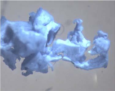


59 Chapter 3. Results 49 (a) (b) (c) (d) (e) (f) (g)
60 Chapter 3. Results Animal Studies Xenotransplantation in Wistar rats Blood count A sample of blood was taken from 36 animals transplanted with HUCPVCs and three control rats who did not receive HUCPVCs, every five days post-transplantation. These samples were used to do a complete blood count and the cell counts from the experimental animals were compared with those from the control animals by the method of generalized estimation equations. For each blood count parameter, a correlation value was calculated to determine the correlation, if any, between the control and experimental values. A p-value was also determined for this correlation parameter based on the null hypothesis that there is no difference between the response variables. These p-values were reported for each blood count parameter in table 3.1. p 0.05 was considered statistically significant, which indicates that the null hypothesis can be rejected. As well for each blood count parameter, the average values (X) and the standard deviation served as descriptive parameters and are shown as time-course plots in figure 3.6. The p-values were not significant ( 0.05) for the white blood cell count (WBC), lymphocyte count (LY#), monocyte count (MON#), platelets (PLT), and mean platelet volume (MPV). All the other parameters had p-values that were significant ( 0.05). In terms of the time course mean values of the parameters that had a p 0.05, the white blood cell count in both the control and experimental animals, increased within five days from the surgery, and remained nearly steady for the remainder of the study period. There was a decrease in the mean white blood cell count in the control animals on day 15 (X control = 5.36 ± /µl vs. X HUCPVC = 8.18 ± /µl), which occured as a result of a larger in two of the three control animals. This decrease promptly returned to the pre-existing levels on day 20 and remained constant until the end of the study period. The experimental animals saw a small drop in mean WBC beginning
61 Chapter 3. Results 51 on day 30 (X HUCPVC = 7.48 ± /µl) and continuing on day 35 (X HUCPVC = 7.25 ± /µl). The mean lymphocyte count followed a similar trend to the WBC, both for the experimental and control animals. This is expected as the lymphocytes made up on average 77.9% of the total WBC. The mean monocyte count in the control and experimental groups saw several perturbations throughout the study period. The control group had the lowest mean monocyte count at 15 days (X control = 0.87 ± /µl) and the highest mean monocyte count at 20 days (X control = 1.77 ± /µl). For the experimental animals, the highest mean monocyte number was seen on day 10 (X HUCPVC = 1.75 ± /µl) and the lowest mean count was seen on day 30 (X HUCPVC = 1.00 ± /µl). The mean granulocyte count in the control group increased beginning on day 5 from basal levels (Day 0: X control = 0.30 ± /µl vs. X HUCPVC = 0.l3 ± /µl) and peaked at day 10 (X control = /µl vs. X HUCPVC = 0.50 ± /µl). It returned close to initial levels by day 15 (X control = vs. X HUCPVC = 0.40 ± /µl) and this was followed by a gradual increase until 35 days which was not seen in the experimental animals. The mean platelet count in the experimental animals decreased initially from 766±172 to 658 ± 195. Conversely, in the control animals, it initially increased from 743 ± 182 to 1089 ± 303 on day 5. A similar increase was seen in the experimental animals however it occured later at 10 days, during which time the platelet count in the control animals began to decrease, reaching its lowest point on day 15 (X HUCPVC = 535 ± 193). Both control and experimental counts leveled off beyond day 20. The mean plasma volume (MPV) also showed variability throughout the study period, with the control animals showing an increase around day 15 that remained until day 30. Contrastingly the experimental mean plasma volume level showed less fluctuation, with the highest value on day 30 (X HUCPVC = 7.65 ± 2.57) and the lowest value on day 40
62 Chapter 3. Results 52 (X HUCPVC = 6.08 ± 1.95). The weights of the rats were also tracked over the course of 40 days, as seen in figure 3.7, and compared to control animals. No significant differences or perturbations in the weights were observed and the animals appeared healthy throughout the study. Table 3.1: p-values in blood count between the experimental group and control rats that received only the CP-PLGA scaffold without HUCPVCs. n=36, rats receiving HUCPVCs. n=3, control rats receiving only scaffold. p-value WBC RBC HGB HCT 1.02E E E-09 MCV MCH MCHC 5.34E E-07 PLT LY# MON# GR# 1.26E-11 RDW MPV Bone healing HUCPVC-seeded constructs were implanted in a femoral defect in the distal metaphysis of male wistar rats. The rate of new bone formation was decreased within the defect area


63 Chapter 3. Results 53 Figure 3.6: Mean blood count parameter time-course values with standard deviations. Experimental, n=36; control, n=3.


64 Chapter 3. Results 54 Figure 3.7: Post-operative weights of animals receiving HUCPVCs+scaffold, and control animals receiving only scaffold. Experimental, n=36; control, n=3. after HUCPVC transplantation compared with controls, as shown by micro computed tomography analysis of the mineral content within the defect seen in figure 3.8a. The difference was statistically significant at seven days (p 0.001) as determined by a student s t-test. This was also apparent by visual inspection of the two-dimensional micro-ct images of femur cross sections, specifically at seven days in figures 3.8b-c. Macroscopic inspection of the femurs at 60 days post surgery (see figures 3.8d-e) revealed that some treated defects had still not completely healed, whereas, control defects showed complete closure. In histological sections (see figure 3.9), a fibrous tissue encapsulation was observed surrounding the HUCPVC treated defects at seven days, and this fibrous tissue persisted surrounding scaffold remnants until 60 days, although it decreased in size throughout the study. This fibrous tissue is collagenous as evidenced by birefringence under polarizing filters (figure 3.10a) and by continuity with new bone when stained with Goldner s trichrome (figure 3.10b). Control defects consisted mainly of osteoid and bone appositional to the scaffold after seven days, with a fibrous tissue distributed throughout the defect. Very little fibrous
65 Chapter 3. Results 55 tissue remained in the defect by 30 days, with some remnants of scaffold remaining. Fibrous tissue was not observed at 60 days in the control defects. In both the control and the experimental animals, the scaffold was resorbed as the study proceeded, and by 60 days it was eventually engulfed in the new trabecular bone. In addition to the impaired healing observed, massive trabecular bone outgrowths appeared towards the distal end of the metaphysis and the epiphyses in one HUCPVC treated femur at 30 days (figure 3.11a), and another at 60 days (figure 3.11c). They also appeared in a control femur at 60 days (figure 3.11b). These growths are unusual and were observed away from the defect site. Survival of human cells and immune cell infiltration Transplanted HUCPVCs were tracked using an anti-human nuclei antibody raised in mouse. In figure 3.12a cells were found near the scaffold in the hematopoietic cell infiltrate occurring in the defect at four days. Some positive staining material (not pictured) appeared diffuse, similar to destroyed nuclei. At seven days, there are very few cells remaining in the extracellular matrix of the defect, as pictured in figure 3.12c. RT-PCR was attempted using the RNA extracted from the femoral defects to detect Figure 3.8 (following page): Healing of femoral defect in Wistar rats transplanted with HUCPVCs. (a) MicroCT measurements of the mineral volume indicates that there is a greater mineral content in the control defects versus the experimental defects, most significantly at 7d. Standard deviations are presented as standard error bars (n=5, for 15d n=4). Asterisks indicates statistically significant difference; *, p (b) Cross sections of femurs taken by micro-ct show greater trabecular bone formation in the cortical space of a control defect (scaffold only) when compared to (c) a HUCPVC treated defect after 7d. (d) A macroscopic view of the femurs from 60d shows complete healing in the control femur as compared to (e) incomplete healing of the defect transplanted with HUCPVCs.

 (c)")


66 Chapter 3. Results 56 (a) (b) (c) (d) (e)
67 Chapter 3. Results 57 human specific genes outline in table 2.3. Results were inconsistent and the extracted RNA appeared to be of poor quality. Femurs from seven days stained with Maximows stain (see figure 3.14b,a,c), used to identify hematopoietic cells in bone marrow, show a significant cell infiltrate in the experimental defect compared to controls. Within this infiltration, in figure 3.14c there are identifiable eosinophils, which suggest that there should also be other immune cells present. CD68 staining, for activated rat macrophages confirms this, showing a greater infiltration of macrophages in the presence of HUCPVCs in the defect site as compared to control scaffolds (see figure 3.14a). Figure 3.9 (following page): Progression of fibrous tissue formation and scaffold resorption in a femoral defect in Wistar rats transplanted with HUCPVCs. (top) H&E cross sections from rat femora transplanted with HUCPVCs+scaffold, (bottom) and control CaP-PLGA scaffolds at 7d, 30d (cross sections) and 60d (longitudinal sections). Faster osteogenesis and scaffold resorption is observed within the femora implanted with empty scaffold. Greater fibrous tissue formation is observed appositional to the scaffolds in the HUCPVC group, persisting until 60d. s - scaffold, example is encircled in yellow; f - fibrous tissue, example is encircled in black, t - trabecular bone; FW = 4.3 mm.

68 Chapter 3. Results 58
 Paired bright field (left) and polarized light images (right) from 10d post-operation defect sections.")
 Goldner s trichrome stain of another 10d section confirms that the extracellular matrix is collagen containing (green, identified by red arrow) and continuous")
69 Chapter 3. Results 59 (a) (b) Figure 3.10: Idenification of fibrous tissue in a femoral defect in Wistar rats transplanted with HUCPVCs at 10 days. (a) Paired bright field (left) and polarized light images (right) from 10d post-operation defect sections. Each show considerable connective tissue matrix which is birefringent, identified by red arrows (thus collagen containing). FW = 858 µm. (b) Goldner s trichrome stain of another 10d section confirms that the extracellular matrix is collagen containing (green, identified by red arrow) and continuous with newly formed bone. Right image is a magnification of inset box; r - FW = 858 µm, l - FW = 343 µm.

 At 30d, trabecular growth is observed mainly within the medulla in femur 23L,")
 At 60d, femur 25R, a control that received only scaffold, reveals a massive")
 Longitudinal section of the same femur shows the outgrowth mainly within the")
at 60d, femur 26R, a femur that received HUCPVCs, reveals an (e) Longitudinal")
70 Chapter 3. Results 60 (a) (b) (c) (d) (e) Figure 3.11: Unusual trabecular outgrowth observed in experimental and control femurs after 30 and 60 days. (a) At 30d, trabecular growth is observed mainly within the medulla in femur 23L, transplanted with HUCPVCs, and confined to a small outgrowth near the epiphysis. (b) At 60d, femur 25R, a control that received only scaffold, reveals a massive outgrowth in the epiphysis, away from the original defect site. (d) Longitudinal section of the same femur shows the outgrowth mainly within the epiphysis. happening through the growth plate. (c)at 60d, femur 26R, a femur that received HUCPVCs, reveals an outgrowth in the epiphysis, away from the original defect site. (e) Longitudinal sections of the same femur at different depths reveal the trabecular growth in the epiphysis of the femur.

 Human nuclear staining (red) at 4 days (c) and 7 days.")
.")

71 Chapter 3. Results 61 (a) (b) (c) (d) Figure 3.12: Human specific staining of HUCPVCs in femoral defects in Wistar rats. (a) Human nuclear staining (red) at 4 days (c) and 7 days. At 4d positive staining cells are near the scaffold, in the cellular infiltration (black arrows). Few HuNu+ cells remain within the extracellular matrix surrounding the scaffold at 7d (one is identified by a black arrow). (b), (d)negative controls, (primary antibody omitted). (a), (c) - FW=343 µm; insets FW=214 µm; (b), (d) - FW=2.14 mm.


 A magnification of (a).")
72 Chapter 3. Results 62 (a) (b) (c) Figure 3.13: Cellular infiltration in HUCPVC-treated defects in Wistar rats. (a) Staining with Maximow s stain for bone marrow reveals a greater hematopoietic celluar infiltration in the presence of HUCPVCs at 7 days post-operatively, compared to (b)empty scaffold control; FW=858 µm. (c) A magnification of (a). Eosiniphils indicated by black arrows; FW=136 µm.
73 Chapter 3. Results 63 Figure 3.14 (following page): CD68 staining in HUCPVC-treated defects in Wistar rats. (a) A greater activated macrophage (brown cells) infiltration is seen in the presence of HUCPVCs at 7 days post-operatively, compared to (c)empty scaffold control; FW=858 µm. (b) A magnification of (a). ED1 + cells indicated by black arrows; FW=343 µm. (d) A magnification of (c) shows fewer ED1 + cells within the defect; FW=343 µm. (e) Negative control (primary antibody omitted); FW=858 µm.

 (c)")


74 Chapter 3. Results 64 (a) (b) (c) (d) (e)
75 Chapter 3. Results Xenotransplantation in athymic rats Tissue engineered constructs, using pooled HUCPVCs from five different donors were cultured as previously described and implanted into a femoral defect in athymic rats (strain: Crl:NIH-Foxn1rnu). This animal model, as compared to the previously used Wistar rats, has a normal B immune system, while functional T lymphocytes are absent. This is similar to the more common NOD/SCID mouse model, which has an impaired ability to make T or B lymphocytes, or activate the complement system, and cannot efficiently fight infections nor reject tumors or transplants. Bone healing The rate of new mineral volume formation in athymic rats was increased within the defect area, as compared to controls, after HUCPVC transplantation. This was demonstrated through morphometry and histological analysis (figure 3.15). Fibrous tissue formation was observed within the HUCPVC defects at four days, which was eventually replaced by new bone after 45 days. The majority of the scaffold was also resorbed by that point (see figure 3.15). HUCPVC-treated defects contain a greater mineral deposition at 15 and 30 days based on micro-ct morphometry (figure 3.16a). When the initial number of seeded cells was increased from 250,000 to 750,000, this enabled an increase in mineral volume when comparing HUCPVC-treated defects to control defects to become statistically significant at 15 days (figure 3.16b). There was no significant difference observed however at 30 days (figure 3.16c)when the initial seeding density was increased to 750,000 cells. Tracking human cells using a fluorescent carbocyanine dye in undecalcified resin sections HUCPVCs were stained with DiR and DiD, fluorescent carbocyanine cell membrane dyes, prior to transplantation. After harvesting the femurs, they were embedded in
76 Chapter 3. Results 66 methylmethacrylate resin, cut, ground, and polished into fine sections. The sections were analyzed by a phase contrast microscope equipped with a fluorescent lamp and optical filters for DiR. In culture plates, fixed cells stained with DiR or DiD and DAPI were detectable as seen in figure Cell membranes were fluorescent, however fluoresence was not detected uniformly throughout the culture as seen in figures 3.17a and 3.17c. Cells stained with DiR were cultured and analysed by flow cytometry in between passaging throughout a 3 week culture period. It was found that the mean fluorescence intensity of the cells decreased almost linearly with increasing population doublings as seen in figure In resin sections from animals transplanted with DiR stained cells, fluorescent cells/signals were not detected in the femoral defects after 15 days as seen in figure In sections from animals transplanted with DiD stained cells, fluorescent cells were also not detected in the femurs due to autofluorescence from the scaffold (not shown). Survival and engraftment of human cells and expression of human osteogenic proteins Paraffin embedded sections of the femurs transplanted with HUCPVCs were stained with anti-human nuclear, anti-human mitochondria, and anti-human osteopontin and osteocalcin. HUCPVCs appeared in a loose connective tissue matrix at seven days (figure 3.19a), which was distinct from the matrix formed by the endogenous rat cells. Some Figure 3.15 (following page): Healing of femoral defect in athymic rats transplanted with HUCPVCs. A femoral defect treated with HUCPVCs at 4d shows infiltration of hematopoietic cells and some fibrous tissue formation. At 7d, fibrous tissue and some regenerated trabecular bone is observed in the HUCPVC-treated defect. At 15d and 45d, fibrous tissue is replaced by trabecular bone, which is eventually resorbed along with the remnants of scaffold; FW = 4.3 mm.

77 Chapter 3. Results 67


.")
78 Chapter 3. Results 68 (a) (b) (c) Figure 3.16: MicroCT analysis of bone healing in athymic rats transplanted with HUCPVCs. (a)microct analysis of femoral defects indicates a greater mineral content in the defect at 15 and 30 days. (b) When the initial cell seeding density was increased from 250,000 to 750,000, the increase in mineral volume at 15 days post-operative is statistically significant, as determined by student s t-test, when compared to a control femur. ** indicates indicates statistical significance (p 0.029, n=5). (b) At 30 days post-operative, however, the difference is no longer statistically significant (p=0.23, n=5).
79 Chapter 3. Results 69 positively staining cells were also observed in newly forming osteoid at the edge of the defect (3.19c), and this HuNu staining was confirmed by detecting positive anti-human mitochondrial stain in the same region(3.19d). At 10 days, the Hunu + cells took on a more rounded morphology and appeared embedded in the new bone (see figure 3.20a). In serial sections, cells in the vicinity of the Hunu + also stained positively for human osteopontin (figure 3.20b). The HuNu staining was also confirmed by observing positive anti-human mitochondrial stain in the same region (figure 3.20c). In serial sections, cells in the vicinity of the human mitochondria + cells also stain for human osteocalcin (figure 3.20e,d). At 15 days, Hunu + cells appeared embedded in trabecular bone near the scaffold remnants. Very few Hunu + cells were detected in the trabecular bone in the defect, relative to the amount of bone that had formed. At 30 days, some osteocytes within the defect stain positively for human nuclear antigen and cells in their vicinity in serial sections, express cytoplasmic human osteocalcin and osteopontin (figure 3.22). Again very few Hunu + cells were detected relative to the size of the defect. At 45 days, Hunu + cells were not found in the bone or extracellular matrix in the defect (not shown). Figure 3.17 (following page): HUCPVCs in culture stained with DiD and DiR. (a) Fluorescence image taken with DiD filter of HUCPVCs in culture stained with DiD. Signal is not uniform throughout. A pseudocolor has been applied to the fluorescence signal using ImageJ software to contrast the dapi staining, FW 2 mm. (b) Higher magnification of (a) taken with DiD filter, FW 120µm. (c) Fluorescence image taken with DiR filter of HUCPVCs in culture stained with DiR, FW 2 mm. (d) Higher magnification of (c) taken with DiR filter, FW 120µm. (e) A plot of the mean fluorescence intensity, as measured by flow cytometry (FL5 filter) of HUCPVCs stained with DiR at variation population doublings after staining. The mean fluorescence intensity decreases in a linear fashion with increased doublings.

 (c)")


80 Chapter 3. Results 70 (a) (b) (c) (d) (e)

 Trans image showing scaffold (black) embedded in")

 Higher magnification trans image of the same section.")
81 Chapter 3. Results 71 (a) (b) (c) (d) Figure 3.18: Resin sections observed by phase contrast microscope under DiR optical filter. (a) Trans image showing scaffold (black) embedded in connective tissue and trabecular bone in defect (b) Corresponding fluorescence image taken with DiR filter shows little signal, FW = mm. (c) Higher magnification trans image of the same section. (d) Corresponding higher magnification fluorescence image, FW = 858 µm.
82 Chapter 3. Results 72 Figure 3.19 (following page): HUCPVCs survive in an athymic rat after seven days. (a) human cells are identified by localization within a loose connective matrix (encircled by black dashed line), distinct from the more dense surrounding endogenous matrix at seven days; FW=858 µm. (c) Hunu + cells are also seen in the newly forming osteoid at the edge of the defect (black arrows) and this is confirmed by (d) positive staining of human mitochondria (black arrows); FW=429 µm. (b) Negative control for (a) (IgG isotype antibody); FW=858 µm. (e) Negative control for (c),(d) (IgG isotype antibody); FW=429 µm.

 (c)")


83 Chapter 3. Results 73 (a) (b) (c) (d) (e)
84 Chapter 3. Results 74 Figure 3.20 (following page): HUCPVCs survive in an athymic rat after 10 days. (a) Hunu + cells appear embedded in the forming bone at 10 days (black arrows). (b) In serial sections, cells are positive for cytoplasmic osteopontin (black arrows); FW=343 µm. (c), staining for human mitochondria in forming bone (black arrows); (d) In serial sections, cells are positive for cytoplasmic osteocalcin (black arrows); FW=429 µm. (e) Another section revealing positive staining of human mitochondria (black arrows); FW=429 µm,( (f) high magnification of (e); FW=172 µm). (g) Negative control for (a),(b) (IgG isotype antibody); FW=429 µm. (h) Negative control for (c),(e),(f),(d) (IgG isotype antibody); FW=429 µm.

 (c)")
 (g)")


85 Chapter 3. Results 75 (a) (b) (c) (d) (e) (f) (g) (h)

 Hunu + cells appear embedded in the forming")
; FW=172 µm.")
86 Chapter 3. Results 76 (a) (b) (c) Figure 3.21: HUCPVCs contribute to new bone in an athymic rat after 15 days. (a) Hunu + cells appear embedded in the forming bone at 15 days (black arrows); FW=429 µm, (b) Magnification of (a); FW=172 µm. (c) Negative control for (a) (IgG isotype antibody); FW=429 µm.
87 Chapter 3. Results 77 Figure 3.22 (following page): HUCPVCs persist in new bone until 30 days. (a) Hunu + osteocytes (identified by black arrows) embedded in trabecular bone, appositional to remnants of scaffold; FW=343 µm. (b) Positive staining of human mitochondria at 30d (black arrows); FW=172 µm. (c) Hunu + and (e) h Osteopontin + positive cells in serial sections at 30d (black arrows); FW=343 µm. (d) Hunu + positive and (f) h Osteocalcin + positive cells in serial sections at 30d (black arrows); FW=343 µm. (g) Negative control for (a),(b)(igg isotype antibody); FW=429 µm. (h) Negative control for (c),(e),(d),(f) (IgG isotype antibody); FW=429 µm.

 (c)")
 (g)")


88 Chapter 3. Results 78 (a) (b) (c) (d) (e) (f) (g) (h)
At the conclusion of this lesson you should be able to:
 Learning Objectives At the conclusion of this lesson you should be able to: Understand the key terms and definitions regarding stem cells Differentiate between the adult and embryonic stem cells Differentiate
Stem cells and tissue engineering
 Stem cells and tissue engineering S. Swaminathan Director Centre for Nanotechnology & Advanced Biomaterials School of Chemical & Biotechnology SASTRA University Thanjavur 613 401 Tamil Nadu Joint Initiative
NPTEL Biotechnology Tissue Engineering. Stem cells
 Stem cells S. Swaminathan Director Centre for Nanotechnology & Advanced Biomaterials School of Chemical & Biotechnology SASTRA University Thanjavur 613 401 Tamil Nadu Joint Initiative of IITs and IISc
Regenerative Medicine and Stem Cell Therapies
 Regenerative Medicine and Stem Cell Therapies Regenerative Medicine Major component of successful regenerated / tissue engineered organs Scaffolds A critical element is the binding of the repopulating
Background. Background. Background. Background. Background 3/4/2013
 3/4/213 Human Very Small Embryonic-like Stem Cells (s) Capacity to Regenerate Bone Tissue: A Potential Cell-based Therapy for Autogenous Bone Grafting Aaron M. Havens, DMD Need to discover an improved
Tissue Engineering. Mesenchymal Stem Cells for. Tissue Engineering
 Tissue Engineering Mesenchymal Stem Cells for Tissue Engineering Reference: Culture of Cells for Tissue Engineering (Culture of Specialized Cells), Chapter 2 Shu-Ping Lin, Ph.D. Date: 03.20.2012 Institute
Lyset BOOST YOUR CELL CULTURE TODAY FOR THE EXPERIMENTS OF TOMORROW
 Lyset BOOST YOUR CELL CULTURE TODAY FOR THE EXPERIMENTS OF TOMORROW Lyset, the human platelet derived supplement for cell culture Among the different alternatives to animal serum, platelet derived preparations
B i o m a t e r i a l s E n g i n e e r i n g
 B i o m a t e r i a l s E n g i n e e r i n g Collagen, a key factor for clinical success INTRODUCTION A REVOLUTIONARY INNOVATION Tecnoss exclusive manufacturing process is able to neutralize the antigenic
About ATCC. Established partner to global researchers and scientists
 Discovering ATCC Primary Immunology Cells - Model Systems to Study the Immune and Cardiovascular Systems James Clinton, Ph.D. Scientist, ATCC July 14, 2016 About ATCC Founded in 1925, ATCC is a non-profit
Cellular Allogeneic Bone Graft
 Cellular Allogeneic Bone Graft Technical Brochure Strips Allograft Chips Allograft Contents Page 1. Properties of Bone Formation...3 2. Stem Cells and Bone Repair...4 3. Multipotent Adult Progenitor (MAPC
Stem Cel s Key Words:
 Stem Cells Key Words: Embryonic stem cells, Adult stem cells, ips cells, self-renewal, differentiation, pluripotent, multipotent, Inner cell mass, Nuclear transfer (Therapeutic cloning), Feeder cells,
BNG 331 Cell-Tissue Material Interactions. Wound Healing I
 BNG 331 Cell-Tissue Material Interactions Wound Healing I Course update LBL 4 Friday; paper posted Need a volunteer to switch groups! Much better job on LBL 3 figure summaries BNG spring seminar 3 this
Tissue Engineered Medical Products
 WORKSHOP 8 Tissue Organizer: Jeremy J. Rawlinson PhD Speakers: Jack E. Lemons, PhD Lawrence J. Bonassar, PhD Mauro R. Alini, PhD Michael J. Yaszemski, MD, PhD Standards for Tissue J. Lemons, University
Stem Cells & Neurological Disorders. Said Ismail Faculty of Medicine University of Jordan
 Stem Cells & Neurological Disorders Said Ismail Faculty of Medicine University of Jordan Outline: - Introduction - Types & Potency of Stem Cells - Embryonic Stem Cells - Adult Stem Cells - ipscs -Tissue
ISCT Telegraft Column: Mesenchymal Stromal Cell (MSC) Product Characterization and Potency Assay Development
 Product Characterization and Potency Assay Development") ISCT Telegraft Column: Mesenchymal Stromal Cell (MSC) Product Characterization and Potency Assay Development University of Wisconsin-Madison, Production Assistance for Cellular Therapies (PACT) Over the
ISCT Telegraft Column: Mesenchymal Stromal Cell (MSC) Product Characterization and Potency Assay Development University of Wisconsin-Madison, Production Assistance for Cellular Therapies (PACT) Over the
Assuring Multipotency of human Mesenchymal Stem Cells (hmsc)
") Assuring Multipotency of human Mesenchymal Stem Cells (hmsc) Introduction Over the past decade, stem cell research has provided new avenues for deeper investigation into tissue repair and aging processes,
Assuring Multipotency of human Mesenchymal Stem Cells (hmsc) Introduction Over the past decade, stem cell research has provided new avenues for deeper investigation into tissue repair and aging processes,
UNIT CELL PROCESSES UNDERLYING TISSUE ENGINEERING AND REGENERATIVE MEDICINE
 Massachusetts Institute of Technology Harvard Medical School Brigham and Women s Hospital VA Boston Healthcare System 2.79J/3.96J/20.441/HST522J UNIT CELL PROCESSES UNDERLYING TISSUE ENGINEERING AND REGENERATIVE
PRIME-XV Cell Therapy Products by
 PRIME-XV Cell Therapy Products by Product List 2017 TRINOVA BIOCHEM now distributes the PRIME-XV Cell Therapy line by Irvine Scientific in Germany, Austria and Switzerland. Irvine Scientific is a worldwide
PRIME-XV Cell Therapy Products by
 PRIME-XV Cell Therapy Products by Product List 2017 TRINOVA BIOCHEM now distributes the PRIME-XV Cell Therapy line by Irvine Scientific in Germany, Austria and Switzerland. Irvine Scientific is a worldwide
Introduction to Cell/ Biomaterial Engineering
 Introduction to Cell/ Biomaterial Engineering Module 3, Lecture 1! 20.109 Spring 2011! Topics for Lecture 1 Introduction to tissue engineering! motivation! basic principles + examples! Introduction to
NEXT GENERATION ECM-BASED ALLOGRAFT TECHNOLOGY:
 NEXT GENERATION ECM-BASED ALLOGRAFT TECHNOLOGY: Potent biological scaffolds strategically control stem cell fate and function, allowing our allografts to harness the regenerative potential of patient s
Introduction to Cell and Biomaterial Engineering! Module 3, Lecture 1!! Spring 2014!
 Introduction to Cell and Biomaterial Engineering! Module 3, Lecture 1!! 20.109 Spring 2014! Topics for Lecture 1! Introduction to tissue engineering! motivation! basic principles! examples! Introduction
YOUR OWN LIFE
 YOUR OWN LIFE Stromal Tissue (ST), a Regenerative Source There is a worldwide consensus that the isolation and collection of regenerative Mesenchymal Stem Cells (MSC s) from differentiated body tissues
Growth factor delivery
 Growth factor delivery S. Swaminathan Director Centre for Nanotechnology & Advanced Biomaterials School of Chemical & Biotechnology SASTRA University Thanjavur 613 401 Tamil Nadu Joint Initiative of IITs
Des cellules-souches dans le poumon : pourquoi faire?
 Des cellules-souches dans le poumon : pourquoi faire? Karl-Heinz Krause Dept. of Pathology and Immunology, Medical Faculty Dept. of Genetic and Laboratory Medicine, University Hospitals Geneva, Switzerland
Adult Stem Cells for Chronic Pain. Dr. John Hughes, DO January 24 th, 2018
 Adult Stem Cells for Chronic Pain Dr. John Hughes, DO January 24 th, 2018 Dr. John Hughes, DO Doctor of Osteopathy From Georgia Arizona College of Osteopathic Medicine - 2007 Aspen Integrative Medicine
Cultrex Rat Mesenchymal Stem Cell Growth and Differentiation Products
 ITM-2011/06/07 Cultrex Rat Mesenchymal Stem Cell Growth and Differentiation Products Background: Mesenc ymal Stem Cells (MSC), also known as marrow stromal cells, are a self - renewing population of multipotent
A NANOFIBROUS HYDROGEL FOR BONE TISSUE ENGINEERING
 A NANOFIBROUS HYDROGEL FOR BONE TISSUE ENGINEERING Umadevi Kandalam, PhD Assistant Professor Department of Pediatric Dentistry College of Dental Medicine Nova Southeastern University Fort Lauderdale, Florida
Adipose rabbit mesenchymal stem cells for the treatment of the chronic scar tissue of the vocal cords
 Adipose rabbit mesenchymal stem cells for the treatment of the chronic scar tissue of the vocal cords Dr. Vasiliki E Kalodimou, Head of Flow Cytometry-Research and Regenerative Medicine Department, IASO-Maternity
Molecular Medicine. Stem cell therapy Gene therapy. Immunotherapy Other therapies Vaccines. Medical genomics
 Molecular Medicine Molecular Medicine Stem cell therapy Gene therapy Tumor therapy Immunotherapy Other therapies Vaccines Genetic diagnostics Medical genomics Medication Diagnostics medicine: old desire
Supplemental Information
 Supplemental Information DLA-matched bone marrow transplantation reverses the immunodeficiency of SCID dogs. Bone marrow transplantation studies were initiated with the goal of reversing the immunodeficiency
Stem cell: a cell capable of 1) tissue plasticity - make different cell types 2) infinite self renewal through asymmetric division
 tissue plasticity - make different cell types 2) infinite self renewal through asymmetric division") Stem cell: a cell capable of 1) tissue plasticity - make different cell types 2) infinite self renewal through asymmetric division stem cell stem cell skin muscle nerve Properties of STEM cells Plasticity
Stem cell: a cell capable of 1) tissue plasticity - make different cell types 2) infinite self renewal through asymmetric division stem cell stem cell skin muscle nerve Properties of STEM cells Plasticity
Nanosystems in regenerative medicine. Jöns Hilborn Materials Chemistry The Ångström Laboratory Uppsala University Sweden
 Nanosystems in regenerative medicine Jöns Hilborn Materials Chemistry The Ångström Laboratory Uppsala University Sweden Outline Motivation for tissue regeneration Cell based approaches Material based
Chapter 8 Healthcare Biotechnology
 Chapter 8 Healthcare Biotechnology Outline: 8.1 Introduction 8.2 Biopharming 8.3 Models of Human Disease 8.4 Detecting and Diagnosing Human Disease 8.5 Monoclonal Antibodies 8.6 Gene Therapy 8.7 Tissue
Contribution and Mobilization of Mesenchymal Stem Cells in a mouse model of carbon tetrachloride-induced liver fibrosis
 Contribution and Mobilization of Mesenchymal Stem Cells in a mouse model of carbon tetrachloride-induced liver fibrosis Yan Liu 1,*, Zhipeng Han 1,*, Yingying Jing 1,*, Xue Yang 1, Shanshan Zhang 1, Chen
SmartPReP 2: The Gold Standard
 The Optimal Platelet Rich Plasma Composition SmartPReP 2: The Gold Standard Harvest Technologies is the leader in developing point-of-care cellular platforms to isolate and concentrate autologous growth
Regulation of advanced blood cell therapies
 Regulation of advanced blood cell therapies www.pei.de Clinical trials using cell-based products Substantially manipulated cells and cells for non-homologous use Quality, safety and non-clinical aspects
Tissue engineering and regenerative medicine an other way of anti-aging? Inducing autologous production of stemcells in human by using PRP
 Tissue engineering and regenerative medicine an other way of anti-aging? Inducing autologous production of stemcells in human by using PRP a change of paradigm and therapeutical perspectives 1 Tissue Engineering
hpsc Growth Medium DXF Dr. Lorna Whyte
 hpsc Growth Medium DXF Dr. Lorna Whyte 27.06.2014 Training from Heidelberg Overview Background: Stem Cells Introduction: Human Pluripotent Stem Cells (hpsc) vs. Adult Stem Cells Promise of PSC Research
Stem Cells: Introduction and Prospects in Regenerative Medicine.
 Stem Cells: Introduction and Prospects in Regenerative Medicine www.gothamgazette.com/.../stemcell/stem_cell.jpg Ode to a Stem Cell, Part II by VCW There once was stem cell stuck in the hood Dividing endlessly,
A review of Dr. Dinakar Golla s clinical research with AdMatrix surgical grafts for soft tissue repair
 TEAMeffort: Using aggressive surgical techniques in combination with AdMatrix (Lattice Biologics acellular dermal scaffold product) to heal difficult and persistent wounds A review of Dr. Dinakar Golla
Production and commercialisation of vascularized and customized bone Clinical trials, market authorisation Pre-clinical trials
 Production and commercialisation of vascularized and customized bone Clinical trials, market authorisation Pre-clinical trials Basic Research: scaffolds, cells/ media, in vivo imaging, modelling www.vascubone.eu
Future of Stem Cell Engineering. Jaeseung Jeong, Ph.D Department of Bio and Brain Engineering KAIST
 Future of Stem Cell Engineering i Jaeseung Jeong, Ph.D Department of Bio and Brain Engineering KAIST Keywords of Stem Cell Engineering g Embryo and Fetus (Foetus) Adult stem cells and embryonic stem cells
Cell-based therapies in tissue engineering and regenerative medicine (TE/RM)
") Cell-based therapies in tissue engineering and regenerative medicine (TE/RM) Pamela Gehron Robey, Ph.D. Craniofacial and Skeletal Diseases Branch National Institutes of Dental and Craniofacial Research
Cell-based therapies in tissue engineering and regenerative medicine (TE/RM) Pamela Gehron Robey, Ph.D. Craniofacial and Skeletal Diseases Branch National Institutes of Dental and Craniofacial Research
Mesenchymal Stromal/Stem Cells
 Mesenchymal Stromal/Stem Cells Peiman Hematti, M.D. Department of Medicine Financial Disclosure: Consulting Fees from Celgene Cellular Therapeutics in 2011 & 2012 1 Caplan used the term Mesenchymal Stem
Introduction to Nanotechnology
 Introduction to Nanotechnology Textbook: Nanophysics and Nanotechnology by: Edward L. Wolf Instructor: H. Hosseinkhani E-mail: hosseinkhani@yahoo.com Classroom: A209 Time: Thursday; 13:40-16:30 PM Office
Physiology Unit 3 HEMATOLOGY
 Physiology Unit 3 HEMATOLOGY In Physiology Today Hematocrit Percentage of blood volume that is erythrocytes 45% in men 42% in women Average blood volume in is 5 L Plasma Large amounts of organic/inorganic
Fundamental properties of Stem Cells
 Stem cells Learning Goals: Define what a stem cell is and describe its general properties, using hematopoietic stem cells as an example. Describe to a non-scientist the current progress of human stem cell
ISTINYE UNIVERSITY INSTITUTE OF HEALTH SCIENCES DEPARTMENT OF STEM CELL AND TISSUE ENGINEERING (THESIS) COURSE DESCRIPTIONS
 COURSE DESCRIPTIONS") ISTINYE UNIVERSITY INSTITUTE OF HEALTH SCIENCES DEPARTMENT OF STEM CELL AND TISSUE ENGINEERING (THESIS) COURSE DESCRIPTIONS 1 st SEMESTER Adult Stem Cell Biology 5 ECTS In this course, the characteristics
ISTINYE UNIVERSITY INSTITUTE OF HEALTH SCIENCES DEPARTMENT OF STEM CELL AND TISSUE ENGINEERING (THESIS) COURSE DESCRIPTIONS 1 st SEMESTER Adult Stem Cell Biology 5 ECTS In this course, the characteristics
Genetics Lecture 19 Stem Cells. Stem Cells 4/10/2012
 Genetics Lecture 19 Stem Cells Stem Cells Much of the excitement about stem cells in the scientific and medical communities comes from their largely untapped and unproven potential for treating human conditions)
PRODUCT REPORT. Premiere Regenerative Medicine.
 PRODUCT REPORT Premiere Regenerative Medicine 01 KEY TERMS Regenerative medicine; Cord blood; Growth factors; Mesenchymal stem cells (MSCs); Vascular enothelial growth factor (VEGF); Fibroblast basic growth
Mesenchymal Stromal Cells Large-scale Culture
 Mesenchymal Stromal Cells Large-scale Culture Adrian Gee Center for Cell & Gene Therapy Baylor College of Medicine Houston, Texas Hanley et al: Efficient manufacturing of therapeutic mesenchymal stromal
Artificial blood vessels
 Artificial blood vessels S. Swaminathan Director Centre for Nanotechnology & Advanced Biomaterials School of Chemical & Biotechnology SASTRA University Thanjavur 613 401 Tamil Nadu Joint Initiative of
Stem Cell Research 101
 Stem Cell Research : A promising type of bioscience research The Stem Cell debate and the impact of the induced pluripotent stem cell procedure Why Is Vocabulary Important? Key terms frame the debate Mature
Results and Discussion
 Growth Kinetics of Human Mesenchymal Stem Cells in a 3L Single-Use, Stirred Tank Bioreactor EMD Millipore Corporation 80 Ashby Rd, Bedford, MA, 01730 www.millipore.com Human mesenchymal stem cells hold
Cell Development, Cell Sources
 Cell Development, Cell Sources 4 4.1 Cell Development The skeletal tissue contains various types of mesenchymal cells, such as osteoblasts, osteocytes, chondroblasts, chondrocytes, myoblasts, adipocytes
Outcomes in Mesenchymal Stem Cell Manufacturing. Athena Russell, MT(AAB) Human Cellular Therapy Laboratory Mayo Clinic Jacksonville, FL
 Human Cellular Therapy Laboratory Mayo Clinic Jacksonville, FL") Outcomes in Mesenchymal Stem Cell Manufacturing Athena Russell, MT(AAB) Human Cellular Therapy Laboratory Mayo Clinic Jacksonville, FL Background HCTL established in 1992 to support BMT programs of Mayo
Outcomes in Mesenchymal Stem Cell Manufacturing Athena Russell, MT(AAB) Human Cellular Therapy Laboratory Mayo Clinic Jacksonville, FL Background HCTL established in 1992 to support BMT programs of Mayo
Cross-Linker Modulation to Maintain Phenotype of RGD-Alginate-Embedded Mesenchymal Stem Cells
 Cross-Linker Modulation to Maintain Phenotype of RGD-Alginate-Embedded Mesenchymal Stem Cells Ashley B. Allen, Hazel Y. Stevens, Robert E. Guldberg. Georgia Institute of Technology, Atlanta, GA, USA. Disclosures:
Introduction to Cell- Biomaterial Engineering!
 Introduction to Cell- Biomaterial Engineering! Module 3, Lecture 1! 20.109 Spring 2010! Topics for Lecture 1!! Introduction to tissue engineerin! motivation"! basic principles + examples"! Introduction
TISSUE ENGINEERING AND REGENERATION: TECHNOLOGIES AND GLOBAL MARKETS
 TISSUE ENGINEERING AND REGENERATION: TECHNOLOGIES AND GLOBAL MARKETS HLC101B August 2014 Yojana Jeevane Project Analyst ISBN: 1-56965-894-3 BCC Research 49 Walnut Park, Building 2 Wellesley, MA 02481 USA
What Are Stem Cells? Where Are These Stem Cells?
 While stem cell therapies may seem to have appeared overnight, they have been practiced for some time prior to the recent burst in new applications and stem cell sources. The most recent developments in
Contents. The Right Surface for Every Cell Extracellular Matrices and Biologically Coated Surfaces ECM Mimetic and Advanced Surfaces...
 Contents The Right Surface for Every Cell... 1 Extracellular Matrices and Biologically Coated Surfaces... 2 Corning Matrigel Matrix... 2 Corning BioCoat Cultureware... 3 ECM Mimetic and Advanced Surfaces...
Erectile and voiding dysfunction: will stem cells solve it all?
 Erectile and voiding dysfunction: will stem cells solve it all? Maarten Albersen MD PhD Dept. of Development and Regeneration Leuven University, Belgium Disclosures No conflicts of interest. ED stem cell
UK +44 (0) CH +41 (0) DE +49 (0) US
 CH +41 (0) DE +49 (0) US") UK +44 (0) 1235 232100- CH +41 (0) 91 604 5522 - DE +49 (0) 69 779099 - US +1 855 267 2464 Featured Product Areas Stem Cell Fate Regulators and Synthetic Retinoid ec23 Recombinant Growth Factor Mimetics
UK +44 (0) 1235 232100- CH +41 (0) 91 604 5522 - DE +49 (0) 69 779099 - US +1 855 267 2464 Featured Product Areas Stem Cell Fate Regulators and Synthetic Retinoid ec23 Recombinant Growth Factor Mimetics
ANAT 3231 Cell Biology Lecture 21 Stem Cells
 ANAT 3231 Cell Biology Lecture 21 Stem Cells Outline What are Stem Cells? Totipotency - Pluripotency - Multipotency What are different sources of Stem Cells? Embryonic vs Adult Pros and Cons for each type
Tissue Engineering: The art of growing body parts. Robby Bowles, Ph.D Cornell University
 Tissue Engineering: The art of growing body parts Robby Bowles, Ph.D Cornell University What is Tissue Engineering? What is Tissue Engineering? TE is an interdisciplinary field that applies the principles
ANAT 2341 Embryology Lecture 18 Stem Cells
 ANAT 2341 Embryology Lecture 18 Stem Cells 29 September 2010 Dr Antonio Lee Neuromuscular & Regenera
Clinical and Research Challenges Associated with Spinal Fusion
 University of Virginia 2016 UVA Orthopaedic Research Retreat April 23, 2016 Clinical and Research Challenges Associated with Spinal Fusion University of Virginia Francis H. Shen, MD Warren G. Stamp Professor
Immunogenicity of Stem Cells in Therapeutic Applications. MDPB-Registry meeting nov 25th Stem cell immunogenicity in therapeutic applications
 Immunogenicity of Stem Cells in Therapeutic Applications MDPB-Registry meeting nov 25th 2016 Stem cell immunogenicity in therapeutic applications Has it been underestimated? Transplantation of any type
10:10-10:22. YIA-1 A study of newly established human peripheral blood monocyte-derived ips cell line used in allergy research 10:22-10:32
 10:10-10:22 YIA-1 A study of newly established human peripheral blood monocyte-derived ips cell line used in allergy research 10:22-10:32 EPA-1 Integration of conventional cell viability assays- recruiting
Strategies for Assessment of Immunotoxicology in Preclinical Drug Development
 Strategies for Assessment of Immunotoxicology in Preclinical Drug Development Rebecca Brunette, PhD Scientist, Analytical Biology SNBL USA Preclinical Immunotoxicology The study of evaluating adverse effects
Tissue Engineering and Regenerative Medicine
 Tissue Engineering and Regenerative Medicine NIH Center for Engineering Complex Tissues (CECT) June 8, 2018 Bhushan Mahadik, Ph.D. Assistant Director, CECT University of Maryland Regenerative Medicine
FDA Regulatory, Compliance and Policy Developments: 361 HCT/Ps
 FDA Regulatory, Compliance and Policy Developments: 361 HCT/Ps September 27, 2018 Presentation by: Elaine H. Tseng Partner FDA and Life Sciences Group King & Spalding Discussion with: Thomas Poché Vice
Pittsburgh Tissue Engineering Initiative Annual Progress Report: 2011 Formula Grant
 Pittsburgh Tissue Engineering Initiative Annual Progress Report: 2011 Formula Grant Reporting Period July 1, 2012 December 31, 2012 Formula Grant Overview The Pittsburgh Tissue Engineering Initiative received
Prof. Steven S. Saliterman. Department of Biomedical Engineering, University of Minnesota
 Department of Biomedical Engineering, University of Minnesota http://saliterman.umn.edu/ Mimicking the fibrillar structure of the extracellular matrix is important for scaffolds. Clinical trails to date
Characterisation of the osteogenic differentiation of human mesenchymal stem cells using Raman spectroscopy Lindsay L. McManus
 Characterisation of the osteogenic differentiation of human mesenchymal stem cells using Raman spectroscopy Lindsay L. McManus IOM3 Young Persons World Lecture Competition São Paulo, Brazil 29 th September
Effects of KAATSU Training on proliferation and differentiation of goat bone marrow mesenchymal stem cells
 Chinese Journal of Laboratory Diagnosis Issued on 25 Aug 2016 Vol. 20, No. 8 P.1240 Effects of KAATSU Training on proliferation and differentiation of goat bone marrow mesenchymal stem cells YANG Yu-hui,
Regenerative Medicine. Speaker Disclosures. What is regenerative medicine? 6/22/2016
 Regenerative Medicine Halland Chen, MD Speaker Disclosures I disclose that I am not a consultant or have any financial interests. 2 What is regenerative medicine? Regeneration of human cells, tissues,
Biocompatibility and War and Peace
 Professor David Williams, D.Sc.,F.R.Eng., Professor and Director of International Affairs, Wake Forest Institute of Regenerative Medicine, North Carolina, USA Editor-in-Chief, Biomaterials President-elect,
Lesson 7A Specialized Cells, Stem Cells & Cellular Differentiation
 Lesson 7A Specialized Cells, Stem Cells & Cellular Differentiation Learning Goals I can explain the concept of cell differentiation and cell specialization. I can explain how the cell structure relates
Differentiation. Ahmed Ihab Abdelaziz MD, PhD Associate Prof. Of Molecular Medicine NewGiza University (NGU)
") Differentiation Ahmed Ihab Abdelaziz MD, PhD Associate Prof. Of Molecular Medicine NewGiza University (NGU) 1 Developmental Genetics Objectives: Explain how a differentiated cell achieves and maintains
Differentiation Ahmed Ihab Abdelaziz MD, PhD Associate Prof. Of Molecular Medicine NewGiza University (NGU) 1 Developmental Genetics Objectives: Explain how a differentiated cell achieves and maintains
1) Determining the best cell sources and scaffold materials for TEHV development.
 Determining the best cell sources and scaffold materials for TEHV development.") Broadly speaking, my primary research interests focus on the development and application of methodologies that can be employed in the basic understanding and optimization of tissue engineered heart valves
Broadly speaking, my primary research interests focus on the development and application of methodologies that can be employed in the basic understanding and optimization of tissue engineered heart valves
Mesenchymal Stem Cell Characterization. Peiman Hematti, M.D. Department of Medicine
 Mesenchymal Stem Cell Characterization Peiman Hematti, M.D. Department of Medicine Terminology has evolved over the years: Colony forming units-fibroblasts (CFU-E) Fibroblast colony forming cells (FCFC)
Developing Targeted Stem Cell Therapeutics for Cancer. Shawn Hingtgen, Ph.D. Assistant Professor UNC Eshelman School of Pharmacy May 22 nd, 2013
 Developing Targeted Stem Cell Therapeutics for Cancer Shawn Hingtgen, Ph.D. Assistant Professor UNC Eshelman School of Pharmacy May 22 nd, 2013 The Challenge of Drug Delivery for Brain Cancer Stem Cells
3.4 DNA sequencing: the Sanger method
 3.4 DNA sequencing: the Sanger method Four separate polymerization reactions are performed 1 3.4 DNA sequencing: the Sanger (dideoxy) method 2 3.4 The polymerase chain reaction 3 3.5 Centrifugation can
Promises and Challenges
 Vladimir Mironov & Richard Visconti Regenerative Medicine: Promises and Challenges What is Regenerative Medicine? Regeneration is a well known biological term and phenomenon. According to the popular Webster
Course Handbook MSc in Bioengineering Tissue Engineering Specialisation
 Course Handbook 2013-2014 MSc in Bioengineering Tissue Engineering Specialisation 1 Course Objectives & Learning Outcomes This programme aims to give a sound and broad basis in tissue engineering. In particular,
WHAT TO KNOW BEFORE GETTING STEM CELLS. Dr. John Hughes, DO Aspen Integrative Medicine June 15th, 2017
 WHAT TO KNOW BEFORE GETTING STEM CELLS Dr. John Hughes, DO Aspen Integrative Medicine June 15th, 2017 Disclaimer: I have no material interest or investment in any stem cell companies or products. I. Introduction
BEH.462/3.962J Molecular Principles of Biomaterials Spring 2003
 Lecture 17: Drug targeting Last time: Today: Intracellular drug delivery Drug targeting Reading: T.J. Wickham, Ligand-directed targeting of genes to the site of disease, Nat. Med. 9(1) 135-139 (2003) Drug
THE SCIENCE OF PLATELET RICH PLASMA
 THE SCIENCE OF PLATELET RICH PLASMA Cheri A. Ong, MD FACS ONG INSTITUTE FOR PLASTIC SURGERY & HEALTH WHAT IS PRP BLOOD PLASMA 55% of the body s today body volume 45% blood cells:- Red blood cells, White
Module 1 Objectives Regenerative Cell Therapy. Module 1. Why Use Regenerative Cell Therapy? What is Regenerative Medicine? Regenerative Therapy Goals
 Module 1 Principles of Regenerative Medicine Module 1 Objectives Regenerative Cell Therapy Learn why to use regenerative cells Learn what they are Learn the relevant mechanisms of action Learn why adipose
Stem cells in Development
 ANAT 2341 Embryology Lab 10 8 Oct 2009 Therapeutic Use of Stem Cells Practical Hurdles & Ethical Issues Stem cells in Development Blastocyst Cord blood Antonio Lee PhD Neuromuscular & Regenerative Medicine
Athought-provoking study, recently published in Nature Reviews Genetics,1
 Australia Athought-provoking study, recently published in Nature Reviews Genetics,1 considered the consequences of an increased exposure to micronutrients. A tricky question, given the widespread, sometimes
THE THERAPEUTIC REALITIES OF STEM CELLS
 Ferghana Partners Group is a Life Sciences investment banking house with offices in London and New York. We specialise in creating and executing effective strategic transactions for pharmaceutical, chemical,
Project Proposal Ultrasound Mediated Tissue Engineering Project Team 21
 Project Proposal Ultrasound Mediated Tissue Engineering Project Team 21 Nicholas Calistri, Aimon Iftikhar, Yvonne Nanakos and Kelly Stratton Project 40 for Dr. Yusuf Khan University of Connecticut Health
Stem cells in Development
 ANAT 2341 Embryology Lab 10 8 Oct 2009 Therapeutic Use of Stem Cells Practical Hurdles & Ethical Issues Stem cells in Development Blastocyst Cord blood Antonio Lee PhD Neuromuscular & Regenerative Medicine
Stem Cells: A Primer National Institutes of Health
 Stem Cells: A Primer National Institutes of Health I. INTRODUCTION Research on stem cells is advancing knowledge about how an organism develops from a single cell and how healthy cells replace damaged
Cells Culture Techniques Marta Czernik
 Cells Culture Techniques 13.03.2018 Marta Czernik Why we need the cell/tissue culture Research To overcome problems in studying cellular behaviour such as: - confounding effects of the surrounding tissues
Stem Cell Research From Bench to Bedside
 Stem Cell Research From Bench to Bedside Stem Cell Dialogues Jan A. Nolta, Ph.D. Professor, Internal Medicine Director, UC Davis Institute for Regenerative Cures October 26, 2010 Stem Cells at UC Davis
Stem Cells: Evidence for Efficacy
 Stem Cells: Evidence for Efficacy Disclosures VA RR&D 1IK2RX001476-01A1 (PI: Harvey Smith) VA RR&D I01 RX002274 (PI: Harvey Smith) Department of Veterans Affairs Medical Center, Philadelphia, PA FDA Orthopaedic