Application of Antibody
|
|
|
- Ethelbert Richards
- 6 years ago
- Views:
Transcription
1 Application of Antibody -Antibody -Western blot -Immunoprecipitation -Immunohistochemical staining -Elisa -Elispot -Flow cytometry -Fluorescenct microscope -EMSA -Affinity purification -Immunotoxin -Mimickry of ligand (activatory, inhibitory) --neutralizing Ab as therapeutics 2013/10/2 Antibodies 1
2 References 1. Antibodies laboratory manual David Lane et al chapter 5,6,14 2. Immunology Kuby et al. chapter 3, /10/2 Antibodies 2
3 Antibody - Classification -Antibody production and diversity -Structure and function -Antibody/antigen interaction -Reagents able to affect Ab 2013/10/2 Antibodies 3
4 Antibody Class Immunoglobulins--- Ig A, IgD, IgE, IgG, IgM. IgG predominates. The light chains have a variable portion that is different in each type of antibodies and is the active portion of the molecule that bind with the antigens. The light chains and the heavy chains are connected by S-S bond. Soluble in water or dilute salt solution. Antibodies can act independently of the rest of the immune sys. 2013/10/2 Antibodies 4
5 Classes of Antibodies Five classes of antibodies (IgA, IgD, IgE, IgG, and IgM) can be distinguished by differences in the heavy chain Differences in the heavy chains (the tail regions) impart distinctive functional properties to the classes of antibodies 2013/10/2 Antibodies 5
6 Classes of Antibodies IgG: Produced during the secondary immune response. Major class secreted into the blood. Activates the complement system. Binds to macrophages and neutrophils inducing phagocytosis. Only antibody to pass from mother to fetus via the placenta. Secreted into the mother s milk. 2013/10/2 Antibodies 6
7 Classes of Antibodies IgM: First antibody produced by B cells. Membrane bound then secreted into the blood in the primary response. Activates the complement system IgD: Produced by activation of resting B cells. Contains same antigen binding sites as IgM. Membrane bound. Not secreted. Function unknown 2013/10/2 Antibodies 7
8 Classes of Antibodies IgA: Principle class of antibodies secreted in saliva, tears, milk, respiratory track, and intestines. IgE: The Fc region binds with high affinity to the surface of mast cells in tissues and basophils in blood. Binding of antigen produces allergic reactions 2013/10/2 Antibodies 8
9 Antibody Production The antigen receptor of a membrane bound antibody molecule is presented on the surface of a resting B cell. Upon activation, B cells proliferate and produce millions of copies of a secreted antibody with the same antigen binding sites. 2013/10/2 Antibodies 9

10 2013/10/2 Antibodies 10
11 Antibody Diversity A human can make at least antibody molecules. Antibodies are proteins encoded by genes. The human genome is thought to contain less than 10 5 genes. How can a human make more antibodies than there are genes in its genome? 2013/10/2 Antibodies 11
12 Antibody Diversity During B cell development, separate gene segments are assembled in a genetic recombination event before the gene is transcribed. Experiments done in 1976 showed that the C-coding and V-coding regions on different parts of the chromosome were spliced together. 2013/10/2 Antibodies 12
13 Combinatorial Diversification In the mouse variable light chain, about 300 different V-regions can be joined to 4 different J segments which are then joined to one C-region. In the mouse variable heavy chain, 500 different V-regions can be joined to 4 J segments and 12 D segments. What is the total number of possible combinations? 2013/10/2 Antibodies 13
14 Combinatorial Diversification Light Variable Chain: 300 x 4 = 1200 Heavy Variable Chain: 500 x 4 x 12 = 24,000 Total possible combinations: 1200 x 24,000 = 28,800, /10/2 Antibodies 14
15 2013/10/2 Antibodies 15
16 2013/10/2 Antibodies 16
17 2013/10/2 Antibodies 17
18 Antibody Structure Consists of four interconnected polypeptide chains. Two heavy chains (H-chains) and joined to two shorter chains (L-chains). These four chains are arranged in the form of a Y ; with the stalk of the Y is called the crystallizable fragment and the top of the Y is known as the antigen-binding fragment. 2013/10/2 Antibodies 18

19 Hv Lv Lc Hc The amino acid sequence and configuration of an antibody were determined in the 1960s by the biochemists Gerald Edelman, an American, and R.R. porter, An English man; for this achievement they shared the 1972 Nobel Prize for physiology or Medicine /10/2 Antibodies 19

20 2013/10/2 Antibodies 20
21 A Fab is the one copy of the variable region of an antibody. 2013/10/2 Antibodies 21
22 2013/10/2 Antibodies 22
23 Antibody Fine Structure Light and heavy chains have variable regions and constant regions (relative to amino acid sequence) The 110 amino acid variable regions are located at the amino-terminal ends and form the antigen binding site Within the variable regions are three hypervariable regions (5 to 10 amino acids) 2013/10/2 Antibodies 23

24 Constant and Variable Regions 2013/10/2 Antibodies 24
25 2013/10/2 Antibodies 25
26 Antibody Domains Both chains are composed of 110 amino acid repeating segments The repeating segments fold independently to form functional domains Each domains is held together by one intrachain disulfide bond The domains evolved by gene duplications 2013/10/2 Antibodies 26

27 2013/10/2 Antibodies 27
28 Three-Dimensional Structure Each domain folds into a very similar three-dimensional structure In the variable domain are three hypervariable loops that form the antigen binding site Changing the length and amino acid sequence of the hypervariable loops does not disturb the threedimensional structure 2013/10/2 Antibodies 28

29 2013/10/2 Antibodies 29

30 2013/10/2 Antibodies 30
31 Structure of Light & Heavy Chains Heavy Chains Genes encoding the constant regions are similarly designated (Cm, Cd, Cg, Ca, Ce) Any individual of a species makes all 5 types of H chains In any one Ab molecule both H chains are identical Several subclasses IgG1, IgG2, IgG3 and IgG4 IgA1 and IgA2 2013/10/2 Antibodies 31
32 Ig Structure Variable Region Part that binds to the epitope Determines the numerous individual specificities Greatest variability in sequence in the N-terminal 110 aa of both the L and H chains Kabat and Wu showed there are three regions of the L and H chains that show the greatest amount of variability hypervariable regions Participate in binding with Ag Form the complementarity-determining regions (CDRs) of the L and H chains Folding brings the the CDR s together Variability in the CDRs provides the diversity in shape and size of the combining site site often takes the form of a cleft or depression thus fit affects affinity and cross-reactivity 2013/10/2 Antibodies 32
33 Variable Region A particular Ab combining site may have the ability to combine with two or more Ags redundancy Cross-reactivity may reduce the number of different Abs needed to defend an individual against the range of Antigenic challenge 2013/10/2 Antibodies 33
34 Immunoglobulin Variants Isotypes 5 different classes of Heavy chains Allotypes Genetic differences between individuals Allelic forms of the same protein Allotypic differences at known loci usually involve changes in only one or two aa in the constant region of a chain Idiotypes The combined antigenic determinants (idiotypes) expressed in the variable region of Ab of an individual that are directed at a particular Ag 2013/10/2 Antibodies 34
35 Immunoglobulin Isotypes Biological Properties of IgG classical Ab Largest % of total Ig and 15% of total protein in serum Equally distributed b/w intra and extra-vascular spaces Four subclasses IgG1, IgG2 and IgG 4 ½ life of approx 23 days longest of all Ig isotypes (IgG3 about 7 days) Most suitable for passive immunization by transfer of ab 2013/10/2 Antibodies 35
36 Antigenic Determinants (Epitopes) The specific site of an antigen that binds to an antibody is called an antigenic determinant or epitope. Most antigens have a variety of epitopes that generate a number of different antibodies that are called polyclonal. A single immune response to an antigen is termed monoclonal. 2013/10/2 Antibodies 36
37 2013/10/2 Antibodies 37
38 Antibody-Antigen Interactions Antibodies have two antigen binding sites that are connected by a hinge region to a common tail region. The bivalent nature of antibodies allows for cross-linking of antigens. The hinge region can expand or contract for flexibility in antigen binding. 2013/10/2 Antibodies 38
39 NATURE OF ANTIGEN-ANTIBODY REACTIONS Lock and Key Concept ---There must be a pretty good fit between the shape of the antigen and shape of the antibody receptorsite. Non-covalent Bonds --- Chemical Bonding( Hydrogen Bonds H-N, H-O) --- Physical Bonding Antibodies are negatively charged whereas antigens are frequently positively charged.( electrostatic) Van der Waals force Hydrophobic Bonding Reversible 2013/10/2 Antibodies 39

40 AFFINITY AND AVIDITY Affinity - Antibody affinity is the strength of the reaction between a single antigenic determinant and a single combining site on the antibody. Affinity is the equilibrium constant that describes the Ag- Ab reaction Avidity - Avidity is a measure of the overall strength of binding of an antigen with many antigenic determinants and multivalent antibodies. 2013/10/2 Antibodies 40

41 SPECIFICITY AND CROSS REACTIVITY Specificity - Specificity refers to the ability of an individual antibody combining site to react with only one antigenic determinant or the ability of a population of antibody molecules to react with only one antigen. Cross reactivity - Cross reactivity refers to the ability of an individual antibody combining site to react with more than one antigenic determinant or the ability of a population of antibody molecules to react with more than one antigen. 2013/10/2 Antibodies 41
42 2013/10/2 Antibodies 42

43 2013/10/2 Antibodies 43
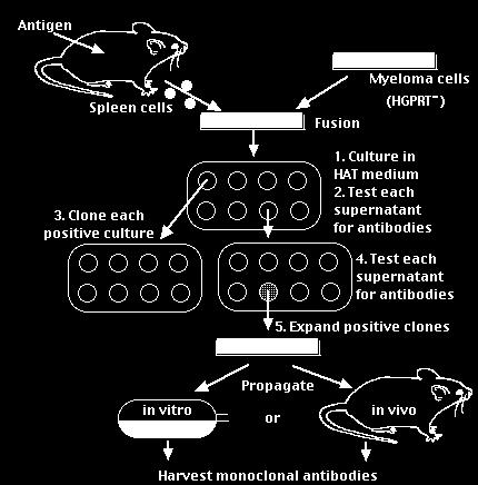
44 Production of monoclonal antibodies -Immunising an animal-usually a mouse -Obtaining immune cells from its spleen -Fusing cells with cancer cells (myeloma) -Tumour of fused cells - hybridoma secretes mab -Cells must grow and form a clone to produce mab Injecting cells into peritoneal cavity of mouse, hybridoma cells multiple and produce fluid (ascites) In-vitro cell culture techniques ages/m/monoclonals.html 2013/10/2 Antibodies 44

45 2013/10/2 Antibodies 45
46 How to choose a region as antigen to immunize animals 2013/10/2 Antibodies 46
47 Deciding which antibody Antibody raised against which portion of the Protein Monoclonal or polyclonal Species cross reactivity Host animal 2013/10/2 Antibodies 47
48 Antibody purification -Most common technique absorption and elution from beads coated with protein A -Protein A binds strongly to sites in 2nd and 3rd constant region of the Fc portion of the immunoglobulin heavy chain -Antibodies bind to protein A chiefly by hydrophobic interactions and are disrupted by transient exposure to low PH -Different classes of IgG vary in sequence in their Fc regions {eg: human IgG1,2,4 high affinity, IgG3, weak} -Unimportant for polyclonal sera, where antibodies against target antigen are distributed throught IgG subclasses -Monoclonal antibodies only one subclass of IgG must be identified prior to purification 2013/10/2 Antibodies 48
49 Terminology Antigen/Immunogen/Antigenic determinants (epitopes) Paratope Priming, boosting, immunization and vaccination Polyclonal antibodies Monoclonal antibodies Memory cells Fab and Fc fragments 2013/10/2 Antibodies 49
50 Western blot/immunoblot : Using Ab to recognize a specific protein 1 0 Ab Ag Enzyme/ Fluorochrome 2 0 Ab 2013/10/2 Antibodies 50
51 I : Western Blotting prepare protein/ag (1) Protein extraction -Total protein standard lysis buffer -Nuclear cytoplasmic extraction (NER-PER, extraction kit, P ierce) To prevent degradation -always centrifuge samples at 4 0 C and -include a protease inhibitor eg Pefabloc (Roche diagnostics) Protein Qantification : Bradford reagent Standard BCA assay (Pierce) 2013/10/2 Antibodies 51
52 I : Western Blotting Electrophoresis (2) Polyacrylamide gels Proteins dissociated: combination of anionic detergent (SDS), reducing agent and heat Polypeptides bind to SDS: ve charge SDS-polypeptide complexes migrate through gel according to size Modifications to polypeptide backbone eg; phosphorylation and glycosylations may have impact 2013/10/2 Antibodies 52
53 I : Western Blotting Electrophoresis (3) SDS-PAGE carried out with discontinuous buffer system Sample and stacking gel Tris-Cl (ph6.8) Resolving gel Tris-Cl (ph8.8) Buffer reservoirs Tris-glycine (ph8.3) - cathode 2013/10/2 Antibodies 53
54 I : Western Blotting Electrophoresis (4) Stacking gel (5%) PH=6.8 Separation gel (>5%) PH=8.8 Constituents : Bis : crosslink monomer of acrylamide SDS : denaturing reagent, TrisCl : Chloride ion front to drive protein into positive electrode. Amonium persulfate : free radical provider TEMED : polymerization stabilizer 2013/10/2 Antibodies 54
55 I : Western Blotting Protein transfer (5) -Wet transfer -Semidry transfer + 3MM paper membrane Gel 3MM paper /10/2 Antibodies 55
56 I : Western Blotting Types of Membranes (6) Nitrocellulose (pore size 0.45 µm) standard membrane Nylon membranes Binds protein with greater affinity, high background Polyvinylidene fluoride (PVDF) high affinity binidiny, pre-wash with methanol Immobilised protein can be visualised with ponceau S (not nylon membranes) 2013/10/2 Antibodies 56
57 I : Western Blotting blocking reagents (7) Western Blotting Horseradish peroxidase 5% non-fat dry milk (Marvel), bovine serum albumin, BLOTTO Alkaline phosphatase 6% casein, 1% polyvinylpyrrolidone, 10mM EDTA in phoshpate-buffered saline, heated to 65 0 C for 1 hour and stored at 4 0 C Immunohistochemistry Sera of animal in which secondary antibody was raised 2013/10/2 Antibodies 57
58 I : Western Blotting Signal Detection (8) Radioiodinated antibodies Antibodies conjugated to enzymes Horseradish peroxidase Alkaline phosphatase Antibodies coupled to biotin Fluorochrome-labelled antibodies : FITC, PE, EC5, PC5 2013/10/2 Antibodies 58
59 I : Western Blotting conjugated enzymes (9) :Hosrseradish peroxidase/alkaline phosphatase 1. Chromogenic Detection: HRP: Diaminobenzidine (DAB)-brown precipitate AP: BCIP/NTP- purple/blue precipitate 2. Chemiluminescent detection HRP: Luminol- oxidised luminol emits blue light AP: AMPPD dephosphorylated product emits light 2013/10/2 Antibodies 59
60 I : Western Blotting Immunodetection (10) 2013/10/2 Antibodies 60

61 2013/10/2 Antibodies 61
62 2013/10/2 Antibodies 62
63 I : Western Blotting Immunoblotting Variables (9) Blocking membranes -5% Blocking buffer (RT or 4 0 C) -incubation time : 60 min ~ O/N Wash 15 min (RT) Primary antibody -concentration :1 mg/ml -time: 1 hour (RT) Wash 15 min (RT) Secondary antibody -concentration :1/10,000 dilution (Sigma) -time: 1 hour (RT) Wash 15 min (RT)+0.5 M NaCl Wash 15 min (RT) Detection: Chromogenic or chemiluminescent 2013/10/2 Antibodies 63
64 Trouble shooting :Non-specific binding -Hyperimmune antisera will contain IgG directed against target antigens and also antibodies directed against other antigens -Antisera may also bind other non-target molecules with low afinity -Antisera can manifest levels of background reactivity that is unacceptably high 2013/10/2 Antibodies 64
65 II. ELISA Enzyme-Linked ImmunoSorbent (ELISA) detecting and quantitating substances such as peptides, proteins, antibodies and hormones. Usually performed on a 96-well plate (Corning s/nunc) Plate coated with capture protein (2-10µg/ml) in buffer Carbonate-Bicarbonate Incubated overnight at 4 0 C Non-specific sites blocked (1% BSA or Super block) Standards/samples added ( µl/well) Antibody directed against antigen of interest conjugated to enzyme HRP/AP Add substrate and detect 2013/10/2 Antibodies 65

66 conjugate bovine serum albumin Second antibodies TNB ntri.tamuk.edu/protocols/ elisa.html 2013/10/2 Antibodies 66


67 Enzyme Linked Immunosorbent Assay (ELISA) based on the measurement of an enzymatic reaction associated Microwell Products with immune complexes The method and principle :By using known amounts of a standard unlabeled antigen one can generate a standard curve relating cpm (Enzyme) bound vs amount of antigen. From this standard curve one can determine the amount of an antigen in an unknown sample. 2013/10/2 Antibodies 67
68 III. Immunohistochemistry Localisation of antigen within Cellular and intracellular localisation Qualitative expression in patient populations; correlations with clinicopathological data 2013/10/2 Antibodies 68
69 Immunohistochemistry variables Cryosections (~7µm) from frozen sections or paraffin embedded sections (~ 5µm) Blocking: endogenous peroxidase H202 ; nonspecific binding with sera from animal in which 20 antibody raised in Concentration of primary antibody (1-5µg) and incubation time 20 antibody, peroxidase conjugated/biotinylated detection with DAB (brown) Counter-stain with haematoxylin (blue) 2013/10/2 Antibodies 69
70 Immunohistochemical localization 2013/10/2 Antibodies 70

71 Tissue microarray 2013/10/2 Antibodies 71
72 IV. Immunofluorescence Co-localisation of two different antigens within tissue Intracellular localisation of antigens 2013/10/2 Antibodies 72
73 Immunofluorescence variables Optimise staining first with immunohistochemistry Primary antibodies 10X concentration Different secondary antibodies with different fluoroscene label Sections mounted in DAKO mounting media, viewed under UV Sections can be mounted with DAPI, stains nuclei blue 2013/10/2 Antibodies 73
74 Flurochrome labelled antibodies When possible use fluorochrome-labeled, affinity purified antibodies Excitation (nm) Emission (nm) Fluorescein (green/yellow) Texas Red (red) R-phycoerythrin (orange/red) 2013/10/2 Antibodies 74
75 Co-localization 2013/10/2 Antibodies 75
76 IV. Immunoprecipitation Antibody directed against a particular antigen is added to cell lysate forming an antibody-antigen complex along with lysis buffer Protein A/G agarose complex is added to the lysate, binding the antigen-antibody complex and precipitating the complex out of solution. After centrifugation, non-specific protein will be removed in the supernatant. The complex is then washed to further remove any non-specific protein binding 2013/10/2 Antibodies 76
77 Application of immunoprecipitation Co-immunoprecipitation is a process that allows the examination of a protein-protein interaction - precipitated out in the complex and detected by Western blotting Activity of immunoprecipitated proteins eg; acetylation assays, DNA footprinting 2013/10/2 Antibodies 77

78 V. EMSA/Gel retardation/(super)band shift assay 2013/10/2 Antibodies 78
79 - +50X Anti-ERa Anti-ERb Anti-SRC1 Anti-SMRT Supershift Estrogen Response Element 2013/10/2 Antibodies 79
80 VI. Antibody (Ab) Microarray A complete microarray-based system for profiling protein expression in biological samples; used to compare two biological samples to measure the relative differences in protein expression. The microarray consists of hundreds of monoclonal antibodies covalently bound in an ordered layout to a glass slide. A protein which can be synthesized in pure form by a single clone (population) of cells. These antibodies can be made in large quantities and have a specific affinity for certain target molecules called antigens which can be found on the surface of cells and those that are malignant. The array can be used as a means to correlate specific proteins with physiological or pathological process of interest, by comparing hundreds of proteins at a time. It is used for toxicity testing, disease investigation, and drug discovery. 2013/10/2 Antibodies 80

81 Factors affecting measurement of Ag/Ab reactions Affinity Avidity Ag:Ab ratio Physical form of the antigen /10/2 Antibodies 81
82 Ab Array Procedure Extraction of total cellular protein from biological samples of interest (eg. Serum samples). Labeling of extracted protein with fluorescent dyes Cy5 and Cy3 (direct labeling, direct labeling with hapten tag, paired Ab sandwich assay). Removal of unbound dye. Incubation of labeled protein with the array. Scanning of the array and the analysis of the results. 2013/10/2 Antibodies 82
83 This procedure is a fluorescence-based analysis; covalently immobilized antibodies are used to capture fluorescently labeled antigens. They do not measure absolute concentrations- instead they provide a relative measure of protein abundance [i.e. the abundance of protein in one sample as compared to another sample]. As part of array development, all antibodies are printed and tested against their specific purified antigen (when available) and against cell lines and tissues samples (for quality control). A reference pool is also used, and similar to the gene expression microarrays, a pool of equal aliquots from each sample to be measured is used, thus ensuring that all proteins from the samples are represented in the reference. 2013/10/2 Antibodies 83

84 2013/10/2 Antibodies 84
 2013/10/2 Antibodies 85")
85 Enzyme linked immusorbent assay (ELISA) 2013/10/2 Antibodies 85
86 Direct Labeling (w/ hapten tag) A convenient method to measure multiple proteins in a complex mixture. All proteins are labeled with either a fluorophore or a hapten tag such as biotin. Advantages: Only one captured antibody per target is required, as compared to the next method- easier to expand detection to new targets for which matched antibody pairs may not be available. Can label different samples with different tags and to co-incubate the samples on the same arrays. Disadvantages: Potential for a high background: all proteins are labeled from the sample, including high concentration proteins such as albumin in serum; nonspecific binding or adsorption of these proteins to Ab could cause interference reduce detection sensitivity or data accuracy. Potential for disruption of antibody-antigen interactions if the labeling reaction severely alters an antigen s binding site. 2013/10/2 Antibodies 86
87 A. Direct Labeling B. Direct Labeling with a hapten tag. C. Paired Ab sandwich assays. 2013/10/2 Antibodies 87
88 Dual Antibody Sandwich Antibodies spotted onto microarray substrates capture specific antigens, and a cocktail of detection antibodies, each antibody matched to one of the spotted antibodies, is incubated on the arrays. Advantages: Quantification of the bound detection antibodies provides a measure of each antigens abundance. Sandwich assays are more sensitive than the direct labeling method because background is reduced through the specific detection of two antibodies instead of one. Disadvantages: The development and validation of assays measuring many targets in parallel is difficult because of the cross reactivity and precipitation when using many detection antibodies. 2013/10/2 Antibodies 88

89 VII. Flow cytometer /FACS 2013/10/2 Antibodies 89

90 2013/10/2 Antibodies 90


91 Flow Cell Injector Tip Sheath fluid Fluorescence signals Focused laser beam 2013/10/2 Antibodies 91
92 Flow Cytometry Optics PMT 4 Flow cell Dichroic Filters PMT 1 Bandpass Filters PMT 2 Laser PMT /10/2 Antibodies 92


93 Optical Filters Dichroic Filter/Mirror at 45 deg Light Source Transmitted Light Reflected light 2013/10/2 Antibodies 93
94 Standard Band Pass Filters White Light Source 630 nm BandPass Filter Transmitted Light nm Light 2013/10/2 Antibodies 94

95 Standard Long Pass Filters 520 nm Long Pass Filter Light Source Transmitted Light >520 nm Light Standard Short Pass Filters 575 nm Short Pass Filter Light Source Transmitted Light <575 nm Light 2013/10/2 Antibodies 95

96 2013/10/2 Antibodies 96
97 THEN THERE WAS FSC AND SSC N U M B E R VOLUME 2013/10/2 Antibodies 97
98 Anti-TCR-gamma-delta-1 PE --> THIS PRODUCES PATTERNS OF CELL CLUSTERS C D CD3+CD4+ CD3-CD4+ CD3+CD4- CD3-CD4- green cyan cyan black CD CD8 TC --> RPCI 2013/10/2 Antibodies 98 LFC
99 VIII. Elispot -Detection of individual cytokine-secreting cells using two high-affinity cytokinespecific antibodies -Capture of secreted cytokines directly at the site of secreting cells -Generation of Spots based on a colorimetric reaction: Spot = Footprint of cells -Spots are permanent and can be quantitated 2013/10/2 Antibodies 99
100 Single cell resolution Advantages High sensitivity(one per million cells) ImmunoSpot plates : synthetic high-proteinbinding membrane No dilution, degradation or absorbtion of cytokines by bystander cells fold more sensitive than ELISA or intracellular cytokine staining True frequency of cells in vivo Easy to set up Rapid and objective analysis of large number of samples using computer-assisted image analysis 2013/10/2 Antibodies 100
101 Technical Aspects ELISPOT Assay 1. Coating the ImmunoSpot plates with Capture antobody 2. Block the Plates Scannnig the plates ImmunoSpot plate scanner Analysis of data ImmunoSpot Software 3. Incubation of cells with stimulating antigens/mitogens usually 24 hours 4. Lysis of Cells Add secondary Detection antobody 5. Detection of antibody-cytokine complex using a chromogen 2013/10/2 Antibodies 101

102 2013/10/2 Antibodies 102


103 HUMAN ELISPOT ASSAY 2013/10/2 Antibodies 103
104 VIII. Enzyme Linked Immunosorbent Assay (ELISA) 1. Based on the measurement of an enzymatic reaction associated with immune complexes. 2. The method and principle :By using known amounts of a standard unlabeled antigen one can generate a standard curve relating cpm (Enzyme) bound vs amount of antigen. From this standard curve one can determine the amount of an antigen in an unknown sample. Coat 1st Ab Ag Wash Coat 2nd Ab* Colorimetry 2013/10/2 Antibodies 104

105 Elisa microplate reader 2013/10/2 Antibodies 105
T-cell response. Taken from NIAID: s.aspx
 T-cell receptor T-cell response 1. Macrophage or dendritic cell digest antigen bacteria, virus 2. Fragments of Ag bind to major histo-compatiblity (MHC) proteins in macrophage. 3. MHC I-Ag fragment expressed
Immunoglobulins. (1 of 2)
") Immunoglobulins (1 of 2) Immunoglobulins (Igs) = antibodies Each B cell synthesizes Igs of single specificity for a specific epitope B cell receptors (BCRs) are the Igs on B cell surface Humoral immunity
Immunoglobulins (1 of 2) Immunoglobulins (Igs) = antibodies Each B cell synthesizes Igs of single specificity for a specific epitope B cell receptors (BCRs) are the Igs on B cell surface Humoral immunity
Immunological Applications. Chapter 8: Background
 Immunological Applications Chapter 8: Background The Immune System Types of Immunity Innate The natural immunity present at birth Acquired A specific response to foreign substances. Some cells remember
Basic Antibody Structure. Multiple myeloma = cancerous plasma cells Monomer = 150,000. Chapter 4. Immunoglobulin Structure and Function
 Chapter 4. Immunoglobulin Structure and Function. Functional Regions. Types of chains. Constant & Variable regions 4. Glycoprotein * * * Heavy chain= 446 aa Light chain= 4aa Each heavy and light chain
Application Note AN001
 Testing hybridoma supernatants with the Spots On Dots Antibody Screening Kit Application Note AN1 Table of Contents Overview... 2 Figure 1. Screening of hybridomas raised against peptide antigens... 3
Lecture 5: 8/31. CHAPTER 5 Techniques in Protein Biochemistry
 Lecture 5: 8/31 CHAPTER 5 Techniques in Protein Biochemistry Chapter 5 Outline The proteome is the entire set of proteins expressed and modified by a cell under a particular set of biochemical conditions.
Immunoglobulins. Structure
 Immunoglobulins Structure Definitions Immunoglobulin is a generic term that refers to a diverse group of molecules found in the blood and tissue fluids They are soluble globulin molecules and they generally
MCB 4211, Fall 2018, Practice Exam 1 Last, First name Student ID # Seat No. ***NOTE: Exam will have 40 multiple choice questions.
 MCB 4211, Fall 2018, Practice Exam 1 Last, First name Student ID # Seat No. ***NOTE: Exam 1 2018 will have 40 multiple choice questions. READ ALL THE CHOICES AND SELECT THE BEST 1. Which of the following
a. Hypoxanthine was present in the media. MCB 4211, Fall 2018, Practice Exam 1 Last, First name Student ID # Seat No.
 MCB 4211, Fall 2018, Practice Exam 1 Last, First name Student ID # Seat No. ***NOTE: Exam 1 2018 will have 40 multiple choice questions. READ ALL THE CHOICES AND SELECT THE BEST 1. Which of the following
Immunoglobulins. Harper s biochemistry Chapter 49
 Immunoglobulins Harper s biochemistry Chapter 49 Immune system Detects and inactivates foreign molecules, viruses, bacteria and microorganisms Two components with 2 strategies B Lymphocytes (humoral immune
Chapter 4 ANTIBODY STRUCTURE AND FUNCTION
 Chapter 4 ANTIBODY STRUCTURE AND FUNCTION Different way to depict an Ig molecule Y In both the heavy and light chain variable regions there is variability at every position and there are hypervariable
Chapter 4. Antigen Recognition by B-cell and T-cell Receptors
 Chapter 4 Antigen Recognition by B-cell and T-cell Receptors Antigen recognition by BCR and TCR B cells 2 separate functions of immunoglobulin (Ig) bind pathogen & induce immune responses recruit cells
Humoral Immune Response. Dr. Iman Hussein Shehata Professor of Medical Microbiology and Immunology
 Humoral Immune Response Dr. Iman Hussein Shehata Professor of Medical Microbiology and Immunology Intended Learning Outcomes By the end of this lesson the student is expected to: 1-Decribe the sequence
Comparing MSD Electrochemiluminescent Detection Technology to Traditional ELISA for Clinical Applications
 Enzyme- linked immunosorbent assays (s) are widely used in clinical settings for detecting substances such as antibodies, peptides, or hormones. However, assays have been gaining in popularity due to advantages
IgG TrueBlot Protocol for Mouse, Rabbit or Goatderived Antibodies - For Research Use Only
 IgG TrueBlot Protocol for Mouse, Rabbit or Goatderived Antibodies - For Research Use Only Introduction The IgG TrueBlot for mouse, rabbit, or goat-derived antibodies represents unique series of respective
Antibody Structure, and the Generation of B-cell Diversity. Chapter 4 5/1/17
 Antibody Structure, and the Generation of B-cell Diversity B cells recognize their antigen without needing an antigen presenting cell Chapter 4 Structure of Immunoglobulins Structure and function Immunoglobulin
It had been determined by several means that the proteins with antibody activity (immunoglobulins) had a molecular weight of approximately 150 kda.
 had a molecular weight of approximately 150 kda.") Immunology Dr. John J. Haddad Chapter 4 Antibody Structure and Function In 1890, Emil von Behring and Shibasaburo Kitasato showed that serum (the straw-colored liquid remaining after blood clots and the
Immunology Dr. John J. Haddad Chapter 4 Antibody Structure and Function In 1890, Emil von Behring and Shibasaburo Kitasato showed that serum (the straw-colored liquid remaining after blood clots and the
THE BASICS OF IMMUNOHISTOCHEMISTRY
 THE BASICS OF IMMUNOHISTOCHEMISTRY Introduction Immunohistochemistry (IHC) identifies specific tissue components by means of a specific antigen/antibody reaction tagged with a visible label. IHC makes
Understanding secondary antibodies
 Understanding secondary antibodies IgG Fragment antigen binding antibodies and isotypes D2 D2 F(ab ) 2 after pepsin cleavage www.abcam.com/secondary_antibody Antibody structure and F(ab) antibodies The
Cdc42 Activation Assay Kit
 A helping hand for your research Product Manual Configuration-specific Monoclonal Antibody Based Cdc42 Activation Assay Kit Catalog Number: 80701 20 assays 1 Table of Content Product Description 3 Assay
BCH 462. Western Blot
 BCH 462 Western Blot Blotting Immunoassay: A test that uses antibody and antigen complexes [immuno-complexes] as a means of generating measurable results. Antigens [Ag]: A substance that when introduced
Arf6 Activation Assay Kit
 A helping hand for your research Product Manual Configuration-specific Monoclonal Antibody Based Arf6 Activation Assay Kit Catalog Number: 82401 20 assays NewEast Biosciences 1 Table of Content Product
Antigens & Antibodies II. Polyclonal antibodies vs Monoclonal antibodies
 A Brief Review of Antibody Structure A Brief Review of Antibody Structure The basic antibody is a dimer of dimer (2 heavy chain-light chain pairs) composed of repeats of a single structural unit known
Protein homology. Antigens & Antibodies I. Administrative issues:
 Administrative issues: Recommended text: Goldsby/Kuby Immunology, 6th edition (Note that Innate Immunity is not adequately covered in the 5th edition.) Text book reading assignments are to supplement the
Immunoglobulins. Light chain ~22-23 KDa whereas the heavy chain ~55-60 KDa
 Immunoglobulins Immunoglobulin (Ig) has a common name which is "Antibody (Ab)", but actually we should say Ig, why? Because the proteins, which are involved, are actually globular proteins "known as globulins"
Serology as a Diagnostic Technique
 Serology as a Diagnostic Technique Characteristics of Any Diagnostic Techniques Any useful detection strategy must be: Specific: yield a positive response for only the target organism or molecule. Sensitive:
Overview of Immunohistochemistry
 Overview of Immunohistochemistry Immunohistochemistry (IHC) combines anatomical, immunological and biochemical techniques to identify discrete tissue components by the interaction of target antigens with
MOLECULAR RECOGNITION
 MOLECULAR RECOGNITION Bioanalytical Methods Classification 1. Biassay: molecular recognition, signal generation and detection in solution or on inert solid phase 2. Biosensor: molecular recognition system
IMMUNOPRECIPITATION (IP)
") 1 IMMUNOPRECIPITATION (IP) Overview and Technical Tips 2 CONTENTS 3 7 8 9 12 13 17 18 19 20 Introduction Factors Influencing IP General Protocol Modifications Of IP Protocols Troubleshooting Contact Us
1 IMMUNOPRECIPITATION (IP) Overview and Technical Tips 2 CONTENTS 3 7 8 9 12 13 17 18 19 20 Introduction Factors Influencing IP General Protocol Modifications Of IP Protocols Troubleshooting Contact Us
PROF. DR. ASMAA HUSSEIN DIRECTOR OF THE MOLECULAR BIOLOGY RESEARCH UNIT
 BY PROF. DR. ASMAA HUSSEIN DIRECTOR OF THE MOLECULAR BIOLOGY RESEARCH UNIT Immunoassay are based on the strong and highly specific interaction occurring between antigens (Ag)) and antibodies (Ab). Ag Ab
Solutions to 7.02 Quiz II 10/27/05
 Solutions to 7.02 Quiz II 10/27/05 Class Average = 83 Standard Deviation = 9 Range Grade % 87-100 A 43 74-86 B 39 55-73 C 17 > 54 D 1 Question 1 (56 points) While studying deep sea bacteria, you discover
RASAQ NURUDEEN OLAJIDE
 RASAQ NURUDEEN OLAJIDE LECTURE CONTENT INTRODUCTION SOUTHERN BLOTTING WESTERN BLOTTING DNA PROFILING (DNA FINGERPRINTING) PARENTAL TESTING PROCEDURES INTRODUCTION Then the Lord said to Cain, Where is your
BIOMAN 2015 IMMUNOASSAYS. Barbara Bielska Northampton Community College Tannersville, PA
 BIOMAN 2015 IMMUNOASSAYS Barbara Bielska Northampton Community College Tannersville, PA 1 Immunoassays An immunoassay is a biochemical test that measures the presence or concentration of a molecules (typically
Rhesus CD16 / FCGR3 ELISA Pair Set
 Rhesus CD16 / FCGR3 ELISA Pair Set Catalog Number : SEK90013 To achieve the best assay results, this manual must be read carefully before using this product and the assay is run as summarized in the General
Rab5 Activation Assay Kit
 A helping hand for your research Product Manual Configuration-specific Monoclonal Antibody Based Rab5 Activation Assay Kit Catalog Number: 83701 20 assays 24 Whitewoods Lane 1 Table of Content Product
SouthernBiotech Custom Services
 SouthernBiotech Custom Services Quality Antibodies for Quality Research Peptide Synthesis for Antibody Production SouthernBiotech provides complete services for production of immunogenic peptides for antibody
SUPPLEMENTAL MATERIAL. Supplemental Methods:
 SUPPLEMENTAL MATERIAL Supplemental Methods: Immunoprecipitation- As we described but with some modifications [22]. As part of another ongoing project, lysate from human umbilical vein endothelial cells
Western blotting technique: principle, procedure and application
 Western blotting technique: principle, procedure and application The term blotting refers to the transfer of biological samples from a gel to a membrane and their subsequent detection on the surface of
RheB Activation Assay Kit
 A helping hand for your research Product Manual Configuration-specific Monoclonal Antibody Based RheB Activation Assay Kit Catalog Number: 81201 20 assays NewEast Biosciences 1 FAX: 610-945-2008 Table
Human Transferrin / TF ELISA Pair Set
 Human Transferrin / TF ELISA Pair Set Catalog Number : SEK11019 To achieve the best assay results, this manual must be read carefully before using this product and the assay is run as summarized in the
IMMUNOCHEMICAL TECHNIQUES
 24 IMMUNOCHEMICAL TECHNIQUES 24.1 INTRODUCTION All vertebrates have advanced immune system. The more complex the organism the more advanced the immune system. The immune system of mammals has evolved over
Attribution: University of Michigan Medical School, Department of Microbiology and Immunology
 Attribution: University of Michigan Medical School, Department of Microbiology and Immunology License: Unless otherwise noted, this material is made available under the terms of the Creative Commons Attribution
IMMUNOPRECIPITATION TROUBLESHOOTING TIPS
 IMMUNOPRECIPITATION TROUBLESHOOTING TIPS Creative Diagnostics Abstract Immunoprecipitation (IP) is the technique of precipitating a protein antigen out of solution using an antibody that specifically binds
A guide to selecting control, diluent and blocking reagents
 Specializing in Secondary Antibodies and Conjugates A guide to selecting control, diluent and blocking reagents Optimize your experimental protocols with Jackson ImmunoResearch Secondary antibodies and
A guide to selecting control, diluent and blocking reagents
 Specializing in Secondary Antibodies and Conjugates A guide to selecting control, diluent and blocking reagents Optimize your experimental protocols with Jackson ImmunoResearch Secondary antibodies and
Chapter 5: Proteins: Primary Structure
 Instant download and all chapters Test Bank Fundamentals of Biochemistry Life at the Molecular Level 4th Edition Donald Voet https://testbanklab.com/download/test-bank-fundamentals-biochemistry-life-molecular-level-
Lab. 7: Serological Tests ELISA. 320 MIC Microbial Diagnosis 320 MBIO PRACTICAL. Amal Alghamdi 2018
 Lab. 7: 320 MIC Microbial Diagnosis Serological Tests ELISA. 320 MBIO PRACTICAL Amal Alghamdi 2018 1 Infection and Immunity Serology is the study of immune bodies in human blood. These are products of
ab Ran Activation Assay Kit
 ab173247 Ran Activation Assay Kit Instructions for Use For the simple and fast measurement of Ran activation. This product is for research use only and is not intended for diagnostic use. Version 1 Last
Mouse PECAM-1/CD31 ELISA Kit (mpecam-elisa)
") Mouse PECAM-1/CD31 ELISA Kit (mpecam-elisa) Cat. No. EK0874 96 Tests in 8 x 12 divisible strips Background Platelet/endothelial cell adhesion molecule-1 (PECAM-1) is an important target GP in DITP. 1 PECAM-1/CD31
Mouse PECAM-1/CD31 ELISA Kit (mpecam-elisa) Cat. No. EK0874 96 Tests in 8 x 12 divisible strips Background Platelet/endothelial cell adhesion molecule-1 (PECAM-1) is an important target GP in DITP. 1 PECAM-1/CD31
Antibodies (Recommended reading: Abbas et al., 4th edition, Chapter 3; Chapter 4; Janeway et al., 5th edition, Chapter 3)
") HST 175 Antibodies (Recommended reading: Abbas et al., 4th edition, Chapter 3; Chapter 4; Janeway et al., 5th edition, Chapter 3) Antibodies protect us from a vast variety of pathogens. Indeed the antibody
HST 175 Antibodies (Recommended reading: Abbas et al., 4th edition, Chapter 3; Chapter 4; Janeway et al., 5th edition, Chapter 3) Antibodies protect us from a vast variety of pathogens. Indeed the antibody
S uf6t<.. f\tj<t1&6t-'l
 Immunagens. An immune response is evoked by a foreign agent called antigen or immunogen. The distinction between these two terms is functional, an antigen is a compound that is capable of binding with
Protein A Agarose Immunoprecipitation Kit
 Protein A Agarose Immunoprecipitation Kit Catalog Number KA0568 20 Reactions Version: 01 Intended for research use only www.abnova.com Table of Contents Introduction... 3 Background... 3 General Information...
Microarray Industry Products
 Via Nicaragua, 12-14 00040 Pomezia (Roma) Phone: +39 06 91601628 Fax: +39 06 91612477 info@lifelinelab.com www.lifelinelab.com Microarray Industry Products Page 10 NBT / BCPIP Chromogenic phosphatase
Anti-HB-EGF (Human) mab
 mab") Page 1 For Research Use Only. Not for use in diagnostic procedures. CODE No. D308-3 Anti-HB-EGF (Human) mab CLONALITY CLONE ISOTYPE QUANTITY SOURCE IMMUNOGEN FORMURATION STORAGE Monoclonal 3H4 Mouse IgG1
Page 1 For Research Use Only. Not for use in diagnostic procedures. CODE No. D308-3 Anti-HB-EGF (Human) mab CLONALITY CLONE ISOTYPE QUANTITY SOURCE IMMUNOGEN FORMURATION STORAGE Monoclonal 3H4 Mouse IgG1
For Research Use Only. Not for use in diagnostic procedures. Anti-NRF2 mab
 Page 1 For Research Use Only. Not for use in diagnostic procedures. Anti-NRF2 mab CODE No. M200-3 CLONALITY CLONE ISOTYPE QUANTITY SOURCE IMMUNOGEN FORMURATION STORAGE Monoclonal 1F2 Mouse IgG1 100 L,
INVESTIGATION OF THE BINDING SPECIFICITY OF IGF-IR USING MONOCLONAL ANTIBODIES
 INVESTIGATION OF THE BINDING SPECIFICITY OF IGF-IR USING MONOCLONAL ANTIBODIES By Mehrnaz Keyhanfar, Pharm.D. A thesis submitted to the University of Adelaide, South Australia in fulfilment of the requirements
RayBio Human IgG1 ELISA Kit
 RayBio Human IgG1 ELISA Kit Catalog #: ELH-IGG1 User Manual Last revised April 15, 2016 Caution: Extraordinarily useful information enclosed ISO 13485 Certified 3607 Parkway Lane, Suite 100 Norcross, GA
Antibody Purification Guide
 Guide Innova Biosciences Guide Innova Biosciences Ltd. Babraham Research Campus, Cambridge, UK, CB22 3AT +44 (0)1223 661000 info@innovabiosciences.com Guide 2 Innova Biosciences specializes in easy to
1. Cross-linking and cell harvesting
 ChIP is a powerful tool that allows the specific matching of proteins or histone modifications to regions of the genome. Chromatin is isolated and antibodies to the antigen of interest are used to determine
SANTA CRUZ BIOTECHNOLOGY, INC.
 TECHNICAL SERVICE GUIDE: Western Blotting 2. What size bands were expected and what size bands were detected? 3. Was the blot blank or was a dark background or non-specific bands seen? 4. Did this same
Gα i Activation Assay Kit
 A helping hand for your research Product Manual Configuration-specific Monoclonal Antibody Based Gα i Activation Assay Kit Catalog Number 80301 20 assays NewEast Biosciences, Inc 1 Table of Content Product
Custom Antibody Services. Antibodies Designed Just for You. HuCAL Recombinant Monoclonal Antibody Generation Service
 Custom Antibody Services Antibodies Designed Just for You HuCAL Recombinant Monoclonal Antibody Generation Service Antibodies Designed Just for You What if there was a technology that would allow you to
Gα 13 Activation Assay Kit
 A helping hand for your research Product Manual Configuration-specific Monoclonal Antibody Based Gα 13 Activation Assay Kit Catalog Number: 80401 20 assays NewEast Biosciences 1 Table of Content Product
Immunological Techniques in Research and Clinical Medicine. Philip L. Cohen, M.D. Chief of Rheumatology, LKSOM 10 March 2016
 Immunological Techniques in Research and Clinical Medicine Philip L. Cohen, M.D. Chief of Rheumatology, LKSOM 10 March 2016 Antibodies Remarkable Tools for Research and Diagnosis You can make an antibody
Antibodies and Antigens In the blood bank
 Antibodies and Antigens In the blood bank 1 Nice game!! http://nobelprize.org/ 2 Karl Landsteiner discovered blood groups in 1901. Awarded Nobel Prize for Physiology or Medicine in 1930 3 Why we study
Hapten - a small molecule that is antigenic but not (by itself) immunogenic.
 immunogenic.") Chapter 4. Antigens Terminology: Antigen: Substances that can be recognized by the surface antibody (B cells) or by the TCR when associated with MHC molecules Immunogenicity VS Antigenicity: Immunogenicity
Chapter 4. Antigens Terminology: Antigen: Substances that can be recognized by the surface antibody (B cells) or by the TCR when associated with MHC molecules Immunogenicity VS Antigenicity: Immunogenicity
IgG1 (Human) ELISA Kit
 ELISA Kit") IgG1 (Human) ELISA Kit Catalog Number KA1730 96 assays Version: 02 Intended for research use only www.abnova.com Table of Contents Introduction... 3 Background... 3 Principle of the Assay... 3 General
IgG1 (Human) ELISA Kit Catalog Number KA1730 96 assays Version: 02 Intended for research use only www.abnova.com Table of Contents Introduction... 3 Background... 3 Principle of the Assay... 3 General
Human C-Reactive Protein / CRP ELISA Pair Set
 Human C-Reactive Protein / CRP ELISA Pair Set Catalog Number : SEK11250 To achieve the best assay results, this manual must be read carefully before using this product and the assay is run as summarized
Segments of the obstructed intestinal loops were fixed in 4% paraformaldehyde
 Supplementary text Supplementary materials and methods Histopathological examination Segments of the obstructed intestinal loops were fixed in 4% paraformaldehyde (PFA) and embedded in paraffin wax with
Immunohistochemistry guide
 Immunohistochemistry guide overview immunohistochemistry Overview Immunohistochemistry is a laboratory technique utilized for the visual detection of antigens in tissue. When working with cells this technique
Mouse IgM ELISpot BASIC
 Mouse IgM ELISpot BASIC Product Code: 3885-2H CONTENTS: Vial 1 (green top) Monoclonal antibody MT6A3 (1.2 ml) Concentration: 0.5 mg/ml Vial 2 (yellow top) Biotinylated monoclonal antibody MT9A2 (100 μl)
Mouse ICAM-1 / CD54 ELISA Pair Set
 Mouse ICAM-1 / CD54 ELISA Pair Set Catalog Number : SEK50440 To achieve the best assay results, this manual must be read carefully before using this product and the assay is run as summarized in the General
Chapter 3. Clonal selection
 Chapter 3. Clonal selection I have called this principle, by which each slight variation, if useful, is preserved, by the term of Natural Selection -Charles Darwin, On the Origin of Species, 1859 4 The
Western blot: proteins separating technique, protocol, theory and trouble shooting
 Journal of Bacteriology & Mycology: Open Access Conceptual Paper Open Access Western blot: proteins separating technique, protocol, theory and trouble shooting Abstract Western blotting is used to visualize
CIHRT Exhibit P-1764 Page 1 IMMUNOHISTOCHEMISTRY ACCURATE LOCALIZATION OF TISSUE OR CELLULAR CONSTITUENTS WITH ANTIBODIES
 CIHRT Exhibit P-1764 Page 1 IMMUNOHISTOCHEMISTRY ACCURATE LOCALIZATION OF TISSUE OR CELLULAR CONSTITUENTS WITH ANTIBODIES CIHRT Exhibit P-1764 Page 2 FUNCTIONAL ROLE OF ANTIBODIES Identify the tissue of
RayBiotech, Inc. Cat # QAM-ISO-G1. Patent Pending Technology. User Manual (Version Sept 2011)
") Quantibody Mouse Immunoglobulin Isotyping Kit -- One step determination of 8 Mouse Immnunoglobulin sub-classes and 2 light chain types in one experiment Patent Pending Technology User Manual (Version Sept
Quantibody Mouse Immunoglobulin Isotyping Kit -- One step determination of 8 Mouse Immnunoglobulin sub-classes and 2 light chain types in one experiment Patent Pending Technology User Manual (Version Sept
SensoLyte Anti-alpha-Synuclein Quantitative ELISA Kit (Human/Mouse/Rat) *Colorimetric*
 *Colorimetric*") Catalog # Kit Size SensoLyte Anti-alpha-Synuclein Quantitative ELISA Kit (Human/Mouse/Rat) *Colorimetric* AS-55550 One 96-well strip plate This kit is optimized to detect human/mouse/rat alpha-synuclein
Catalog # Kit Size SensoLyte Anti-alpha-Synuclein Quantitative ELISA Kit (Human/Mouse/Rat) *Colorimetric* AS-55550 One 96-well strip plate This kit is optimized to detect human/mouse/rat alpha-synuclein
Electrophoresis and transfer
 Electrophoresis and transfer Electrophoresis Cation = positively charged ion, it moves toward the cathode (-) Anion = negatively charged ion, it moves toward the anode (+) Amphoteric substance = can have
Cat. # MK138. For Research Use. Rat IgG EIA Kit. Product Manual. v1012
 Cat. # MK138 For Research Use Product Manual Table of Contents I. Description... 3 II. Principle... 3 III. Kit Components... 4 IV. Materials Required but not Provided... 4 V. Storage... 4 VI. Intended
Human CEACAM6 ELISA Pair Set
 Human CEACAM6 ELISA Pair Set ( CD66c ) Catalog Number : SEK10823 To achieve the best assay results, this manual must be read carefully before using this product and the assay is run as summarized in the
Human CD21 ELISA Pair Set
 Human CD21 ELISA Pair Set Catalog Number : SEKA10811 To achieve the best assay results, this manual must be read carefully before using this product and the assay is run as summarized in the General ELISA
Human SLAMF6 / Ly108 ELISA Pair Set
 Human SLAMF6 / Ly108 ELISA Pair Set Catalog Number : SEK11945 To achieve the best assay results, this manual must be read carefully before using this product and the assay is run as summarized in the General
Human ICAM-2 / CD102 ELISA Pair Set
 Human ICAM-2 / CD102 ELISA Pair Set Catalog Number : SEK10332 To achieve the best assay results, this manual must be read carefully before using this product and the assay is run as summarized in the General
Human IL10RB ELISA Pair Set ( CRFB4 )
") Human IL10RB ELISA Pair Set ( CRFB4 ) Catalog Number : SEK10945 To achieve the best assay results, this manual must be read carefully before using this product and the assay is run as summarized in the
Human IL10RB ELISA Pair Set ( CRFB4 ) Catalog Number : SEK10945 To achieve the best assay results, this manual must be read carefully before using this product and the assay is run as summarized in the
Cyno Monkey IgG Antigen ELISA Kit
 Cyno Monkey IgG Antigen ELISA Kit Catalog No: ICYIGGKT Lot No: SAMPLE INTENDED USE This cynomolgus macaque (Macaca fascicularis) monkey Immunoglobulin G (IgG) antigen assay is intended for the quantitative
Rat IGF-1 ELISA Kit (rigf-1-elisa)
") Rat IGF-1 ELISA Kit (rigf-1-elisa) Cat. No. EK0377 96 Tests in 8 x 12 divisible strips Background Insulin-like growth factor 1 (IGF-1), also known as somatomedin C, is a polypeptide protein hormone similar
Rat IGF-1 ELISA Kit (rigf-1-elisa) Cat. No. EK0377 96 Tests in 8 x 12 divisible strips Background Insulin-like growth factor 1 (IGF-1), also known as somatomedin C, is a polypeptide protein hormone similar
ab IgG Mouse ELISA Kit
 ab151276- IgG Mouse ELISA Kit Instructions for Use For the quantitative measurement of Mouse IgG concentrations in serum and cultured media This product is for research use only and is not intended for
Human IgG ELISpot BASIC
 Human IgG ELISpot BASIC Product Code: 3850-2H CONTENTS: Vial 1 (yellow top) Monoclonal antibodies MT91/145 (1.2 ml) Concentration: 0.5 mg/ml Vial 2 (green top) Biotinylated monoclonal antibodies MT78/145
OpenStax-CNX module: m Antibodies * OpenStax. Abstract
 OpenStax-CNX module: m44823 1 Antibodies * OpenStax This work is produced by OpenStax-CNX and licensed under the Creative Commons Attribution License 3.0 By the end of this section, you will be able to:
Mouse SerpinF2 ELISA Pair Set
 Mouse SerpinF2 ELISA Pair Set Catalog Number : SEK50167 To achieve the best assay results, this manual must be read carefully before using this product and the assay is run as summarized in the General
BIL 256 Cell and Molecular Biology Lab Spring, Development of the Immune System
 BIL 256 Cell and Molecular Biology Lab Spring, 2007 Development of the Immune System Background Information I. Serum Proteins Blood is a remarkable tissue containing cellular elements (erythrocytes, leukocytes
Flow Cytometry - The Essentials
 Flow Cytometry - The Essentials Pocket Guide to Flow Cytometry: 1. Know your Cytometer 2. Understanding Fluorescence and Fluorophores 3. Gating Process 4. Controls 5. Optimization 6. Panel Building 7.
For quantitative detection of mouse IGF-1 in serum, body fluids, tissue lysates or cell culture supernatants.
 Mouse IGF-1 ELISA Kit (migf-1-elisa) Cat. No. EK0378 96 Tests in 8 x 12 divisible strips Background Insulin-like growth factor 1 (IGF-1), also known as somatomedin C, is a polypeptide protein hormone similar
ALP (alkaline phosphatase) calibrators were analyzed manually in microtiter plates to find the linearity range by following this protocol:
 calibrators were analyzed manually in microtiter plates to find the linearity range by following this protocol:") Exam Mol 3008 May 2009 Subject 1 (15p) ALP (alkaline phosphatase) calibrators were analyzed manually in microtiter plates to find the linearity range by following this protocol: Reaction solutions: 50
Exam Mol 3008 May 2009 Subject 1 (15p) ALP (alkaline phosphatase) calibrators were analyzed manually in microtiter plates to find the linearity range by following this protocol: Reaction solutions: 50
Anti-Asian Sea bass (Lates calcarifer) IgM monoclonal antibody labelled with horseradish peroxidase. Product no: C2-HRP
 IgM monoclonal antibody labelled with horseradish peroxidase. Product no: C2-HRP") Anti-Asian Sea bass (Lates calcarifer) IgM monoclonal antibody labelled with horseradish peroxidase Product no: C2-HRP Product Description This monoclonal antibody (Mab) reacts with Asian Sea bass (Lates
Anti-Asian Sea bass (Lates calcarifer) IgM monoclonal antibody labelled with horseradish peroxidase Product no: C2-HRP Product Description This monoclonal antibody (Mab) reacts with Asian Sea bass (Lates
Porcine IL-12/IL-23 p40 ELISA kit
 Porcine IL-12/IL-23 p40 ELISA kit Catalog number: NB-E50014 (96 wells) The kit is designed to quantitatively detect the levels of Porcine IL-12/IL-23 p40 in cell culture supernatants. FOR RESEARCH USE
Product Information. Before you begin. Component A 1 vial of 30 ul vial of 300 ul each Glycerol. Tris
 Glowing Products for Science Mix-n-Stain Antibody Labeling Kits Size: 1 labeling per kit Storage: -20 o C Stability: Stable for at least 1 year from date of receipt when stored as recommended. Components:
Human Junctional Adhesion Molecule A / JAM-A ELISA Pair Set
 Human Junctional Adhesion Molecule A / JAM-A ELISA Pair Set Catalog Number : SEKA10198 To achieve the best assay results, this manual must be read carefully before using this product and the assay is run
ELISPOT and FLUOROSPOT kits
 ELISPOT and FLUOROSPOT kits Interleukins Interferons Granzymes and perforins TNF superfamily ligands and receptors Apoptosis markers And many more... ELISPOT and FLUOROSPOT: a cell-based assay to assess
Human IgG Antigen ELISA Kit
 Human IgG Antigen ELISA Kit Catalog No: IHUIGGKT Lot No: SAMPLE INTENDED USE This human immunoglobulin G antigen assay is intended for the quantitative determination of total human IgG antigen in serum,