Sulforaphane promotes ER stress, autophagy, and cell death: implications for cataract surgery
|
|
|
- Bertram Day
- 6 years ago
- Views:
Transcription
1 JMolMed DOI /s ORIGINAL ARTICLE Sulforaphane promotes ER stress, autophagy, and cell death: implications for cataract surgery Hanruo Liu 1,2 & Andrew JO Smith 2 & Simon SR Ball 2 & Yongping Bao 3 & Richard P Bowater 2 & Ningli Wang 1 & I. Michael Wormstone 2 Received: 7 October 2016 /Revised: 17 December 2016 /Accepted: 20 December 2016 # The Author(s) This article is published with open access at Springerlink.com Abstract Posterior capsule opacification (PCO) commonly develops following cataract surgery and is a wound-healing response that can ultimately lead to secondary visual loss. Improved management of this problem is required. The isothiocyanate, sulforaphane (SFN), is reported to exert cytoprotective and cytotoxic actions, and the latter may be exploited to treat/ prevent PCO. SFN concentrations of 10 μm and above significantly impaired wound-healing in a human lens capsular bag model. A similar pattern of response was also seen with a human lens cell line, FHL124. SFN treatment promoted increased expression of endoplasmic reticulum (ER) stress genes, which also corresponded with protein expression. Evidence of autophagy was observed in response to SFN as determined by increased microtubule-associated protein 1A/ 1B-light chain 3 (LC3)-II levels and detection of autophagic vesicles. This response was disrupted by established autophagy inhibitors chloroquine and 3-MA. SFN was found to promote MAPK signaling, and inhibition of ERK activation using U0126 prevented SFN-induced LC3-II elevation and vesicle formation. SFN also significantly increased levels of reactive oxygen species. Taken together, our findings suggest that SFN is capable of reducing lens cell growth and viability and thus could serve as a putative therapeutic agent for PCO. * I. Michael Wormstone i.m.wormstone@uea.ac.uk Beijing Institute of Ophthalmology, Beijing Tongren Hospital, Capital Medical University, Beijing, China School of Biological Sciences, Norwich Research Park, University of East Anglia, Norwich NR4 7TJ, UK Norwich Medical School, University of East Anglia, Norwich NR4 7TJ, UK Key message & SFN reduces lens epithelial cell growth, migration, and viability. & SFN can promote ER stress and autophagy in lens cells. & SFN promotes MAPK signaling, and inhibition of MEK can suppress SFN-induced autophagy. & ER stress and autophagy in lens cells are likely promoted by ROS production. & SFN may help prevent posterior capsule opacification after cataract surgery. Keywords Sulforaphane. ER stress. Autophagyflux. Lens. Posterior capsule opacification Introduction Cataract renders millions of people blind throughout the world. Despite recent advances in putative cataract treatments, the only currently accepted means of resolving the problem is through surgical intervention [1, 2]. Cataract removal is the most common surgical procedure in the world and is a huge drain on healthcare providers [3, 4]. Posterior capsule opacification (PCO) is the most common complication of cataract surgery and likely the most common cause of nonrefractive decreased postoperative vision [5, 6]. PCO occurs in a significant proportion of cataract surgery patients within 5 years postoperatively, depending on age, geographic location, and the type of intraocular lens (IOL) placed during cataract surgery [2, 7]. PCO reflects the wound-healing process of the lens epithelial cells (LECs) that remain in the capsular bag after cataract surgery. Residual LECs within the capsular bag rapidly grow and proliferate across the posterior lens capsule, which can encroach upon the visual axis and
2 J Mol Med cause a secondary reduction in vision quality [8]. Treatment of PCO is usually straightforward and effective, using the neodymium:yag (Nd:YAG) laser to cut an opening in the posterior lens capsule, thus clearing the visual axis and restoring vision [2]. However, disruption of the posterior capsule due to complications of cataract surgery results in a relative increase in the occurrence of complications such as an elevation in intraocular pressure, retinal cystoid macular edema, glaucoma, intraocular lens damage, iritis, endophthalmitis, and retinal detachment [9]. Nd:YAG treatment is the second most common corrective surgery, which provides further cost to healthcare providers not to mention the reduced quality of life experienced by the patient as PCO develops. Thus, PCO is an important problem, and thus, improved management of this condition is required. A number of strategies have been proposed but are yet to reach the clinic. These include mechanical approaches, which strive to remove all LECs during surgery or by altering shape and materials of the IOL designs [10, 11]. Different pharmaceutical methods to prevent PCO by removingordestroyingresiduallecshavealsobeenproposedthat can either arrest growth, prevent matrix contraction, or destroy the entire lens cell population [12 15]. In the present study, we investigated sulforaphane [1- isothiocyanato-4-(methylsulfinyl)-butane, sulforaphane (SFN)] in the prevention of PCO. SFN is an organic isothiocyanate that is derived from glucosinolates found in cruciferous vegetables [16]. SFN is an intriguing molecule as it is reported to play a role in both cytoprotection and cytotoxicity. These contrasting outcomes are governed by concentration, such that a threshold is eventually reached that is associated with reduced cell viability and death. In the lens, we have previously shown that low micromolar concentrations of SFN do not reduce cell viability or promote cell death [17]. At these lower concentrations, Nrf2 signaling is activated, which leads to increased expression of antioxidant response proteins and thus better prepares cells to manage oxidative stress. Similar responses have also been observed in other cells and tissue [16]. SFN was also found to prevent oxidative stress-induced opacity of cultured whole lenses [17]. With respect to cytotoxic actions of SFN, it has been reported to inhibit tumor growth in many in vivo models by inducing cell cycle arrest and instigating apoptosis [18, 19]. Also, SFN has been proposed to reduce proliferation and promote cell death via ROS generation [20, 21]. In the present study, we aimed to assess whether SFN could reduce cell growth and promote cell death using a human tissue culture model and a human lens cell line, using them as a tool to identify mechanisms that drive these different cellular outcomes. In particular, we elected to investigate the potential involvement of endoplasmic reticulum (ER) stress pathways [22, 23] and autophagy [24, 25]. ER stress is reported to be enhanced by SFN [26, 27]. Similarly, SFN can also initiate autophagy in several cell types [28], which in addition to its housekeeping role can also contribute to cell death in certain circumstances [29]. Our investigation revealed a cytotoxic action of SFN in our human lens capsular bag model, which is an excellent predictive tool for clinical outcomes. Using a human lens cell line, we also identified that SFN can promote ER stress and autophagy in human lens cells. In the case of autophagy, SFN-mediated MAPK signaling appears to play an important role. Materials and methods Human lens capsular bag preparation Simulated cataract operations were performed to create capsular bags from human donor lenses [30] that were obtained with informed consent and used in accordance with the tenets of the Declaration of Helsinki. Approval for the study and experimental protocols (04/Q0102/57) was granted by a national research ethics committee under the Health Research Authority (UK). Using an insulin needle, the anterior capsule was breached approximately 3 mm from the equator, and an incision was made from that point to the center of the capsule. By tugging the flap, created by this incision, with surgical forceps, a continuous curvilinear capsulorhexis was created, such that a disc of anterior capsule was removed, leaving an opening approximately 5 mm in diameter. The resultant window enabled the lens fiber mass to be removed by hydroexpression. Residual fibers were removed by joint irrigation with Hartmann s solution and aspiration. The resultant capsular bag was then dissected free of the zonules and secured on a sterile 35-mm polymethylmethacrylate (PMMA) Petri dish. Eight entomological pins (Anglian Lepidopterist Supplies, Hindolveston, Norfolk, UK) were inserted through the edge of the capsule to retain its circular shape. Capsular bags were maintained in 1.5 ml Eagle s minimum essential medium (EMEM) (Sigma-Aldrich, Poole, UK) and incubated at 35 C in a 5% CO 2 atmosphere. Preparations were exposed to 0, 1, 10, or 100 μm SFN for the first 24 h of culture and then maintained in unsupplemented EMEM for the remaining experimental period (end point at 30 days). Ongoing observations were made using a Nikon phase-contrast microscope (Nikon, Tokyo, Japan). FHL124 human lens cell line FHL124 is a nonvirally transformed cell line generated from human capsule-epithelial explants, showing a 99.5% homology (in transcript profile) with the native lens epithelium [31]. FHL124 cells were routinely cultured at 35 C in a humidified atmosphere of 95% air and 5% CO 2,inEMEMsupplemented with 5% fetal calf serum (FCS) (Gibco, Paisley, UK) and 50 μg/ml gentamicin (Sigma-Aldrich). FHL124 cells were
3 JMolMed seeded on the following: 35-mm tissue culture dishes (30,000/ dish for Western blot, qrt-pcr, TEM, and scratch migration assay), coverslips (10,000) for immunocytochemistry, and 96- well plates (5000/well for MTS (3-(4,5-dimethylthiazol-2-yl)- 5-(3-carboxymethoxyphenyl)-2-(4-sulfophenyl)-2H-tetrazolium) assay and ROS detection assay (Promega, Madison, WI) and lactate dehydrogenase (LDH) assay (Roche). Cell viability A cell proliferation assay (CellTiter 96 AQueous; Promega) was used in accordance with the manufacturer s instructions to assess FHL124 cell viability. This assay is a colorimetric method for determining the number of viable cells in proliferation. The assay is based on the cellular conversion of a tetrazolium salt (MTS) into a formazan product. The resultant absorbance is directly proportional to the number of viable cells in culture. Absorbance was measured at 490 nm with a spectrophotometric plate reader (FLUOstar Omega plate reader; BMG Labtech). Cell death assay A nonradioactive cytotoxicity assay (CytoTox 96R; Roche, Welwyn Garden City, UK) was used to measure the release of LDH from cultured human lens cells. The procedure followed the manufacturer s protocol. The plate was read at 490 nm with a FLUOstar Omega plate reader (BMG Labtech). Scratch wound assay FHL124 cells in each well were allowed to grow to 95% confluence. Cells were then placed in unsupplemented EMEM for a 24-h period. A scratch was made through the sheet of cells using a plastic pipette tip. Photomicrographs were taken immediately after the scratch was made and after 24 h of incubation in experimental conditions. ImageJ1.45s analysis software (available in the public domain at was then used to quantify the initial scratch area and the final area of the scratch. TaqMan qrt-pcr qrt-pcr reactions were performed using an ABI prism 7700 Sequence Detection System (Applied Biosystems, Warrington, UK) under the following conditions: 50 C for 2 min, 95 C for 10 min, and then 40 cycles, each consisting of 15 s at 95 C and 1 min at 60 C. Each reaction was performed in 25 μl and contained reverse transcribed RNA, primers, and probes (sequences for primers and probes are given in Table 1) and TaqMan PCR master mix (Applied Biosystems). Primers and probes were bought as predesigned TaqMan probe and primer sets provided by Applied Biosystems. The threshold cycle (Ct) values, defined as the point at which the fluorescent Table 1 signal is recorded as statistically above background, were obtained using the 7500 Fast system software (Applied Biosystems). Immunoblotting Predesigned TaqMan probe/primer sets for genes of interest Gene name Protein encoded Ref seq TaqMan primer/ probe set AFT6 AFT6 NM_ Hs _m1 ERN1 IRE1 NM_ Hs _m1 EIF2AK3 EIF2α NM_ Hs _m1 HSPA5 BiP NM_ Hs _m1 Cell lysates from FHL124 cells were prepared using Daub s lysis buffer supplemented with 1 mm phenylmethylsulfonyl fluoride (PMSF) and 10 μg/ml aprotinin for 20 min on ice and centrifuged at 16,060 g for 10 min. The protein content was determined by the BCA assay (Bio-Rad, Hemel Hempstead, UK) so that equal amounts of protein per sample were loaded onto 8% SDS polyacrylamide gels and transferred to PVDF membrane using a semidry transfer cell. The membrane was blocked with PBS containing 5% nonfat dry milk and 0.1% Tween-20, hybridized with primary antibody (anti-lc3, (Sigma-Aldrich, Poole, Dorset); anti-erk, anti- JNK, anti-p38, anti-β-actin (Cell Signaling Technology, Beverly, MA, USA), anti-eif-2α, anti-bip/grp78 (BioSource International, Rockville, MD); anti-ire1, anti- ATF6 (Abcam, Cambridge, UK)) followed by incubation with secondary antibody (Amersham Biosciences, Bucks, UK). Proteins were detected using the ECL plus blotting analysis system (Amersham Biosciences). Transmission electron microscopy Cultured FHL124 were treated with 100 μm SFN for 24 h (n = 4) to fixation with 3% gluteraldehyde in 0.1 M phosphate buffer (ph 7.2) and 2% paraformaldehyde (PFA) for 24 h. Following fixation, samples were post-fixed in 1% OsO 4 in the same buffer for 30 min. Appropriate areas for thin sectioning were cut at 70 nm and stained with saturated 2% uranyl acetate and 2% lead citrate before examination on a transmission electron microscope (Libra 120, Carl Zeiss) at 120 kv. Immunofluorescence Cells were maintained in unsupplemented EMEM for 24 h before being placed in experimental conditions for selected periods. Cells were fixed with 4% formaldehyde in PBS for 30 min and permeabilized with PBS containing 0.5% Triton X-100 for 30 min. Preparations were washed three times for
4 J Mol Med 5 min in PBS containing 0.02% w/v BSA and 0.05% v/v IGEPAL. Nonspecific sites were blocked with normal goat or donkey serum (1:50 in 1% w/v BSA in PBS). Following removal of the blocking buffer, rabbit polyclonal primary antibody against microtubule-associated protein 1A/1B-light chain 3 (LC3) (Sigma-Aldrich, Poole, Dorset) diluted 1:200 was applied overnight at 4 C. Cells were subsequently washed with PBS and placed in ALEXA-488 conjugated secondary antibody (1:250; Invitrogen) for 1 h at room temperature. The stained preparations were again washed extensively and mounted on microscope slides with Hydromount mounting medium (National Diagnostics, Hull, UK). Images were viewed using fluorescence microscopy (Axioplan 2; Zeiss), and applicable images were quantified using ImageJ1.45s analysis software (available in the public domain at ROS detection assay ROS levels were measured using a cellular reactive oxygen species detection assay (Abcam, UK) that uses the cell permeant reagent 2,7 -dichlorofluorescin diacetate (DCFDA) to measure hydroxyl, peroxyl, and other ROS activities. This was in accordance with manufacturer instructions. The cells were washed with buffer before being stained with 20 μm DCFDA for 45 min at 37 C, and then washed with buffer again before addition of EMEM and test compounds. The fluorescence (excitation/emission was 485/ 535 nm, respectively) was then measured following a 2-h incubation. Statistical analysis A Student s t test analysis was performed using Excel software (Microsoft, Redmond, WA) to determine any statistical difference between two groups. One-way ANOVA with Tukey s post hoc analysis was used to assess multiple groups when all or many pairwise comparisons were of interest. Oneway ANOVA with Dunnett s post hoc analysis was used to assess all groups compared against the control group. A 95% confidence interval was used to assess significance. Results SFN can reduce lens cell coverage and promote cell death Capsular bags maintained in standard serum-free culture conditions demonstrated progressive cell growth across denuded regions of the anterior capsule, the outer anterior capsule, and, importantly, the previously cell-free posterior capsule. At day 8, cells could be clearly seen on the central posterior capsule. The level of growth in capsular bag preparations treated with 1 μm SFN for the first 24-h period of culture was similar to control preparations (Fig. 1a, b). Cells were also observed on the central posterior capsule with 10 μm SFN, but growth was retarded (Fig. 1a, b). Limited coverage of the central posterior capsule was seen with 100 μm SFN, and indeed, the cells on the anterior capsule appeared distressed at this time point (Fig. 1a, b). Following 30 days of culture (end point), control capsular bags exhibited complete coverage of the posterior capsule (Fig. 1c, d). Exposure to 1 μm SFN for the first 24 h of culture had negligible effect on cell coverage by day 30 (Fig. 1c, d). Capsular bags exposed to 10 μm SFN for 24 h demonstrated a marked reduction in cells growing on the posterior capsule, and 24-h treatment with 100 μm SFN leads to widespread cell death and completely inhibited coverage of the posterior capsule (Fig. 1c, d). Previous work has shown that SFN can reduce cell viability and induce death (by apoptosis) of FHL124 cells [17]; this was determined using the ApoTox-Glo Triplex assay (Promega). In the current study, we verified this response using different biochemical assays. The MTS assay was used to assess cell viability, and it was found that SFN reduced cell viability in a concentration-dependent manner, such that significant differences from control were seen with treatments at 10 μm and above (Fig. 1e). Cell damage/death was observed using the LDH assay, and in this case, significant increases in LDH levels were detected with 10-, 30-, and 100-μM treatments (Fig.1f). We further assessed the impact of SFN on wound-healing of FHL124 cells using the scratch assay method. Following wound formation, control and 1 μm SFN-treated cells were seen to close the denuded region at similar rates. However, significant inhibition of wound closure was observed with 10-, 30-, and 100-μM SFN treatments (Fig. 1g). The findings with the cell line demonstrate that this system is a relevant model to study SFN in relation to PCO and lens cell behavior. We therefore employed this biological tool to further investigate the mechanisms governed by SFN that could contribute to abrogation in the wound-healing response. Promotion of ER stress pathways by SFN In order to investigate the putative involvement of ER stress in SFN-mediated events, real-time PCR and Western blot analysis were employed. FHL124 cells were treated with 10, 30, and 100 μm SFN for 24 h. Real-time PCR revealed increases in all four ER stress gene products tested in FHL124 cells after exposure to SFN for 24 h (Fig. 2). The level of the inositolrequiring enzyme 1 (IRE1) gene (ERN1) in lens cells was significantly increased within 24 h of exposure to 30 μm SFN (Fig. 2d). All genes were significantly elevated at 100 μm SFN (Fig. 2a d). At the protein level, binding immunoglobulin protein (BiP), activating transcription factor 6 (ATF6), eukaryotic translation initiation factor 2α (EIF2α), and IRE1 were significantly upregulated in response to a
, capsulorhexis (arrowed), and outer anterior capsule (AC) captured after a 8andc 30 days.")

5 JMolMed Fig. 1 The effect of SFN on cell survival and growth of human lens cells. Modified dark-field images of four human capsular bag quarters showing the posterior capsule (PC), capsulorhexis (arrowed), and outer anterior capsule (AC) captured after a 8andc 30 days. Cell coverage on the posterior capsule was quantified from dark-field images at b 8 and d 30 days. Data are presented as mean ± SEM (n =4).Asterisk indicates a significant difference between treated and control groups. The effects of SFN exposure to FHL124 cells over a 24-h period on e cell viability, f cell death, and g migration assessed using the MTS, LDH, and scratch wound assays, respectively. The data are presented as mean ± SEM of four independent experiments. Asterisk represents a significant difference between untreated control and treatment groups (p 0.05; ANOVA with Dunnett s post hoc test) Fig. 2 SFN can induce ER stress in FHL124 human lens epithelial cells. Gene expression of a EIF2AK3, b ATF6, c HSPA5, and d ERN1 following 24-h exposure to SFN concentrations, detected by real-time PCR. Values were normalized to 18S gene expression. The data represent mean ± SEM (n =4).Asterisk represents a significant difference between untreated control and treated groups (p 0.05; ANOVA with Dunnett s post hoc test). Detection of ER stress proteins in response to SFN was determined using Western blot methods. e Representative blots showing BiP, ATF6, IRE1, EIF2α, and β-actin levels within FHL124 cells; product bands were observed at 78, 75, 107, 36, and 45 kda, respectively. f Quantitative data derived from band intensities; the protein band intensities for BiP, ATF6, IRE1, and EIF2α were normalized to β-actin. Data are presented as mean ± SEM (n = 4).Asterisk indicates a significant difference between treated and nontreated control groups (p 0.05; ANOVA with Dunnett s post hoc test)

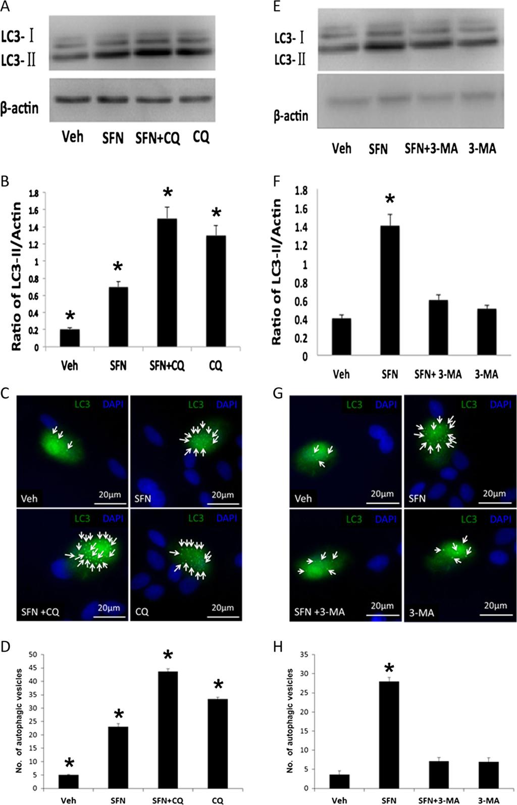
6 J Mol Med 24-h 100-μM SFN treatment. When cells were exposed to 30 μm SFN, there were significant increases in IRE1 and EIF2α expression (Fig. 2e, f). SFN can promote autophagy in lens cells To examine whether SFN could induce autophagy in LEC cells, FHL124 cells were treated with 1, 10, and 100 μm SFN for 24 h. The level of LC3, a well-known protein associated with autophagosome membranes, was analyzed by Western blot. While expression of LC3-I was consistent in all treatment groups, the level of LC3-II in cells treated with SFN was significantly increased compared to control cells without SFN treatment (Fig. 3a, b). To provide further evidence of autophagy in response to SFN, we examined cells using transmission electron microscopy(fig. 3c) and immunofluorescence (Fig. 3d, e). Using these methods, we could observe increased numbers of autophagosomes in cells treated with 10 and 100 μm SFN. The increase of LC3-II could result from several events, including an interruption in autophagosome lysosome fusion, inhibiting lysosome-mediated proteolysis or raising the lysosomal ph [29]. To further clarify whether the increased level of LC3-II by SFN treatment was caused by interrupting the autophagosome lysosome Fig. 4 Chloroquine and 3-MA disrupt SFN-induced autophagy responses in FHL124 human lens epithelial cells. LC3 levels detected using Western blot methods and autophagic vesicle formation in response to 100 μm SFN treatment in the presence and absence of 50 μm chloroquine (a d) and 500 μm 3-MA(e h). Western blots detected products for LC3-I and LC3-II at 18 and 16 kda; a band corresponding to β-actin was detected at 45 kda. Data are presented as mean ± SEM (n = 3).Asterisk indicates a significant difference from all other groups (p 0.05; ANOVA with Tukey s post hoc test) fusion or not, we treated FHL124 cells with 100 μm SFN in the presence of 50 μm chloroquine (CQ) which is a classic lysosomal inhibitor, for 6 h (Fig. 4a d). Using this approach, we observed a significant increase in LC3- II levels relative to control when SFN and CQ were added alone. Application of SFN and CQ together led to a further significant increase. These results were also mirrored using immunofluorescence, which demonstrated significant changes in autophagic vesicle numbers with SFN and CQ, which were further elevated with co-treatment. To further assess the hypothesis that SFN can induce autophagy in FHL124 cells, 3-methyladenine (3-MA) was utilized as an early-stage autophagy inhibitor. Treatment with 100 μm SFN induced significant increases in LC3-II levels and autophagosome numbers. These responses were significantly suppressed by co-treatment with 500 μm 3-MA(Fig.4e h). Fig. 3 SFN can initiate autophagy in FHL124 human lens epithelial cells. LC3-I and LC3-II levels were detected using Western blot methods following 24-h exposure to increasing concentrations of SFN (a, b); a representative blot showing products for LC3-I and LC3-II at 18 and 16 kda (a) and quantitative data (b) are displayed. Data are presented as mean ± SEM (n =4).Asterisk indicates a significant difference between treated and nontreated control groups (p 0.05; ANOVA with Dunnett s post hoc test). Transmission electron micrographs demonstrate the ultrastructure of cells treated with 100 μm SFN(b). A number of autophagic vesicles can be clearly seen with SFN treatment. Fluorescence micrographs showing LC3 distribution in association with autophagic vesicles in control and 100 μm SFN-treated cells (c, d); representative images (c) and quantitative data pooled from three separate experiments (d) are presented. Data are presented as mean ± SEM. Asterisk indicates a significant difference between the treated group and untreated controls (p 0.05; ANOVA with Dunnett s post hoc test)
7 JMolMed
; representative blots showing products at 44/42 kda (phospho")
8 J Mol Med Fig. 5 MAPK signaling involvement in SFN-induced autophagyinfhl124human lens epithelial cells. The effects of 100 μm SFN on p38, JNK, and ERK phosphorylation in conjunction with LC3-II levels detected by Western blot methods (a, b); representative blots showing products at 44/42 kda (phospho and total ERK1/2), 46 kda (phospho and total JNK1), 38 kda (phospho and total p38), and 45 kda (β-actin) (a) and pooled quantitative data (b) are presented. Data are presented as mean ± SEM (n =4). Asterisk indicates a significant difference between treated and nontreated control groups (p 0.05; ANOVAwith Dunnett s post hoc test). The influence of inhibiting ERK phosphorylation (using 5 μm U0126)onSFN induced LC3-II expression (c, d) and autophagic vesicles (e, f). Western blots detected products for LC3-I and LC3-II at 18 and 16 kda; phospho and total ERK1/2 were detected at 44/ 42 kda; a band corresponding to β-actin was detected at 45 kda. Data are presented as mean ± SEM (n =3).Asterisk indicates a significant difference from all other groups (p 0.05; ANOVA with Tukey s post hoc test) MAPK signaling is critical for SFN-induced autophagy The mitogen-activated protein kinases (MAPKs) have been reported to promote autophagy as downstream mediators of ROS [28]. Therefore, we analyzed the level of several phosphorylated MAPKs including p38, c-jun N-terminal kinase (JNK), and ERK by immunoblotting to examine if MAPKs are involved in SFN-induced autophagy in FHL124 cells. One hundred micromolars SFN did not induce a detectable change in the level of phospho-specific p38 at any time point tested. A significant but relatively weak increase in pjnk level was observed at the 24-h time point. In contrast, levels of phosphorylated ERK1/2 (perk) in cells treated with SFN (100 μm) showed significant increases at 6-, 12-, and 24-h time points (Fig. 5a, b). This increase in ERK activation preceded detectable changes in LC3-II levels (Fig. 5a, b). These results indicate that ERK activation might be involved in SFN-induced autophagy. We therefore tested this notion by disrupting ERK activation using the MEK inhibitor U0126 (Fig. 5c f). FHL124 cells were pretreated with U0126 (5 μm) for 30 min before SFN treatment. Application of U0126 significantly reduced SFN-induced increase in LC3- II levels and autophagic vesicle incidence. Finally, it has been suggested that SFN-induced activation of ERK is mediated by ROS. We therefore determined whether SFN could induce ROS levels in FHL124 cells. Application of 100 μm SFN was found to significantly increase ROS relative to controls (Fig. 6). Discussion The influence of SFN on the physiology of human cells has been studied extensively in recent years, but this work has
.Asterisk indicates a significant difference between treated and nontreated control groups (p 0.05; Student s t test) largely focused on cancer [16, 32].")
9 JMolMed Fig. 6 SFN can increase ROS production in FHL124 human lens epithelial cells. The effect of 100 μm SFN on ROS production was detected2hfollowinginitialexposure.dataarepresentedas mean ± SEM (n = 4).Asterisk indicates a significant difference between treated and nontreated control groups (p 0.05; Student s t test) largely focused on cancer [16, 32]. However, the functional role of SFN in the prevention of PCO, which affects a large proportion of cataract patients, has not been investigated. In the present study, it was therefore deemed an important opportunity to test the hypothesis that SFN can inhibit the development of PCO. Evaluation of SFN in a human lens capsular bag model supported this notion, and the effective concentrations required mirrored results obtained in a human lens epithelial cell line, FHL124. This therefore suggested that the readily available cell line could be employed to establish mechanisms that underpin SFN-induced effects. The present study demonstrates that SFN can induce ER stress, which was observed by induction of ER stress gene and protein expression. An important role of the ER is to sense environmental and physiological stresses. This response is mediated by activation and repression of pathways resulting in specific functional outcomes. The ER is the site of protein synthesis, where folding and trafficking are initiated, and also the mediator of internal and external stresses [22]. ER stress can arise because unfolded or misfolded proteins are produced within the ER. Normal levels of unfolded proteins can be counteracted by a number of ER chaperone proteins, but if the level continues to increase, for example, through prolonged oxidative or osmotic stress, then it is detected by the molecule BiP, which in turn activates one or more of three stress pathway initiators (perk, IRE1, and ATF6). Once a prolonged stress is sensed, then a range of external pathways are initiated and cell death through apoptosis can result [23]. Application of the classical ER stress molecule thapsigargin leads to an inhibition of lens cell growth and, ultimately, to cell death through apoptosis [33, 34]. It has also been reported that diabetic and oxidative stresses to lens cells can give rise to an ER stress response, seen as an increase in BiP production and caspase activation [35]. Arsenic trioxide (As 2 O 3 ) is also known to promote ER stress and can reduce lens cell viability [36]. Combinatorial treatment with SFN and As 2 O 3 or other known ER stressors such as thapsigargin has been employed in the management of different cancers [26, 27] and therefore might provide a promising therapeutic approach for PCO. It has been suggested that SFN can increase ROS production [20, 28], and in the current study, we confirm that lens cells show elevated ROS levels in response to SFN. Interestingly, as discussed above, SFN exposure in combination with As 2 O 3 can result in a dramatic increase in levels of ROS compared to treatment with either agent alone [26]. SFN, alone or with As 2 O 3, decreased intracellular glutathione (GSH) content. Furthermore, addition of the free radical scavenger N-acetyl-L-cysteine (NAC) rescued cells from As 2 O 3 / isothiocyanate-mediated cytotoxicity [26]. As As 2 O 3 has been associated with ER stress in the lens and investigated for its potential benefits in the prevention of PCO [36], this suggests that SFN deserves further investigation in combination with As 2 O 3 in the treatment of PCO and it will be interesting to establish the role of ROS in SFN-induced ER stress. Autophagy is a highly controlled process that can be affected by several conditions such as metabolic stress, ER stress, oxidative stress, and hypoxia [37]. The process of autophagy involves the formation of double-membraned vesicles (autophagosomes), which encapsulate the cytoplasm and organelles and fuse with lysosomes, leading to degradation of the contents of the vesicle. It has been associated with specific pathologies including cancer, liver disease, and neurodegeneration by a growing number of studies [38]. Brennan et al. [39] established that autophagy occurs in normal human lens cells, with 42 autophagy genes expressed and a number of autophagosomal proteins detected in both the lens epithelium and fibers. The housekeeping role of autophagy may be particularly important in normal lens maintenance, homeostasis, and fiber differentiation [40, 41]. The loss of lens-specific autophagy-related 5 (Atg5) has been reported to result in age-related cataract formation, and Pik3c3/Vps34 genes are shown to leading cortical cataract in lens [42]. In the present study, we clearly show that SFN is capable of promoting autophagy flux, which occurs in association with reduced viability and increased cell death. With respect to PCO, previous work using in vitro canine lens capsular bags has indicated that cyclosporine A (CsA)- induced cell loss is attributed to an autophagy-related mode of cell death rather than classical apoptosis [43]. While autophagy is typically linked to general maintenance of cells, its relationship with cell death is beginning to emerge [29] and further investigation of the links between cell death and autophagy in response to SFN will be of interest in the future. ERK is a member of the MAPK family that is involved in many aspects of cell biological functions such as proliferation,
10 J Mol Med migration, differentiation, and death [44, 45]. MAPKs such as p38, ERK, and JNK have been found to have downstream effects of ROS in autophagy induction [46, 47]. Here, we showed that SFN can induce ROS and activate ERK, which in turn facilitates autophagy in human lens epithelial cells. It is speculated that SFN generates ROS, which could activate ERK. A link between ROS and ERK activation is recognized, and it is proposed that this event could occur through several putative mechanisms [48]. One possibility is the activation of upstream growth factor receptors, such as EGF receptor, which have been reported to be activated in response to oxidative stress [49]. EGFR is expressed in both FHL124 cells and the native lens. It is also possible that ROS could alter protein structure of signaling molecules and in doing so promote signaling activity [48]. It is also feasible that MAPK phosphatases, the negative regulators of MAPK signaling, are deactivated and degraded by ROS [48]. Removal of this negative regulator would therefore enable persistent activation of ERK to take place and may best explain the results observed. Signaling mediated through ERK is often associated with growth and survival, but it is also linked to death pathways [45]. It has been suggested that ERK activity can play a role in mediating extrinsic and intrinsic apoptotic pathways. In the case of SFN treatment, sustained ERK activity is likely to facilitate promotion of the intrinsic apoptotic pathway that is associated with release of pro-apoptotic factors from the mitochondrion that will lead to caspase-9 activation, which will then activate caspase3/7 [45]. The role of ERK in autophagy is also very interesting. Autophagy itself serves an important role in cell maintenance but in some cases can itself lead to cell death [29, 45]. The mode of cell death could be through conventional apoptosis, but it is also often reported that a distinct autophagy-induced cell death can arise that not only has some features common with conventional apoptosis but also presents distinct morphological changes leading to death. It will be of great interest to tease out the relationship between ROS, ERK, and cell death using free radical scavengers and MEK inhibitors to achieve this. Considering the data as a whole, it is possible to speculate on the sequence of events that may take place following SFN application that ultimately lead to cell death. The primary initiating event is likely to be the generation of free radicals within the cell that will result in an oxidative stress that triggers an ER stress response and promote ERK activity. These two events are both known to stimulate autophagy, and, thus, increased breakdown of cellular material will result. ER stress is associated with reduced protein synthesis, and, thus, a restricted capacity to replace damaged organelles will compromise the cells and trigger death pathways. Sustained ERK activity is likely to deplete mitochondrial numbers, which will in turn activate caspase-mediated cell death. Similarly, a sustained ER stress could also promote caspase-mediated cell death pathways. In summary, we have shown that SFN can reduce lens cell viability in both a human lens cell line and a tissue culture model, which has implications for the treatment of PCO. Moreover, we have demonstrated for the first time that SFN can promote ER stress and autophagy in lens cell. SFNinduced autophagy requires MAPK signaling, and we speculate that both ER stress and autophagy are regulated by ROS production. Acknowledgements We would like to thank all members of the Eye Research Group at the University of East Anglia and Prof. Jenny Gill (UEA) for advice relating to statistical analysis. We also wish to thank Prof. Xiuhua Wan for advice and support. We would also like to express our gratitude to the staff of the East Anglian Eye Bank and Beijing Tongren Eye Bank. We greatly appreciate funding support from the Humane Research Trust (UK), the Beijing New Star of Science and Technology Fund (H ), the Fund of Work Committee for Women and Children of China State Department ( ), the National Natural Science Fund Projects of China ( ), and the Beijing Scholars of Beijing Municipal Government. Compliance with ethical standards Human donor lenses were obtained with informed consent and used in accordance with the tenets of the Declaration of Helsinki. Approval for the study and experimental protocols (04/Q0102/57) was granted by a national research ethics committee under the Health Research Authority (UK). Conflict of interest interest. The authors declare that they have no conflict of Financial support The Humane Research Trust (UK), the Beijing New Star of Science and Technology Fund (H ), the Fund of Work Committee for Women and Children of China State Department ( ), the National Natural Science Fund Projects of China ( ), and the Beijing Scholars of Beijing Municipal Government Open Access This article is distributed under the terms of the Creative Commons Attribution 4.0 International License ( creativecommons.org/licenses/by/4.0/), which permits unrestricted use, distribution, and reproduction in any medium, provided you give appropriate credit to the original author(s) and the source, provide a link to the Creative Commons license, and indicate if changes were made. References 1. Wormstone IM, Eldred JA (2016) Experimental models for posterior capsule opacification research. Exp Eye Res 142: Wormstone IM, Wang L, Liu CS (2009) Posterior capsule opacification. Exp Eye Res 88(2): Brown GC, Brown MM, Menezes A, Busbee BG, Lieske HB, Lieske PA (2013) Cataract surgery cost utility revisited in 2012: a new economic paradigm. Ophthalmology 120: Smith AF, Klotz A, Wormstone IM (2016) Improving the drug development process by reducing the impact of adverse events: the case of cataracts considered. Drug Discov Today 21(3): Schaumberg DA, Dana MR, Christen WG, Glynn RJ (1998) A systematic overview of the incidence of posterior capsule opacification. Ophthalmology 105:
11 JMolMed 6. Apple DJ, Solomon KD, Tetz MR, Assia EI, Holland EY, Legler UF, Tsai JC, Castaneda VE, Hoggatt JP, Kostick AM (1992) Posterior capsule opacification. Surv Ophthalmol 37: Mootha VV, Tesser R, Qualls C (2004) Incidence of and risk factors for residual posterior capsule opacification after cataract surgery. J Cataract Refract Surg 30: Wormstone IM (2002) Posterior capsule opacification: a cell biological perspective. Exp Eye Res 74: Kohnen T, Baumeister M, Kook D, Klaproth OK, Ohrloff C (2009) Cataract surgery with implantation of an artificial lens. Dtsch Arztebl Int 106:695 U Nishi O, Nishi K, Menapace R, Akura J (2001) Capsular bending ring to prevent posterior capsule opacification: 2 year follow-up. J Cataract Refract Surg 27: Eldred JA, Spalton D, Wormstone IM (2014) An in vitro evaluation of the Anew Zephyr(R) open bag IOL in the prevention of posterior capsule opacification using a human capsular bag model. Invest Ophth Vis Sci 55: Eldred JA, McDonald M, Wilkes HS, Spalton DJ, Wormstone IM (2016) Growth factor restriction impedes progression of wound healing following cataract surgery: identification of VEGF as a putative therapeutic target. Sci Rep 6: Wormstone IM, Del Rio-Tsonis K, McMahon G, Tamiya S, Davies PD, Marcantonio JM, Duncan G (2001) FGF: an autocrine regulator of human lens cell growth independent of added stimuli. Invest Ophth Vis Sci 42: Wormstone IM, Tamiya S, Anderson I, Duncan G (2002) TGF-beta2-induced matrix modification and cell transdifferentiation in the human lens capsular bag. Invest Ophth Vis Sci 43: Duncan G, Wang L, Neilson GJ, Wormstone IM (2007) Lens cell survival after exposure to stress in the closed capsular bag. Invest Ophth Vis Sci 48: Juge N, Mithen RF, Traka M (2007) Molecular basis for chemoprevention by sulforaphane: a comprehensive review. Cell Mol Life Sci 64: Liu H, Smith AJ, Lott MC, Bao Y, Bowater RP, Reddan JR, Wormstone IM (2013) Sulforaphane can protect lens cells against oxidative stress: implications for cataract prevention. Invest Ophth Vis Sci 54: Matsui TA, Murata H, Sakabe T, Sowa Y, Horie N, Nakanishi R, Sakai T, Kubo T (2007) Sulforaphane induces cell cycle arrest and apoptosis in murine osteosarcoma cells in vitro and inhibits tumor growth in vivo. Oncol Rep 18: Cheung KL, Kong AN (2010) Molecular targets of dietary phenethyl isothiocyanate and sulforaphane for cancer chemoprevention. AAPS J 12: Xiao D, Powolny AA, Antosiewicz J, Hahm ER, Bommareddy A, Zeng Y, Desai D, Amin S, Herman- Antosiewicz A, Singh SV (2009) Cellular responses to cancer chemopreventive agent D,L-sulforaphane in human prostate cancer cells are initiated by mitochondrial reactive oxygen species. Pharmaceut Res 26: Lee YJ, Lee SH (2011) Sulforaphane induces antioxidative and antiproliferative responses by generating reactive oxygen species in human bronchial epithelial BEAS-2B cells. J Korean Med Sci 26: Boyce M, Yuan J (2006) Cellular response to endoplasmic reticulum stress: a matter of life or death. Cell Death Differ 13: Hetz C (2012) The unfolded protein response: controlling cell fate decisions under ER stress and beyond. Nat Rev Mol Cell Bio 13: Kaur J, Debnath J (2015) Autophagy at the crossroads of catabolism and anabolism. Nat Rev Mol Cell Bio 16(8): Frost LS, Mitchell CH, Boesze-Battaglia K (2014) Autophagy in the eye: implications for ocular cell health. Exp Eye Res 124: Doudican NA, Bowling B, Orlow SJ (2010) Enhancement of arsenic trioxide cytotoxicity by dietary isothiocyanates in human leukemic cells via a reactive oxygen species-dependent mechanism. Leukemia Res 34: Doudican NA, Wen SY, Mazumder A, Orlow SJ (2012) Sulforaphane synergistically enhances the cytotoxicity of arsenic trioxide in multiple myeloma cells via stress-mediated pathways. Oncol Rep 28: Jo C, Kim S, Cho SJ, Choi KJ, Yun SM, Koh YH, Johnson GV, Park SI (2014) Sulforaphane induces autophagy through ERK activation in neuronal cells. FEBS Lett 588: Marino G, Niso-Santano M, Baehrecke EH, Kroemer G (2014) Self-consumption: the interplay of autophagy and apoptosis. Nat Rev Mol Cell Bio 15: Liu CS, Wormstone IM, Duncan G, Marcantonio JM, Webb SF, Davies PD (1996) A study of human lens cell growth in vitro. A model for posterior capsule opacification. Invest Ophth Vis Sci 37: Wormstone IM, Tamiya S, Eldred JA, Lazaridis K, Chantry A, Reddan JR, Anderson I, Duncan G (2004) Characterisation of TGF-beta2 signalling and function in a human lens cell line. Exp Eye Res 78: Jackson SJ, Singletary KW (2004) Sulforaphane inhibits human MCF-7 mammary cancer cell mitotic progression and tubulin polymerization. J Nutr 134: Duncan G, Wormstone IM, Liu CS, Marcantonio JM, Davies PD (1997) Thapsigargin-coated intraocular lenses inhibit human lens cell growth. Nat Med 3: Wang LX, Wormstone IM, Reddan JR, Duncan G (2005) Growth factor receptor signalling in human lens cells: role of the calcium store. Exp Eye Res 80: Mulhern ML, Madson CJ, Danford A, Ikesugi K, Kador PF, Shinohara T (2006) The unfolded protein response in lens epithelial cells from galactosemic rat lenses. Invest Ophth Vis Sci 47: Zhang H, Duncan G, Wang L, Liu P, Cui H, Reddan JR, Yang BF, Wormstone IM (2007) Arsenic trioxide initiates ER stress responses, perturbs calcium signalling and promotes apoptosis in human lens epithelial cells. Exp Eye Res 85: Huang J, Lam GY, Brumell JH (2011) Autophagy signaling through reactive oxygen species. Antioxid Redox Sign 14: Choi AM, Ryter SW, Levine B (2013) Autophagy in human health and disease. N Engl J Med 368: Brennan LA, Kantorow WL, Chauss D, McGreal R, He S, Mattucci L, Wei J, Riazuddin SA, Cvekl A, Hejtmancik JF, Kantorow M (2012) Spatial expression patterns of autophagy genes in the eye lens and induction of autophagy in lens cells. Mol Vis 18: Costello MJ, Brennan LA, Basu S, Chauss D, Mohamed A, Gilliland KO, Johnsen S, Menko AS, Kantorow M (2013) Autophagy and mitophagy participate in ocular lens organelle degradation. Exp Eye Res 116: Basu S, Rajakaruna S, Reyes B, Van Bockstaele E, Menko AS (2014) Suppression of MAPK/JNK-MTORC1 signaling leads to premature loss of organelles and nuclei by autophagy during terminal differentiation of lens fiber cells. Autophagy 10: Morishita H, Eguchi S, Kimura H, Sasaki J, Sakamaki Y, Robinson ML, Sasaki T, Mizushima N (2013) Deletion of autophagy-related 5 (Atg5) and Pik3c3 genes in the lens causes cataract independent of programmed organelle degradation. J Biol Chem 288: Chandler HL, Gervais KJ, Lutz EA, Curto EM, Matusow RB, Wilkie DA, Gemensky-Metzler AJ (2015) Cyclosporine A prevents
12 J Mol Med ex vivo PCO formation through induction of autophagy-mediated cell death. Exp Eye Res 134: Ramos JW (2008) The regulation of extracellular signalregulated kinase (ERK) in mammalian cells. Int J Biochem Cell B 40: Cagnol S, Chambard JC (2010) ERK and cell death: mechanisms of ERK-induced cell death apoptosis, autophagy and senescence. FEBS J 277: Pattingre S, Bauvy C, Codogno P (2003) Amino acids interfere with the ERK1/2-dependent control of macroautophagy by controlling the activation of Raf-1 in human colon cancer HT-29 cells. J Biol Chem 278: Pattingre S, Petiot A, Codogno P (2004) Analyses of Galphainteracting protein and activator of G-protein-signaling-3 functions in macroautophagy. Method Enzymol 390: Son Y, Cheong YK, Kim NH, Chung HT, Kang DG, Pae HO (2011) Mitogen-activated protein kinases and reactive oxygen species: how can ROS activate MAPK pathways? J Signal Transduct 2011: Nakashima I, Takeda K, Kawamoto Y, Okuno Y, Kato M, Suzuki H (2005) Redox control of catalytic activities of membrane-associated protein tyrosine kinases. Arch Biochem Biophys 434:3 10
Apoptosis And Anti-tumor Effect Induced By Mtor Inhibitor And Autophagy Inhibitor In Human Osteosarcoma Cells
 Apoptosis And Anti-tumor Effect Induced By Mtor Inhibitor And Autophagy Inhibitor In Human Osteosarcoma Cells Ryosuke Horie. Kagawa University of medecine, Kita-gun, Japan. Disclosures: R. Horie: None.
Cataract is a consequence of the aging of the lens and is the. A Fully Human In Vitro Capsular Bag Model to Permit Intraocular Lens Evaluation.
 Lens A Fully Human In Vitro Capsular Bag Model to Permit Intraocular Lens Evaluation Lucy J. Dawes, 1,2 Christopher D. Illingworth, 3 and I. Michael Wormstone 1 PURPOSE. To establish a fully human in vitro
Cataract is a consequence of the ageing of the lens and is the
 Lens An In Vitro Evaluation of the Anew Zephyr Open-Bag IOL in the Prevention of Posterior Capsule Opacification Using a Human Capsular Bag Model Julie A. Eldred, 1 David J. Spalton, 1,2 and I. Michael
Cataract is a consequence of the ageing of the lens and is the
 Lens An In Vitro Evaluation of the Anew Zephyr Open-Bag IOL in the Prevention of Posterior Capsule Opacification Using a Human Capsular Bag Model Julie A. Eldred, 1 David J. Spalton, 1,2 and I. Michael
Supplementary Figure legends. Supplementary Methods
 Supplementary Methods Transmission electron microscopy. For transmission electron microscopy (TEM), the cell populations were rinsed with 0.1 Sorensen s buffer (ph 7.5), fixed in 2.5% glutaraldehyde for
CHAPTER 4 IN VITRO CYTOTOXICITY ASSAY ON GOLD NANOPARTICLES WITH DIFFERENT STABILIZING AGENT
 81 CHAPTER 4 IN VITRO CYTOTOXICITY ASSAY ON GOLD NANOPARTICLES WITH DIFFERENT STABILIZING AGENT 4.1 INTRODUCTION The nanoparticles have been shown to adhere to cell membranes (Ghitescu and Fixman 1984)
This Document Contains:
 This Document Contains: 1. In-Cell Western Protocol II. Cell Seeding and Stimulation Supplemental Protocol III. Complete Assay Example: Detailing the Seeding, Stimulation and Detection of the A431 Cellular
An In Vitro Human Lens Capsular Bag Model Adopting a Graded Culture Regime to Assess Putative Impact of IOLs on PCO Formation
 Lens An In Vitro Human Lens Capsular Bag Model Adopting a Graded Culture Regime to Assess Putative Impact of IOLs on PCO Formation Julie A. Eldred, 1 Jiyun Zheng, 2 Sulin Chen, 2 and I. Michael Wormstone
< Supporting Information >
 SUPPORTING INFORMATION 1 < Supporting Information > Discovery of autophagy modulators through the construction of high-content screening platform via monitoring of lipid droplets Sanghee Lee, Eunha Kim,
Autophagy/Cytotoxicity Dual Staining Kit
 Autophagy/Cytotoxicity Dual Staining Kit Catalog Number KA1299 96 assays Version: 03 Intended for research use only www.abnova.com Table of Contents Introduction... 3 Background... 3 Principle of the Assay...
IKK is a therapeutic target in KRAS-induced lung cancer with disrupted p53 activity
 IKK is a therapeutic target in KRAS-induced lung cancer with disrupted p5 activity H6 5 5 H58 A59 H6 H58 A59 anti-ikkα anti-ikkβ anti-panras anti-gapdh anti-ikkα anti-ikkβ anti-panras anti-gapdh anti-ikkα
sirna Transfection Into Primary Neurons Using Fuse-It-siRNA
 sirna Transfection Into Primary Neurons Using Fuse-It-siRNA This Application Note describes a protocol for sirna transfection into sensitive, primary cortical neurons using Fuse-It-siRNA. This innovative
Cell viability. Cell viability was examined by MTT assay (Sigma-Aldrich).
.") Supplementary Materials Supplementary materials and methods Cell culture. Primary human dermal fibroblasts (DFs) were isolated from full-thickness skin samples. Tissue samples were dissected into small
Supplementary Materials Supplementary materials and methods Cell culture. Primary human dermal fibroblasts (DFs) were isolated from full-thickness skin samples. Tissue samples were dissected into small
PARP-1 (cleaved) Human In-Cell ELISA Kit (IR)
 Human In-Cell ELISA Kit (IR)") ab110215 PARP-1 (cleaved) Human In-Cell ELISA Kit (IR) Instructions for Use For the quantitative measurement of Human PARP-1 (cleaved) concentrations in cultured adherent and suspension cells. This product
ab110215 PARP-1 (cleaved) Human In-Cell ELISA Kit (IR) Instructions for Use For the quantitative measurement of Human PARP-1 (cleaved) concentrations in cultured adherent and suspension cells. This product
Post-translational modification
 Protein expression Western blotting, is a widely used and accepted technique to detect levels of protein expression in a cell or tissue extract. This technique measures protein levels in a biological sample
Reagents and cell culture Mcl-1 gene expression: real-time quantitative RT-PCR In vitro PP2A phosphatase assay Detection of Mcl-1 in vivo
 Reagents and cell culture Antibodies specific for caspase 3, PARP and GAPDH were purchased from Cell Signaling Technology Inc. (Beverly, MA). Caspase inhibitor z-vad-fmk and ROS scavenger N-acetyl-Lcysteine
Autophagy/Cytotoxicity Dual Staining Kit
 Autophagy/Cytotoxicity Dual Staining Kit Item No. 600140 Customer Service 800.364.9897 * Technical Support 888.526.5351 www.caymanchem.com TABLE OF CONTENTS GENERAL INFORMATION 3 Materials Supplied 4 Precautions
Sulfenylated Protein Cell-Based Detection Kit
 Sulfenylated Protein Cell-Based Detection Kit Item No. 600320 www.caymanchem.com Customer Service 800.364.9897 Technical Support 888.526.5351 1180 E. Ellsworth Rd Ann Arbor, MI USA TABLE OF CONTENTS GENERAL
ab Autophagy/ Cytotoxicity Dual Staining Kit
 ab133075 Autophagy/ Cytotoxicity Dual Staining Kit Instructions for Use For the study of the regulation of autophagy and cytotoxicity at the cellular level. This product is for research use only and is
ApoTrack Cytochrome c Apoptosis ICC Antibody Kit
 ab110417 ApoTrack Cytochrome c Apoptosis ICC Antibody Kit Instructions for Use For the Immunocytochemistry analysis of cytochrome c and a mitochondrial marker (Complex Vα) in apoptotic cells and non-apoptotic
Aldehyde Site Detection Kit
 Aldehyde Site Detection Kit Catalog Number KA1295 96 assays Version: 04 Intended for research use only www.abnova.com Table of Contents Introduction... 3 Background... 3 Principle of the Assay... 3 General
B. ADM: C. D. Apoptosis: 1.68% 2.99% 1.31% Figure.S1,Li et al. number. invaded cells. HuH7 BxPC-3 DLD-1.
 A. - Figure.S1,Li et al. B. : - + - + - + E-cadherin CK19 α-sma vimentin β -actin C. D. Apoptosis: 1.68% 2.99% 1.31% - : - + - + - + Apoptosis: 48.33% 45.32% 44.59% E. invaded cells number 400 300 200
For identifying inhibitors and activators of mitochondrial biogenesis in adherent cultured cells.
 ab110216 MitoBiogenesis TM In-Cell ELISA Kit (IR) Instructions for Use For identifying inhibitors and activators of mitochondrial biogenesis in adherent cultured cells. This product is for research use
Heparan Sulphate in Breast Cancer Low, Y.L.A 1 and Yip, C.W.G 2
 Heparan Sulphate in Breast Cancer Low, Y.L.A 1 and Yip, C.W.G 2 Department of Anatomy, Yong Loo Lin School of Medicine, National University of Singapore MD10, 4 Medical Drive, Singapore 117597 ABSTRACT
Supplementary Online Material
 Material and Methods Supplementary Online Material Reagents and antibodies Wortmannin, JNK inhibitor II (Anthra[1,9-cd]pyrazol-6(2H)-one 1,9-pyrazoloanthrone), SB 2358, and PD 9859 were purchased from
Supplemental Materials and Methods
 Supplemental Materials and Methods In situ hybridization In situ hybridization analysis of HFE2 and genin mrna in rat liver tissues was performed as previously described (1). Briefly, the digoxigenin-labeled
USER GUIDE. Introduction. Catalog No. C10730, C10731
 USER GUIDE CellEvent Caspase-3/7 Red Detection Reagent Catalog No. C10730, C10731 Pub. No. MAN0016090 Rev. B.0 Table 1. Contents and storage Material C10730 Amount C10731 Concentration Storage* CellEvent
System. Dynamic Monitoring of Receptor Tyrosine Kinase Activation in Living Cells. Application Note No. 4 / March
 System Application Note No. 4 / March 2008 Dynamic Monitoring of Receptor Tyrosine Kinase Activation in Living Cells www.roche-applied-science.com Dynamic Monitoring of Receptor Tyrosine Kinase Activation
Please read manual carefully before starting experiment
 RayBio Cell-Based Human/Mouse/Rat ERK1/2, JNK, p38 MAPK Phosphorylation ELISA Sampler Kit For the semi-quantitative detection of phosphorylated human, mouse, or rat ERK1/2 (Thr202/Tyr204), JNK (Thr183/Tyr185),
Autophagy/Cytotoxicity Dual Staining Kit
 Autophagy/Cytotoxicity Dual Staining Kit Catalog Number KA1299 96 assays Version: 05 Intended for research use only www.abnova.com Table of Contents Introduction... 3 Background... 3 Principle of the Assay...
SUPPLEMENTARY INFORMATION
 DOI: 10.1038/ncb2386 Figure 1 Src-containing puncta are not focal adhesions, podosomes or endosomes. (a) FAK-/- were stained with anti-py416 Src (green) and either (in red) the focal adhesion protein paxillin,
In-Cell Western Kits I and II
 Odyssey and Aerius Infrared Imaging Systems In-Cell Western Assay Kits I and II Published November, 2006. The most recent version of this protocol is posted at http://biosupport.licor.com/protocols.jsp
Sarker et al. Supplementary Material. Subcellular Fractionation
 Supplementary Material Subcellular Fractionation Transfected 293T cells were harvested with phosphate buffered saline (PBS) and centrifuged at 2000 rpm (500g) for 3 min. The pellet was washed, re-centrifuged
TRIPLE (Insulin, Glucagon and EGFP) Immunofluorescence Staining Protocol in Pancreas Woogyun Choi 1, Randal J. Kaufman 2 and Sung Hoon Back 3*
 Immunofluorescence Staining Protocol in Pancreas Woogyun Choi 1, Randal J. Kaufman 2 and Sung Hoon Back 3*") TRIPLE (Insulin, Glucagon and EGFP) Immunofluorescence Staining Protocol in Pancreas Woogyun Choi 1, Randal J. Kaufman 2 and Sung Hoon Back 3* 1 School of Biological Sciences, University of Ulsan, Ulsan,
TRIPLE (Insulin, Glucagon and EGFP) Immunofluorescence Staining Protocol in Pancreas Woogyun Choi 1, Randal J. Kaufman 2 and Sung Hoon Back 3* 1 School of Biological Sciences, University of Ulsan, Ulsan,
USER GUIDE. Introduction. Catalog No. C10423, C10723
 USER GUIDE CellEvent Caspase-3/7 Green Detection Reagent Catalog No. C10423, C10723 Pub. No. MAN0003556 Rev. B.0 Table 1. Contents and storage Material C10423 Amount C10723 Concentration Storage* CellEvent
Supplementary Figure 1 Phosphorylated tau accumulates in Nrf2 (-/-) mice. Hippocampal tissues obtained from Nrf2 (-/-) (10 months old, 4 male; 2
 mice. Hippocampal tissues obtained from Nrf2 (-/-) (10 months old, 4 male; 2") Supplementary Figure 1 Phosphorylated tau accumulates in Nrf2 (-/-) mice. Hippocampal tissues obtained from Nrf2 (-/-) (10 months old, 4 male; 2 female) or wild-type (5 months old, 1 male; 11 months old,
Supplementary Figure 1 Phosphorylated tau accumulates in Nrf2 (-/-) mice. Hippocampal tissues obtained from Nrf2 (-/-) (10 months old, 4 male; 2 female) or wild-type (5 months old, 1 male; 11 months old,
Segments of the obstructed intestinal loops were fixed in 4% paraformaldehyde
 Supplementary text Supplementary materials and methods Histopathological examination Segments of the obstructed intestinal loops were fixed in 4% paraformaldehyde (PFA) and embedded in paraffin wax with
Supplementary Figure 1, Wiel et al
 Supplementary Figure 1, Wiel et al Supplementary Figure 1 ITPR2 increases in benign tumors and decreases in aggressive ones (a-b) According to the Oncomine database, expression of ITPR2 increases in renal
ab Hypoxic Response Human Flow Cytometry Kit
 ab126585 Hypoxic Response Human Flow Cytometry Kit Instructions for Use For measuring protein levels by flow cytometry: hypoxia-inducible factor 1-alpha (HIF1A) and BCL2/adenovirus E1B 19 kda proteininteracting
Gα i Activation Assay Kit
 A helping hand for your research Product Manual Configuration-specific Monoclonal Antibody Based Gα i Activation Assay Kit Catalog Number 80301 20 assays NewEast Biosciences, Inc 1 Table of Content Product
ApoTrack Cytochrome c Apoptosis ICC Antibody
 ab110417 ApoTrack Cytochrome c Apoptosis ICC Antibody Instructions for Use For the Immunocytochemistry analysis of cytochrome c and a mitochondrial marker (Complex Vα) in apoptotic cells and nonapoptotic
For Western blot analyses, cells were lysed in RIPA buffer (50 mm Tris-HCl ph 7.2,
 Western blot assay For Western blot analyses, cells were lysed in RIPA buffer (50 mm Tris-HCl ph 7.2, 150 mm NaCl, 1% NP40, 0.1% SDS, 0.5% DOC, 1 mm PMSF, 25 mm MgCl 2, and supplemented with a phosphatase
Cell Viability and Senescence Detection Kits
 Cell Viability and Senescence Detection Kits Cell viability and proliferation assays Growth factor/cytokine assays Cell culture condition optimization Cell number determination Multiwell and automation
T H E J O U R N A L O F C E L L B I O L O G Y
 Supplemental material Thompson et al., http://www.jcb.org/cgi/content/full/jcb.200909067/dc1 T H E J O U R N A L O F C E L L B I O L O G Y Figure S1. Modification-specific antibodies do not detect unmodified
LI-COR, Odyssey, Aerius, IRDye and In-Cell Western are registered trademarks or trademarks of LI-COR Biosciences Inc.
 PROTOCOL PARP-1 (cleaved) In-Cell ELISA Kit (IR) 185 Millrace Drive, Suite 3A Eugene, Oregon 9743 MSA43 Rev. DESCRIPTION An In-Cell ELISA kit for measuring cleaved PARP-1 (89kDa fragment) in human adherent
MitoBiogenesis In-Cell ELISA Kit (Colorimetric)
") PROTOCOL MitoBiogenesis In-Cell ELISA Kit (Colorimetric) DESCRIPTION 1850 Millrace Drive, Suite 3A Eugene, Oregon 97403 MS643 Rev.2 For identifying inhibitors and activators of mitochondrial biogenesis
PROTOCOL MitoBiogenesis In-Cell ELISA Kit (Colorimetric) DESCRIPTION 1850 Millrace Drive, Suite 3A Eugene, Oregon 97403 MS643 Rev.2 For identifying inhibitors and activators of mitochondrial biogenesis
Supplementary material and methods
 Inhibitory effect of caffeic acid on ADP-induced thrombus formation and platelet activation involves mitogen-activated protein kinases Yu Lu 1,2,3,#, Quan Li 3,4,#, Yu-Ying Liu 3,4, Kai Sun 3,4, Jing-Yu
Supplementary Information (Ha, et. al) Supplementary Figures Supplementary Fig. S1
 Supplementary Figures Supplementary Fig. S1") Supplementary Information (Ha, et. al) Supplementary Figures Supplementary Fig. S1 a His-ORMDL3 ~ 17 His-ORMDL3 GST-ORMDL3 - + - + IPTG GST-ORMDL3 ~ b Integrated Density (ORMDL3/ -actin) 0.4 0.3 0.2 0.1
Supplementary Information (Ha, et. al) Supplementary Figures Supplementary Fig. S1 a His-ORMDL3 ~ 17 His-ORMDL3 GST-ORMDL3 - + - + IPTG GST-ORMDL3 ~ b Integrated Density (ORMDL3/ -actin) 0.4 0.3 0.2 0.1
Please read manual carefully before starting experiment
 RayBio Cell-Based Human/Mouse/Rat EGFR (Multi-site) Phosphorylation ELISA Sampler Kit For the semi-quantitative detection of phosphorylated human, mouse, or rat EGFR (Tyr845), (Tyr992), (Tyr1068), (Activated)
In-Cell Western Assay
 In-Cell Western Assay Complete Sample Protocol for Measuring IC 50 of Inhibitor U0126 in NIH3T3 Responding to Acidic Fibroblast Growth Factor (afgf-1) Developed for: Aerius, Odyssey Classic, Odyssey CLx,
ApoTrack Cytochrome c Apoptosis ICC Antibody Kit: 2 color immunocytochemistry of cytochrome c and mitochondria.
 PROTOCOL ApoTrack Cytochrome c Apoptosis ICC Antibody Kit 1850 Millrace Drive, Suite 3A Eugene, Oregon 97403 MSA07 Rev.1 DESCRIPTION ApoTrack Cytochrome c Apoptosis ICC Antibody Kit: 2 color immunocytochemistry
Autophagy Assays (LC3B immunofluorescence, LC3B western blot, acridine orange assay) Xin Zhang and Qingsong Liu *
 Xin Zhang and Qingsong Liu *") Autophagy Assays (LC3B immunofluorescence, LC3B western blot, acridine orange assay) Xin Zhang and Qingsong Liu * High Magnetic Field Laboratory, Chinese Academy of Sciences, Hefei, Anhui, China *For correspondence:
Autophagy Assays (LC3B immunofluorescence, LC3B western blot, acridine orange assay) Xin Zhang and Qingsong Liu * High Magnetic Field Laboratory, Chinese Academy of Sciences, Hefei, Anhui, China *For correspondence:
ER stress and autophagy: new players in the mechanism of action and drug resistance of SUPPLEMENTAL DATA
 ER stress and autophagy: new players in the mechanism of action and drug resistance of the cyclin-dependent kinase inhibitor SUPPLEMENTAL DATA METHODS Confocal immunofluorescence microscopy of fixed cells:
Cytotoxicity of Botulinum Neurotoxins Reveals a Direct Role of
 Supplementary Information Cytotoxicity of Botulinum Neurotoxins Reveals a Direct Role of Syntaxin 1 and SNAP-25 in Neuron Survival Lisheng Peng, Huisheng Liu, Hongyu Ruan, William H. Tepp, William H. Stoothoff,
Data Sheet. TCR activator / PD-L1 - CHO Recombinant Cell line Cat. #: 60536
 Data Sheet TCR activator / PD-L1 - CHO Recombinant Cell line Cat. #: 60536 Product Description Recombinant CHO-K1 cells constitutively expressing human PD-L1 (Programmed Cell Death 1 Ligand 1, CD274, B7
Cdc42 Activation Assay Kit
 A helping hand for your research Product Manual Configuration-specific Monoclonal Antibody Based Cdc42 Activation Assay Kit Catalog Number: 80701 20 assays 1 Table of Content Product Description 3 Assay
2-step or indirect immunofluorescence 1. Substrate on which cells are plated: plastic vs. glass; coating vs. non
 Variables in standard immunostaining protocol 2-step or indirect immunofluorescence 1. Substrate on which cells are plated: plastic vs. glass; coating vs. non 2. Plating density: sparse vs. confluent 3.
Data Sheet. Hippo Pathway TEAD Reporter MCF7 Cell Line Catalog #: 60618
 Data Sheet Hippo Pathway TEAD Reporter MCF7 Cell Line Catalog #: 6618 Background The Hippo pathway regulates cell proliferation and cell death. It is activated by high cell density and cell stress to stop
SUPPLEMENTAL MATERIALS SIRTUIN 1 PROMOTES HYPEROXIA-INDUCED LUNG EPITHELIAL DEATH INDEPENDENT OF NRF2 ACTIVATION
 SUPPLEMENTAL MATERIALS SIRTUIN PROMOTES HYPEROXIA-INDUCED LUNG EPITHELIAL DEATH INDEPENDENT OF NRF ACTIVATION Haranatha R. Potteti*, Subbiah Rajasekaran*, Senthilkumar B. Rajamohan*, Chandramohan R. Tamatam,
Cell death analysis using the high content bioimager BD PathwayTM 855 instrument (BD
 Supplemental information Materials and Methods: Cell lines, reagents and antibodies: Wild type (A3) and caspase-8 -/- (I9.2) Jurkat cells were cultured in RPMI 164 medium (Life Technologies) supplemented
RNA was isolated using NucleoSpin RNA II (Macherey-Nagel, Bethlehem, PA) according to the
 according to the") Supplementary Methods RT-PCR and real-time PCR analysis RNA was isolated using NucleoSpin RNA II (Macherey-Nagel, Bethlehem, PA) according to the manufacturer s protocol and quantified by measuring the
Supplementary Methods RT-PCR and real-time PCR analysis RNA was isolated using NucleoSpin RNA II (Macherey-Nagel, Bethlehem, PA) according to the manufacturer s protocol and quantified by measuring the
Spironolactone ameliorates PIT1-dependent vascular osteoinduction in klotho-hypomorphic mice
 Spironolactone ameliorates PIT1-dependent vascular osteoinduction in klotho-hypomorphic mice Supplementary Material Supplementary Methods Materials Spironolactone, aldosterone and β-glycerophosphate were
DNA damage - AP site- Assay Kit (Cell-Based)
") ab133076 DNA damage - AP site- Assay Kit (Cell-Based) Instructions for Use For the detection of aldehyde sites in cells. This product is for research use only and is not intended for diagnostic use. 1
ab133076 DNA damage - AP site- Assay Kit (Cell-Based) Instructions for Use For the detection of aldehyde sites in cells. This product is for research use only and is not intended for diagnostic use. 1
Supplementary Material - Methods
 Novel Protein-Protein Interactions in the Schizophrenia interactome Supplementary Material - Methods Experimental validations of predicted interactions Table S1-1: Protein pairs that were validated by
Assays for studying mitochondrial health and function
 APPLICATION NOTE Fluorescence labeling and detection Assays for studying mitochondrial health and function Introduction Mitochondria play a critical role in maintaining normal cellular activities. Mitochondria
Supporting Information
 Supporting Information Stavru et al. 0.073/pnas.357840 SI Materials and Methods Immunofluorescence. For immunofluorescence, cells were fixed for 0 min in 4% (wt/vol) paraformaldehyde (Electron Microscopy
In Vitro Angiogenesis Assay Kit
 In Vitro Angiogenesis Assay Kit Catalog Number KA1323 100 assays Version: 02 Intended for research use only www.abnova.com Table of Contents Introduction... 3 Background... 3 Principle of the Assay...
For Research Use Only. Not for use in diagnostic procedures. Anti-NRF2 mab
 Page 1 For Research Use Only. Not for use in diagnostic procedures. Anti-NRF2 mab CODE No. M200-3 CLONALITY CLONE ISOTYPE QUANTITY SOURCE IMMUNOGEN FORMURATION STORAGE Monoclonal 1F2 Mouse IgG1 100 L,
Data Sheet. MAPK/ERK Signaling Pathway SRE Reporter HEK293 Cell Line Catalog #: 60406
 Data Sheet MAPK/ERK Signaling Pathway SRE Reporter HEK293 Cell Line Catalog #: 60406 Description The MAPK/ERK signaling pathway is a major participant in the regulation of cell growth and differentiation.
Short hairpin RNA (shrna) against MMP14. Lentiviral plasmids containing shrna
 against MMP14. Lentiviral plasmids containing shrna") Supplemental Materials and Methods Short hairpin RNA (shrna) against MMP14. Lentiviral plasmids containing shrna (Mission shrna, Sigma) against mouse MMP14 were transfected into HEK293 cells using FuGene6
Supplemental Materials and Methods Short hairpin RNA (shrna) against MMP14. Lentiviral plasmids containing shrna (Mission shrna, Sigma) against mouse MMP14 were transfected into HEK293 cells using FuGene6
TECHNICAL BULLETIN. Color. Fig.1. Cell-Based protein phosphorylation procedure
 Cell-Based ELISA Sampler Kit for detecting phospho-erk1/2 (pthr 202 /ptyr 204 ), phospho-jnk (pthr 183 /ptyr 185 ), and phospho-p38 MAPK (pthr 180 /ptyr 182 ) in cultured cell lines adequate for 192 assays
Tumor tissues or cells were homogenized and proteins were extracted using
 SUPPLEMENTAL MATERIALS AND METHODS Western Blotting Tumor tissues or cells were homogenized and proteins were extracted using T-PER tissue protein extraction buffer. Protein concentrations were determined
Supplementary Methods
 Supplementary Methods Reverse transcribed Quantitative PCR. Total RNA was isolated from bone marrow derived macrophages using RNeasy Mini Kit (Qiagen), DNase-treated (Promega RQ1), and reverse transcribed
Instructions For Research Use Only. Not For Use In Diagnostic Procedures
 Instructions For Research Use Only. Not For Use In Diagnostic Procedures 3D Culture Cell Harvesting Kit For harvesting and lysate preparation from 3D Culture for Western analysis Sufficient reagents for
ab CFSE Fluorescent Cell Labeling Kit
 ab113853 CFSE Fluorescent Cell Labeling Kit Instructions for Use For the durable fluorescent labeling of live cells for fluorescent microscopy and flow cytometry, population growth studies and within sample
Using Sapphire700 Stain and DRAQ5 Stain for Cell Number Normalization
 Using Sapphire700 Stain and DRAQ5 Stain for Cell Number Normalization Developed for: Aerius, Odyssey Classic, Odyssey CLx, and Odyssey Sa Infrared Imaging Systems Please refer to your manual to confirm
Table of contents. 1. Description Principle Features Kit component Storage... 3
 Table of contents 1. Description... 2 2. Principle... 2 3. Features... 3 4. Kit component... 3 5. Storage... 3 6. Protocol 6-1. Reagent and instrument required other than this kit... 3 6-2. Protocol...
WST-1 Cell Proliferation Assay Kit
 WST-1 Cell Proliferation Assay Kit Item No. 10008883 Customer Service 800.364.9897 * Technical Support 888.526.5351 www.caymanchem.com TABLE OF CONTENTS GENERAL INFORMATION 3 Materials Supplied 4 Precautions
Exploring Protein Phosphorylation with RayBio Antibody Arrays and ELISAs
 Exploring Protein Phosphorylation with RayBio Antibody Arrays and ELISAs Valerie Sloane Jones, Ph.D.* 1, Hao Tang, Ph.D. 1, Ruo-Pan Huang, M.D., Ph.D. 1,2 1 RayBiotech, Inc., 3607 Parkway Lane, Suite 100,
Please read manual carefully before starting experiment
 RayBio Cell-Based Human/Mouse/Rat JNK (Thr183/Tyr185) Phosphorylation ELISA Kit For the semi-quantitative detection of phosphorylated human, mouse or rat JNK (Thr183/Tyr185) and total JNK in adherent whole
Fig. S1. Effect of p120-catenin overexpression on the interaction of SCUBE2 with E-cadherin. The expression plasmid encoding FLAG.
 Fig. S1. Effect of p120-catenin overexpression on the interaction of SCUBE2 with E-cadherin. The expression plasmid encoding FLAG.SCUBE2, E-cadherin.Myc, or HA.p120-catenin was transfected in a combination
Gα 13 Activation Assay Kit
 A helping hand for your research Product Manual Configuration-specific Monoclonal Antibody Based Gα 13 Activation Assay Kit Catalog Number: 80401 20 assays NewEast Biosciences 1 Table of Content Product
Development of an AlphaLISA MEK1 Kinase Assay Using Full-Length ERK2 Substrate
 TECHNICAL NOTE Development of an AlphaLISA MEK1 Kinase Assay Using Full-Length ERK2 Substrate AlphaLISA Technology Author: Jeanine Hinterneder, PhD PerkinElmer, Inc. Hopkinton, MA Introduction The mitogen-activated
TECHNICAL BULLETIN. MEK Activity Assay Kit. Product Code CS0490 Storage Temperature 20 C
 MEK Activity Assay Kit Product Code CS0490 Storage Temperature 20 C TECHNICAL BULLETIN Product Description The MAP kinase kinases (MAPKK, mitogen-activated protein kinase kinase, also termed MEK) are a
SUPPORTING INFORMATION
 Electronic Supplementary Material (ESI) for Nanoscale. This journal is The Royal Society of Chemistry 2015 SUPPORTING INFORMATION Chemical Sporulation and Germination: Cytoprotective Nanocoating of Individual
SelectFX Alexa Fluor 488 Peroxisome Labeling Kit
 SelectFX Alexa Fluor 488 Peroxisome Labeling Kit Catalog no. S34201 Table 1. Contents and storage information. Material Amount Concentration Storage Stability SelectFX Alexa Fluor 488 Peroxisome Labeling
Cancer cells that survive radiation therapy acquire HIF-1 activity and translocate toward tumor blood vessels Supplementary Information
 Cancer cells that survive radiation therapy acquire HIF-1 activity and translocate toward tumor blood vessels Supplementary Information 1. Supplementary Figure S1-S10: Pages 2-11 2. Supplementary References:
Effects of Tetrahedral DNA Nanostructures on Autophagy in. Chondrocytes
 Electronic Supplementary Material (ESI) for Chemical Communications. This journal is The Royal Society of Chemistry 2018 Effects of Tetrahedral DNA Nanostructures on Autophagy in Chondrocytes Sirong Shi,
Modeling Cardiac Hypertrophy: Endothelin-1 Induction with qrt-pcr Analysis
 icell Cardiomyocytes Application Protocol Modeling Cardiac Hypertrophy: Endothelin-1 Induction with qrt-pcr Analysis Introduction Cardiac hypertrophy is characterized by several different cellular changes,
CytoSelect LDH Cytotoxicity Assay Kit
 Product Manual CytoSelect LDH Cytotoxicity Assay Kit Catalog Number CBA- 241 960 assays FOR RESEARCH USE ONLY Not for use in diagnostic procedures Introduction The measurement and monitoring of cell cytotoxicity
Improved Immunoblot by Brief Application of Low Dose Paraformaldehyde
 ISSN 1007-7626 CN 11-3870 / Q http / /cjbmb bjmu edu cn Chinese Journal of Biochemistry and Molecular Biology 2013 12 29 12 1187 ~ 1193 1 1 2 * 1 310012 2 310053 protein immunoblot Western blot 0 4% 0
FRAUNHOFER IME SCREENINGPORT
 FRAUNHOFER IME SCREENINGPORT Detection technologies used in drug discovery Introduction Detection technologies in drug discovery is unlimited only a biased snapshot can be presented Differences can presented
Hydroxyl Radical Antioxidant Capacity [HORAC] Assay
![Hydroxyl Radical Antioxidant Capacity [HORAC] Assay](/thumbs/93/111541697.jpg "Hydroxyl Radical Antioxidant Capacity [HORAC] Assay") Package Insert Hydroxyl Radical Antioxidant Capacity [HORAC] Assay 96 Wells For Research Use Only v. 1.0 Eagle Biosciences, Inc. 20A NW Blvd., Suite 112, Nashua, NH 03063 Phone: 866-419-2019 Fax: 617-419-1110
Package Insert Hydroxyl Radical Antioxidant Capacity [HORAC] Assay 96 Wells For Research Use Only v. 1.0 Eagle Biosciences, Inc. 20A NW Blvd., Suite 112, Nashua, NH 03063 Phone: 866-419-2019 Fax: 617-419-1110
Sulfenylated Protein Cell-Based Detection Kit. Item No
 Sulfenylated Protein Cell-Based Detection Kit Item No. 600320 TABLE OF CONTENTS GENERAL INFORMATION 3 Materials Supplied 4 Precautions 4 If You Have Problems 4 Storage and Stability INTRODUCTION 5 Background
Data Sheet PD-1 / NFAT - Reporter - Jurkat Recombinant Cell Line Catalog #: 60535
 Data Sheet PD-1 / NFAT - Reporter - Jurkat Recombinant Cell Line Catalog #: 60535 Product Description Recombinant Jurkat T cell expressing firefly luciferase gene under the control of NFAT response elements
Measuring Cell Proliferation and Cytotoxicity using the ATPlite 1step System and the VICTOR Nivo Multimode Plate Reader
 PPLICTION NOTE Multimode Detection uthors: Maria Kuzikov Roman Kanke Fraunhofer IME ScreeningPort Hamburg, Germany Measuring Cell Proliferation and Cytotoxicity using the TPlite 1step System and the VICTOR
Alpha or beta human chorionic gonadotropin knockdown decrease BeWo cell fusion by
 Alpha or beta human chorionic gonadotropin knockdown decrease BeWo cell fusion by down-regulating PKA and CREB activation Sudha Saryu Malhotra 1, Pankaj Suman 2 and Satish Kumar Gupta 1 * 1 Reproductive