vector company modification/annotation insert
|
|
|
- Abigail Moore
- 6 years ago
- Views:
Transcription
1 Supplemental information Plasmids Table 1. List of constructs used in the study. vector company modification/annotation insert pegfp-c1 Mikhaylova et al Caln1 (NM_ ) pegfp-c1 Caln1 ΔC (aa 1-190) pegfp-c1 Caln1 CT (aa ) pegfp-c1 Caln1 23aa (aa ) pegfp-c1 Caln1 17aa ( ) pegfp-c1 Mikhaylova et al Caln2 (NM_ ) pegfp-c1 EGFP substituted by VC155 with HA-tag as linker Caln1 pegfp-c1 BD Biosciences, Heidelberg, EGFP substituted by VN173 with myc-tag as linker Caln1 pegfp-c1 Germany EGFP substituted by Rluc Caln1 pegfp-n1 Caln1 pegfp-n1 Mikhaylova et al NCS-1 (NM_ ) pegfp-n1 EGFP substituted by VC155 with HA-tag as linker Caln1 pegfp-n1 EGFP substituted by VN173 with myc-tag as linker Caln1 peyfp-c1 Caln1 peyfp-c1 Navarro et al Calmodulin peyfp-n1 Caln1 ptagrfp-c1 Evrogen,Moscow, Russia Caln1 pcdna3.1 Caln1 pcdna3.1 Caln2 pet-sumo Invitrogen, Darmstadt, Germany Mikhaylova et al Caln1 pet-sumo Caln1 ΔC pet-sumo Caln1 CT pmal-c2x NEB, Frankfurt, Germany Mikhaylova et al Caln1 vector company annotation pac-gfpc1-sec61beta Addgene plasmid by Tom Rapoport pbifc-vc155 Addgene, Cambridge USA Addgene plasmid by Chang-Deng Hu pbifc-vn173 Addgene plasmid by Chang-Deng Hu prluc-gfp2 PerkinElmer, Rodgau, Germany pdsred-monomer-golgi BD Biosciences, Heidelberg, Germany vector from annotation peyfp-trc40 F.Vilardi and B. Dobberstein, Vilardi et al Heidelberg, Germany Favaloro et al pgex-trc40 pvsv-g-gfp M.M. Kessels and B. Qualmann Antibodies Primary antibodies used in this study: Anti-ß-Actin mouse (A5441, SIGMA, Hamburg, Germany), Anti-Asna1 mouse (ab54843, Abcam, Cambridge, UK), Anti- CaBP7 (N-19) (sc-86350, Santa Cruz Biotechnology, Heidelberg, Germany), Anti- Calneuron-1 rabbit ( AP, Acris, Herford, Germany), Anti-Calneuron1 rabbit (produced in the lab and characterized previously), Anti-GFP mouse (MMS-118R, HiSS Diagnostics, Freiburg, Germany), Anti-GFP rabbit (Abcam, Cambridge, UK), Anti-GFP rabbit (generated in the lab), Anti-GM130 rabbit (ab , ab52649, Abcam, Cambridge, UK), Anti-Syntaxin 6 rabbit (110062, Synaptic Systems, Göttingen, Germany), Anti-TGN38 mouse (610899, BD Bioscience, Heidelberg, Germany). Secondary antibodies were:goat anti-mouse Immunoglobulins HRP conjugated (P0447, Dako, Hamburg, Germany), goat anti-rabbit IgG HRP conjugated (#7074, Cell Signaling, Frankfurt am Main, Germany), Alexa Fluor 568 goat antimouse IgG, Alexa Fluor 568 goat anti-rabbit IgG (A11031, A11036, Invitrogen,
2 2 Darmstadt, Germany), Cy5-conjugated AffiniPure goat anti-mouse IgG ( , Dianova, Hamburg, Germany). Subcellular fractionation HeLa cells were harvested and homogenized in a buffer containing 10 mm HEPES- NaOH, ph 7.4, 150 mm NaCl, 2mM MgCl 2, 100 µm DTT and protease inhibitors (Complete TM, Roche). The cell homogenate (H) was centrifuged at 1000xg for 10 min to spin down the nuclei and cell debris, followed by the ultracentrifugation of the supernatant (SN1) at 400,000 x g for 2 hrs to pellet down the microsomal fraction containing fragments of ER, PM, Golgi and vesicular structures (P2). The protein concentration of the resulting supernatant (SN2) as well as that of the fractions, H, SN1 and P2 were determined by amido-black assays and 20 µg protein of each of these fractions was loaded on SDS-PAGE in replicates. The gels were then subjected to immunoblotting using either anti-cabp7 (N-19) rabbit polyclonal antibody (Santa Cruz Biotechnology Inc., 1:200 dilution), anti-syntaxin 6 rabbit polyclonal antibody (Synaptic Systems,1:1000 dilution) or anti-β-actin mouse monoclonal antibody (Sigma, 1: 2000 dilution). Golgi localization assay To compare the efficiency of Golgi localization for the five different EGFP- Calneuron-1 constructs, COS-7 cells were transfected with the desired plasmids for 24 hours, followed by an ICC with GM130 (1:1000) and TGN38 (1:500). The analysis of the cells was done using the ImageJ software (NIH, USA). Therefore maximum intensity projections of the obtained z-stacks were created on which for each cell 3 ROI s (1 st the nucleus; 2 nd the non nucleus overlapping GM130- immunoreactive area as Golgi; 3 rd the complete cell area subtracted by 1 st and 2 nd ) were defined. EGFP fluorescence was measured as Integrated density for the complete cell ROI and the Golgi ROI. The ratio of Golgi/complete cell were plotted for each EGFP-Calneuron-1 construct and compared to EGFP transfected cells. BRET assays HEK-293T cells were transiently co-transfected with a constant amount of cdna encoding for the protein fused to Rluc and increasing amounts of cdna corresponding to the protein fused to YFP. To quantify protein-yfp expression cells (20 µg protein) were distributed in 96-well microplates (black plates with a transparent bottom) and fluorescence was read in a FLUOstar Optima Fluorimeter (BMG Labtechnologies, Offenburg, Germany) using an excitation filter at 400nm. Protein-fluorescence expression was determined as fluorescence of the sample minus the fluorescence of cells expressing the BRET donor alone. For BRET measurements, cell suspensions (20 µg protein) were distributed in 96-well microplates (Corning 3600, white plates; Sigma) and 5µM coelenterazine H (Molecular Probes, Eugene, OR) was added. After 1 minute, the readings were collected using a Mithras LB 940 that allows the integration of the signals detected in the short-wavelength filter at 485 nm ( nm) and the long-wavelength filter at 530 nm ( nm). To quantify protein-rluc luminescence readings were also collected after 10 minutes of adding coelenterazine H. The net BRET is defined as [(long-wavelength emission)/(short-wavelength emission)]-cf where Cf corresponds to [(longwavelength emission)/(short-wavelength emission)] for the donor construct expressed alone in the same experiment. BRET is expressed as mili BRET units, mbu (net BRET x 1000). Digitonin permeabilization assay COS-7 cells were co-transfected with equal amounts of pegfp-c1-calneuron-1 and ptagrfp-c1. 24 hours after transfection cells were washed with KD buffer (125 mm
3 NaCl, 2,5 mm KCl, 2 mm MgSO 4, 2 mm Ca 2+, 10 mm glucose, 30 mm Hepes, ph 7.3) and placed under the microscope. After baseline recording, 40 µm of digitonin was added to the buffer. Images were acquired with 10 sec per frame. To obtain digitonin extracted fraction of cytosol for immunoblotting COS-7 cells were transfected with pegfp-c1-calneuron-1 or pegfp-c1-calneuron-1_δc. 24 hours after cells were washed with KD buffer and incubated for 30 or 60 seconds with a new KD buffer containing 40 µm of digitonin. Control cells were kept with a regular KD buffer for 60 sec. After this treatment KD buffer was collected and cells were harvested and analysed by immunoblotting with anti-gfp mouse, anti-syntaxin 6 rabbit and anti-β-actin mouse antibody. 3
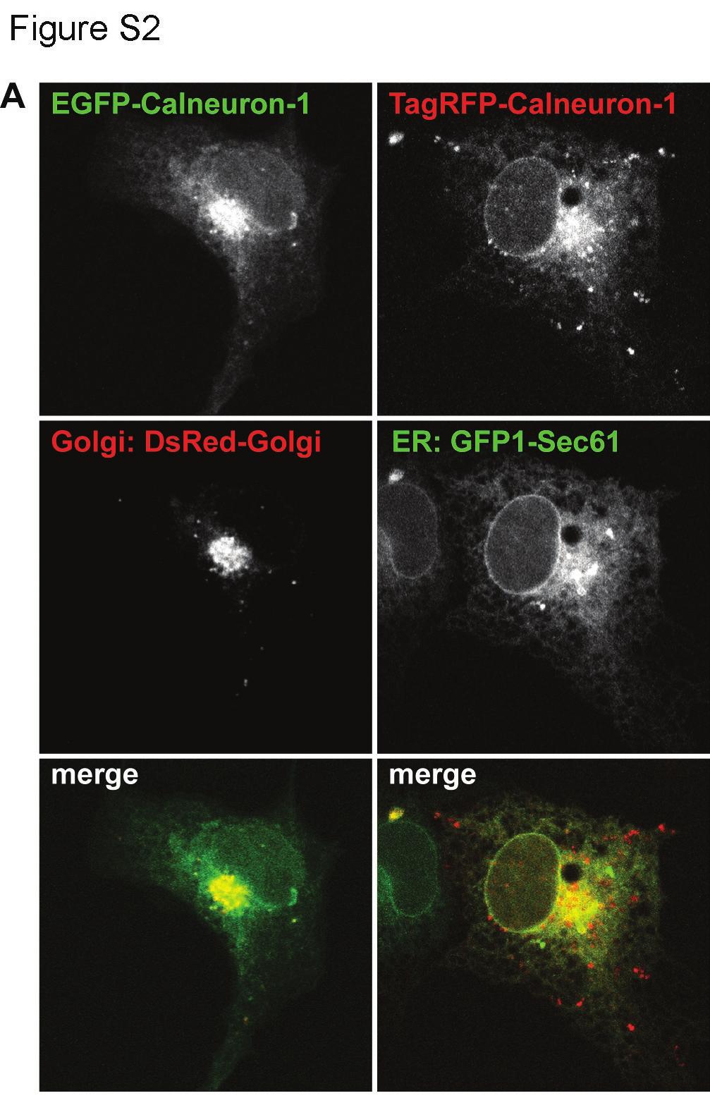
4 4 Supplemental Figures Figure S1. Overexpressed Calneuron-1 is mainly present in the membrane-bound form with a small fraction in cytosol. (A) Live imaging of COS-7 cells co-transfected with EGFP-Calneuron-1 and tagrfp. 5 min after the baseline recording cells were pemeabilized with 40 µm of digitonin. Images were taken with 20 sec per frame. (B) Examples of fluorescence intensity curves before and after digitonin treatment, measured in the Golgi, the cytosol, the nucleus and the area outside of the cell. Permeabilization and diffusion of soluble cytosolic proteins was confirmed by the loss in TagRFP fluorescence quickly after the addition of digitonin. EGFP-Calneuron-1 fluorescence was not changed at the perinuclear Golgi area but was reduced in the nucleus and cytosol. (C) Digitonin permeabilization assay in COS-7 cells transfected with full length EGFP-Calneuron-1 or the deletion construct lacking the C-terminal transmembrane region. Growth medium was replaced by KD buffer and cells were either untreated (for the control group) or treated with 40 µm of digitonin for 30 or 60 seconds. Then the buffer was collected and centrifuged (SN) in order to remove cell debris. COS-7 cells attached to the wells were harvested in the same volume of fresh KD buffer (P). Both the SN and P fractions from each experiment were subjected to SDS-PAGE and analyzed with anti-gfp, anti-syntaxin 6 and anti-β-actin antibody. EGFP-Calneuron-1_ΔC is found in SN fraction already after 30 seconds of digitonin treatment whereas the full length EGFP-Calneuron-1 is detectable only after 60 seconds. Figure S2. EGFP-Calnueuron-1 co-localizes with the Golgi marker DsRed- Monomeric-Golgi (left panel). The reticular distribution of Calneuron-1 subfraction probably represents the protein localized at the ER (visualized by co-transfection with the ER marker GFPC1-Sec61β / right panel). Figure S3. Characterization of the antibody used fort he PLA assay. Anti-GFP mouse, anti-asna-1 mouse and anti-calneuron-1 rabbit (ProteinTech) antibodies were tested on COS-7 cells overexpressing or lacking the respective proteins that were fused to a fluorescent tag (GFP/RFP/YFP). Immunostaining was observed only in cells that overexpress the respective protein. The staining pattern of untagged Calneuron (pcdna-caln1) observed with the anti-caln1 rabbit antibody is comparable to the GFP fluorescence pattern of pegfp-caln1. Figure S4. (A) GFP-Calneuron-1 constructs expressed in COS-7 cells are also showing higher molecular weight complexes compared to GFP in a semi-native PAGE. Complete denaturation of the sample results in a pattern comparable to the pull down inputs of Fig. 4C. (B) Semi-native PAGE for different GFP-tagged constructs of Calneuron-1 solubilized in the native loading dye. Next to the appearance of the bands corresponding to the molecular weight of each monomeric construct, double (indicated by *) size bands can be detected in all GFP-Calneuron-1 constructs. Especially the full length Calneuron-1 construct seems to be to a majority in at least a dimmer form. The weak binding of the full length construct to ASNA-1 in the pull down experiment shown in Fig. 4C can be therefore a result of a relatively small pool of monomeric Calneuron-1. (C) Calibration of Superdex 200 column using protein standards in the presence of 1 mm EDTA (left) and 1 mm Ca 2+ (right).

5
6

7
8
Segments of the obstructed intestinal loops were fixed in 4% paraformaldehyde
 Supplementary text Supplementary materials and methods Histopathological examination Segments of the obstructed intestinal loops were fixed in 4% paraformaldehyde (PFA) and embedded in paraffin wax with
Plasmid DNA transfection of SW480 human colorectal cancer cells with the Biontex K2 Transfection System
 Plasmid DNA transfection of human colorectal cancer cells with the Biontex K2 Transfection System Stephanie Hehlgans and Franz Rödel, Department of Radiotherapy and Oncology, Goethe- University Frankfurt,
Post-expansion antibody delivery, after epitope-preserving homogenization.
 Supplementary Figure 1 Post-expansion antibody delivery, after epitope-preserving homogenization. (a, b) Wide-field fluorescence images of Thy1-YFP-expressing mouse brain hemisphere slice before expansion
Supplementary information to accompany: A novel role for the DNA repair gene Rad51 in Netrin-1 signalling
 Supplementary information to accompany: A novel role for the DNA repair gene Rad51 in Netrin-1 signalling Glendining KA 1, Markie D 2, Gardner RJM 4, Franz EA 3, Robertson SP 4, Jasoni CL 1 Supplementary
This Document Contains:
 This Document Contains: 1. In-Cell Western Protocol II. Cell Seeding and Stimulation Supplemental Protocol III. Complete Assay Example: Detailing the Seeding, Stimulation and Detection of the A431 Cellular
Dolphin-Chemi Plus. Aim: To visualise and evaluate the performance of chemiluminescent immunoblots using Wealtec s Dolphin-Chemi plus image system
 Application Note 03 Dolphin-Chemi plus 8/22/2007 Dolphin-Chemi Plus Aim: To visualise and evaluate the performance of chemiluminescent immunoblots using Wealtec s Dolphin-Chemi plus image system INTRODUCTION
Supplemental Data. LMO4 Controls the Balance between Excitatory. and Inhibitory Spinal V2 Interneurons
 Neuron, Volume 61 Supplemental Data LMO4 Controls the Balance between Excitatory and Inhibitory Spinal V2 Interneurons Kaumudi Joshi, Seunghee Lee, Bora Lee, Jae W. Lee, and Soo-Kyung Lee Supplemental
ab GFP ELISA Kit Instructions for Use For the quantitative measurement of GFP protein expression
 ab117992 GFP ELISA Kit Instructions for Use For the quantitative measurement of GFP protein expression This product is for research use only and is not for diagnostic use. intended www.abcam.com Table
Technical Note Detection of post-immunoprecipitation proteins by Western blot using the Quick Western Kit IRDye 680RD
 Technical Note Detection of post-immunoprecipitation proteins by Western blot using the Quick Western Kit IRDye 680RD Developed for: Aerius, Odyssey Classic, Odyssey CLx and Odyssey Sa Imaging Systems
Supplementary Figures and Legends
 Supplementary Figures and Legends Figure S1. Tests of the optical alignment and focal properties of the confocal microscope. (A) Images of the optical cross-section of fluorescent microspheres differing
Viral RNAi suppressor reversibly binds sirna to. outcompete Dicer and RISC via multiple-turnover
 Supplementary Data Viral RNAi suppressor reversibly binds sirna to outcompete Dicer and RISC via multiple-turnover Renata A. Rawlings 1,2, Vishalakshi Krishnan 2 and Nils G. Walter 2 * 1 Biophysics and
X2-C/X1-Y X2-C/VCAM-Y. FRET efficiency. Ratio YFP/CFP
 FRET efficiency.7.6..4.3.2 X2-C/X1-Y X2-C/VCAM-Y.1 1 2 3 Ratio YFP/CFP Supplemental Data 1. Analysis of / heterodimers in live cells using FRET. FRET saturation curves were obtained using cells transiently
SANTA CRUZ BIOTECHNOLOGY, INC.
 TECHNICAL SERVICE GUIDE: Western Blotting 2. What size bands were expected and what size bands were detected? 3. Was the blot blank or was a dark background or non-specific bands seen? 4. Did this same
supplementary information
 DOI: 1.138/ncb1839 a b Control 1 2 3 Control 1 2 3 Fbw7 Smad3 1 2 3 4 1 2 3 4 c d IGF-1 IGF-1Rβ IGF-1Rβ-P Control / 1 2 3 4 Real-time RT-PCR Relative quantity (IGF-1/ mrna) 2 1 IGF-1 1 2 3 4 Control /
Respiratory distress and early neonatal lethality in Hspa4l/Hspa4 double mutant mice
 Respiratory distress and early neonatal lethality in Hspa4l/Hspa4 double mutant mice Belal A. Mohamed, Amal Z. Barakat, Torsten Held, Manar Elkenani, Christian Mühlfeld, Jörg Männer, and Ibrahim M. Adham
SOD1 as a Molecular Switch for Initiating the Homeostatic ER Stress Response under Zinc Deficiency
 Molecular Cell, Volume 52 Supplemental Information SOD1 as a Molecular Switch for Initiating the Homeostatic ER Stress Response under Zinc Deficiency Kengo Homma, Takao Fujisawa, Naomi Tsuburaya, Namiko
Supplementary Table 1. The Q-PCR primer sequence is summarized in the following table.
 Supplementary Table 1. The Q-PCR primer sequence is summarized in the following table. Name Sequence (5-3 ) Application Flag-u ggactacaaggacgacgatgac Shared upstream primer for all the amplifications of
Azure Biosystems Western Blotting Workflow
 Azure Biosystems Western Blotting Workflow PROBE PLAN SEPARATE ANALYZE VISUALIZE PLAN Plan your experiment and choose your detection method Chemiluminescent Western Blotting The most common method for
One-step split GFP staining for sensitive protein detection and localization in mammalian cells
 Supplementary Materials For: One-step split GFP staining for sensitive protein detection and localization in mammalian cells Lara Kaddoum 1,3, Eddy Magdeleine 1,3, Geoffrey S. Waldo 4, Etienne Joly 1,3,
Short hairpin RNA (shrna) against MMP14. Lentiviral plasmids containing shrna
 against MMP14. Lentiviral plasmids containing shrna") Supplemental Materials and Methods Short hairpin RNA (shrna) against MMP14. Lentiviral plasmids containing shrna (Mission shrna, Sigma) against mouse MMP14 were transfected into HEK293 cells using FuGene6
Supplemental Materials and Methods Short hairpin RNA (shrna) against MMP14. Lentiviral plasmids containing shrna (Mission shrna, Sigma) against mouse MMP14 were transfected into HEK293 cells using FuGene6
Supplementary Figure S1. Immunodetection of full-length XA21 and the XA21 C-terminal cleavage product.
 Supplementary Information Supplementary Figure S1. Immunodetection of full-length XA21 and the XA21 C-terminal cleavage product. Total protein extracted from Kitaake wild type and rice plants carrying
Supplemental Online Material. The mouse embryonic fibroblast cell line #10 derived from β-arrestin1 -/- -β-arrestin2 -/-
 #1074683s 1 Supplemental Online Material Materials and Methods Cell lines and tissue culture The mouse embryonic fibroblast cell line #10 derived from β-arrestin1 -/- -β-arrestin2 -/- knock-out animals
Supporting Information
 Supporting Information Shao et al. 10.1073/pnas.1504837112 SI Materials and Methods Immunofluorescence and Immunoblotting. For immunofluorescence, cells were fixed with 4% paraformaldehyde and permeabilized
phab Amine and Thiol Reactive Dyes for Antibody Internalization Studies Nidhi Nath, Ph.D. Group Leader, Protein Analysis Promega Corporation
 phab Amine and Thiol Reactive Dyes for Antibody Internalization Studies Nidhi Nath, Ph.D. Group Leader, Protein Analysis 1 Outline 1. phab Dyes 2. Protocols for conjugating phab Dyes to antibodies 3. Applications:
Kinase Reaction and Alkylation Protocol
 Kinase Reaction and Alkylation Protocol Protocol for the treatment of substrates prior to detection by Thiophosphate Ester antibodies This product is for research use only and is not intended for diagnostic
TECHNICAL BULLETIN. In Vitro Bacterial Split Fluorescent Protein Fold n Glow Solubility Assay Kits
 In Vitro Bacterial Split Fluorescent Protein Fold n Glow Solubility Assay Kits Catalog Numbers APPA001 In Vitro Bacterial Split GFP "Fold 'n' Glow" Solubility Assay Kit (Green) APPA008 In Vitro Bacterial
mcherry Monoclonal Antibody (16D7) Catalog Number M11217 Product data sheet
 Catalog Number M11217 Product data sheet") Website: thermofisher.com Customer Service (US): 1 800 955 6288 ext. 1 Technical Support (US): 1 800 955 6288 ext. 441 mcherry Monoclonal Antibody (16D7) Catalog Number M11217 Product data sheet Details
Website: thermofisher.com Customer Service (US): 1 800 955 6288 ext. 1 Technical Support (US): 1 800 955 6288 ext. 441 mcherry Monoclonal Antibody (16D7) Catalog Number M11217 Product data sheet Details
Supporting Information
 Supporting Information Copper and zinc ions specifically promote non-amyloid aggregation of the highly stable human γ-d crystallin Liliana Quintanar, 1,* José A. Domínguez-Calva, 1 Eugene Serebryany, 2
AFFINITY HIS-TAG PURIFICATION
 DESCRIPTION Nickel NTA Agarose Cartridges 5ml are used for purification of histidine-tagged proteins in native or denaturing conditions. This cartridge can be used with an automated chromatography system,
1. Cross-linking and cell harvesting
 ChIP is a powerful tool that allows the specific matching of proteins or histone modifications to regions of the genome. Chromatin is isolated and antibodies to the antigen of interest are used to determine
Supplemental Information. A Versatile Tool for Live-Cell Imaging. and Super-Resolution Nanoscopy Studies. of HIV-1 Env Distribution and Mobility
 Cell Chemical Biology, Volume 24 Supplemental Information A Versatile Tool for Live-Cell Imaging and Super-Resolution Nanoscopy Studies of HIV-1 Env Distribution and Mobility Volkan Sakin, Janina Hanne,
Test Your Plate Reader Set-up Before Using LanthaScreen Eu Assays
 Test Your Plate Reader Set-up Before Using LanthaScreen Eu Assays Purpose This LanthaScreen Eu Microplate Reader Test provides a method to verify the ability of your fluorescent plate reader to detect
PROCEDURE FOR USE NICKEL NTA Magnetic Agarose Beads (5%)
") 1 AFFINITY HIS-TAG PURIFICATION PROCEDURE FOR USE NICKEL NTA Magnetic Agarose Beads (5%) DESCRIPTION Nickel NTA Magnetic Agarose Beads are products that allow rapid and easy small-scale purification of
1 AFFINITY HIS-TAG PURIFICATION PROCEDURE FOR USE NICKEL NTA Magnetic Agarose Beads (5%) DESCRIPTION Nickel NTA Magnetic Agarose Beads are products that allow rapid and easy small-scale purification of
FACS Blue LacZ beta Galactosidase detection kit
 ab189815 FACS Blue LacZ beta Galactosidase detection kit Instructions for Use For the detection of beta-galactosidase using Enzyme or FACS Assay This product is for research use only and is not intended
CytoPainter Golgi Staining Kit Green Fluorescence
 ab139483 CytoPainter Golgi Staining Kit Green Fluorescence Instructions for Use Designed for the detection of Golgi bodies by microscopy This product is for research use only and is not intended for diagnostic
3. Results. 3.1 Generation of HEK293 cell clones stably expressing ETA and ETB receptors
 3. Results 3.1 Generation of HEK293 cell clones stably expressing ETA and ETB receptors To investigate the dimerisation of the endothelin receptor subtypes HEK293 cells were stably transfected with plasmids
Supplemental information
 Supplemental information - Control samples (200 subjects) - Immunohistochemistry of rat brain - Immunocytochemistry on neuronal cultures - Immunocompetition assay - Immunoprecipitation - Immunocytochemistry
The preparation of native chromatin from cultured human cells.
 Native chromatin immunoprecipitation protocol The preparation of native chromatin from cultured human cells. All solutions need to be ice cold. Sucrose containing solutions must be made up fresh on the
INOS. Colorimetric Cell-Based ELISA Kit. Catalog #: OKAG00807
 INOS Colorimetric Cell-Based ELISA Kit Catalog #: OKAG00807 Please read the provided manual entirely prior to use as suggested experimental protocols may have changed. Research Purposes Only. Not Intended
Product Information. Before you begin. Component A 1 vial of 30 ul vial of 300 ul each Glycerol. Tris
 Glowing Products for Science Mix-n-Stain Antibody Labeling Kits Size: 1 labeling per kit Storage: -20 o C Stability: Stable for at least 1 year from date of receipt when stored as recommended. Components:
AFFINITY HIS-TAG PURIFICATION
 DESCRIPTION Resins are products that allow batch or column purifications. This product is supplied as a suspension in 50% aqueous suspension containing 30 vol % ethanol. INSTRUCTIONS The resins are adapted
ab Vimentin Human Profiling ELISA Kit
 ab173190 Vimentin Human Profiling ELISA Kit Instructions for Use For the measurement of total Vimentin protein in Human samples. This product is for research use only and is not intended for diagnostic
GM130 Is Required for Compartmental Organization of Dendritic Golgi Outposts
 Current Biology, Volume 24 Supplemental Information GM130 Is Required for Compartmental Organization of Dendritic Golgi Outposts Wei Zhou, Jin Chang, Xin Wang, Masha G. Savelieff, Yinyin Zhao, Shanshan
Confocal immunofluorescence microscopy
 Confocal immunofluorescence microscopy HL-6 and cells were cultured and cytospun onto glass slides. The cells were double immunofluorescence stained for Mt NPM1 and fibrillarin (nucleolar marker). Briefly,
ab MDR Assay Kit (Fluorometric)
") ab112142 MDR Assay Kit (Fluorometric) Instructions for Use For detecting MDR pump activities in cells using our proprietary fluorescence probe. This product is for research use only and is not intended
ab112142 MDR Assay Kit (Fluorometric) Instructions for Use For detecting MDR pump activities in cells using our proprietary fluorescence probe. This product is for research use only and is not intended
How to run Alpha assay: How to setup an Alpha assay Make your own assay!
 How to run Alpha assay: How to setup an Alpha assay Make your own assay! 1 2009 PerkinElmer AlphaLISA kits - recommendations before starting the assay Samples: Phenol red and hemoglobin: choose AlphaLISA
Calcium Assay Kit. Technical Data Sheet. Product Information. Description. Storage. Materials not included
 BD Technical Data Sheet Calcium Assay Kit Product Information Catalog Number: 640176 Size Reagents for 10 plates Components: Calcium Indicator, 1 vial, lyophilized 10X Signal Enhancer, 10 ml 1X Calcium
SensoLyte Anti-alpha-Synuclein Quantitative ELISA Kit (Human/Mouse/Rat) *Colorimetric*
 *Colorimetric*") Catalog # Kit Size SensoLyte Anti-alpha-Synuclein Quantitative ELISA Kit (Human/Mouse/Rat) *Colorimetric* AS-55550 One 96-well strip plate This kit is optimized to detect human/mouse/rat alpha-synuclein
Catalog # Kit Size SensoLyte Anti-alpha-Synuclein Quantitative ELISA Kit (Human/Mouse/Rat) *Colorimetric* AS-55550 One 96-well strip plate This kit is optimized to detect human/mouse/rat alpha-synuclein
To isolate single GNS 144 cell clones, cells were plated at a density of 1cell/well
 Supplemental Information: Supplemental Methods: Cell culture To isolate single GNS 144 cell clones, cells were plated at a density of 1cell/well in 96 well Primaria plates in GNS media and incubated at
Myers Lab ChIP-seq Protocol v Modified January 10, 2014
 Myers Lab ChIP-seq Protocol V011014 1 Contact information: Dr. Florencia Pauli Behn HudsonAlpha Institute for Biotechnology 601 Genome Way Huntsville, AL 35806 Telephone: 256-327-5229 Email: fpauli@hudsonalpha.org
Cycles of vascular plexus formation within the nephrogenic zone of the developing mouse kidney
 1 Supplementary text and data for: 2 3 4 5 Cycles of vascular plexus formation within the nephrogenic zone of the developing mouse kidney Authors: David A. D. Munro 1*, Peter Hohenstein 2, and Jamie A.
GFP CCD2 GFP IP:GFP
 D1 D2 1 75 95 148 178 492 GFP CCD1 CCD2 CCD2 GFP D1 D2 GFP D1 D2 Beclin 1 IB:GFP IP:GFP Supplementary Figure 1: Mapping domains required for binding to HEK293T cells are transfected with EGFP-tagged mutant
Supplementary Information
 Journal : Nature Biotechnology Supplementary Information Targeted genome engineering in human cells with RNA-guided endonucleases Seung Woo Cho, Sojung Kim, Jong Min Kim, and Jin-Soo Kim* National Creative
ab Hypoxic Response Human Flow Cytometry Kit
 ab126585 Hypoxic Response Human Flow Cytometry Kit Instructions for Use For measuring protein levels by flow cytometry: hypoxia-inducible factor 1-alpha (HIF1A) and BCL2/adenovirus E1B 19 kda proteininteracting
Supporting Online Material, Matsumoto et al.
 Supporting Online Material, Matsumoto et al. Material and Methods Library. Poly(A) + mrna was purified from RAW264.7 cells stimulated with murine IFN-γ (100 units/ml) and bacterial LPS (100 ng/ml) for
OPPF-UK Standard Protocols: Mammalian Expression
 OPPF-UK Standard Protocols: Mammalian Expression Joanne Nettleship joanne@strubi.ox.ac.uk Table of Contents 1. Materials... 3 2. Cell Maintenance... 4 3. 24-Well Transient Expression Screen... 5 4. DNA
Calcein AM Cell Viability Kit
 Instructions For Research Use Only. Not For Use In Diagnostic Procedures Calcein AM Cell Viability Kit Catalog# 4892-010-K 1000 Tests* * Calculated based on using 1 μm final concentration of Calcein AM;
The Human Protein PRR14 Tethers Heterochromatin to the Nuclear Lamina During Interphase and Mitotic Exit
 Cell Reports, Volume 5 Supplemental Information The Human Protein PRR14 Tethers Heterochromatin to the Nuclear Lamina During Interphase and Mitotic Exit Andrey Poleshko, Katelyn M. Mansfield, Caroline
LacZ beta Galactosidase Intracellular Detection Kit
 ab189816 LacZ beta Galactosidase Intracellular Detection Kit Instructions for Use For the detection of beta-galactosidase using Microplate or FACS Assay This product is for research use only and is not
EGFR (Phospho-Ser695)
") Assay Biotechnology Company www.assaybiotech.com Tel: 1-877-883-7988 Fax: 1-877-610-9758 EGFR (Phospho-Ser695) Colorimetric Cell-Based ELISA Kit Catalog #: OKAG02090 Please read the provided manual entirely
Assay Biotechnology Company www.assaybiotech.com Tel: 1-877-883-7988 Fax: 1-877-610-9758 EGFR (Phospho-Ser695) Colorimetric Cell-Based ELISA Kit Catalog #: OKAG02090 Please read the provided manual entirely
CF Dyes Next Generation Fluorescent Dyes Secondary antibody
 CF Dyes Next Generation Fluorescent Dyes Secondary antibody OZYME 10 AVENUE AMPÈRE - CS 30268-78053 ST QUENTIN EN YVELINES CEDEX Tél. : 01 34 60 24 24 - Fax : 01 34 60 92 12 - www.ozyme.fr/info CF Dyes
IMMUNOPRECIPITATION TROUBLESHOOTING TIPS
 IMMUNOPRECIPITATION TROUBLESHOOTING TIPS Creative Diagnostics Abstract Immunoprecipitation (IP) is the technique of precipitating a protein antigen out of solution using an antibody that specifically binds
ab Cell Viability Assay Kit Fluorometric Dual Green/Red
 ab112121 Cell Viability Assay Kit Fluorometric Dual Green/Red Instructions for Use For detecting cell viability in suspension and adherent cells by using dual proprietary green and red fluorescence probes.
Nodes of regulation in cellular systems
 Nodes of regulation in cellular systems cell membrane signal transduction ligands receptors oligomerization transport signal transduction modified protein Golgi transcription factor transport ER transport
ab Serum Albumin Human SimpleStep ELISA Kit
 ab179887 Serum Albumin Human SimpleStep ELISA Kit Instructions for Use For the quantitative measurement of Serum Albumin in human serum, plasma and cell culture supernatants. This product is for research
ab Fluo-8 No Wash Calcium Assay Kit
 ab112129 Fluo-8 No Wash Calcium Assay Kit Instructions for Use For detecting calcium in cells by using our proprietary fluorescence probe. This product is for research use only and is not intended for
For identifying inhibitors and activators of mitochondrial biogenesis in adherent cultured cells.
 ab110216 MitoBiogenesis TM In-Cell ELISA Kit (IR) Instructions for Use For identifying inhibitors and activators of mitochondrial biogenesis in adherent cultured cells. This product is for research use
ab65354 Superoxide Dismutase Activity Assay kit (Colorimetric)
") Version 9 Last updated 11 January 2018 ab65354 Superoxide Dismutase Activity Assay kit (Colorimetric) For the measurement of Superoxide Dismutase Activity in various samples. This product is for research
Version 9 Last updated 11 January 2018 ab65354 Superoxide Dismutase Activity Assay kit (Colorimetric) For the measurement of Superoxide Dismutase Activity in various samples. This product is for research
over time using live cell microscopy. The time post infection is indicated in the lower left corner.
 Title of file for HTML: Supplementary Information Description: Supplementary Figures and Supplementary Table Title of file for HTML: Supplementary Movie 1 Description: Fusion of NBs. BSR cells were infected
Identification of Microprotein-Protein Interactions via APEX Tagging
 Supporting Information Identification of Microprotein-Protein Interactions via APEX Tagging Qian Chu, Annie Rathore,, Jolene K. Diedrich,, Cynthia J. Donaldson, John R. Yates III, and Alan Saghatelian
Supplementary Information
 Supplementary Information Extended Loop Region of Hcp1 is Critical for the Assembly and Function of Type VI Secretion System in Burkholderia pseudomallei Yan Ting Lim, Chacko Jobichen, Jocelyn Wong, Direk
FLUORESCENT PEPTIDES. Outstanding Performance and Wide Application Range
 FLUORESCENT PEPTIDES Peptides and amino acids labeled with and Tide Quencher TM We offer peptides and amino acids tagged with fluorescent dyes. They meet highest demands in fluorescence intensity and photo-stability,
ab Beta Galactosidase Detection Kit (Fluorometric) Instructions for Use For monitoring β-galactosidase activity in cells.
 Instructions for Use For monitoring β-galactosidase activity in cells.") ab176721 Beta Galactosidase Detection Kit (Fluorometric) Instructions for Use For monitoring β-galactosidase activity in cells. This product is for research use only and is not intended for diagnostic
ab176721 Beta Galactosidase Detection Kit (Fluorometric) Instructions for Use For monitoring β-galactosidase activity in cells. This product is for research use only and is not intended for diagnostic
1 ml gel corresponds to ml of 75% (v/v) Glutathione Agarose suspension.
 Glutathione Agarose suspension.") 1 AFFINITY GST PURIFICATION Procedure for Use Glutathione Agarose 4 Resin DESCRIPTION Glutathione Agarose Resin is used to purify recombinant derivatives of glutathione S-transferases or glutathione binding
1 AFFINITY GST PURIFICATION Procedure for Use Glutathione Agarose 4 Resin DESCRIPTION Glutathione Agarose Resin is used to purify recombinant derivatives of glutathione S-transferases or glutathione binding
Fluo-8 Medium Removal Calcium Assay Kit
 ab112128 Fluo-8 Medium Removal Calcium Assay Kit Instructions for Use For detecting calcium in cells by using our proprietary fluorescence probe This product is for research use only and is not intended
Porcine Transferrin Receptor(TFR) ELISA Kit
 ELISA Kit") Porcine Transferrin Receptor(TFR) ELISA Kit Catalog No. CSB-E13481p (96T) This immunoassay kit allows for the in vitro quantitative determination of porcine TFR concentrations in serum, plasma. Expiration
Porcine Transferrin Receptor(TFR) ELISA Kit Catalog No. CSB-E13481p (96T) This immunoassay kit allows for the in vitro quantitative determination of porcine TFR concentrations in serum, plasma. Expiration
Supplemental Information. PARP1 Represses PAP and Inhibits Polyadenylation during Heat Shock
 Molecular Cell, Volume 49 Supplemental Information PARP1 Represses PAP and Inhibits Polyadenylation during Heat Shock Dafne Campigli Di Giammartino, Yongsheng Shi, and James L. Manley Supplemental Information
Protocol for induction of expression and cell lysate production
 Protocol for induction of expression and cell lysate production AV-04 Doxycyclin induction and cell lysate 1.0 Introduction / Description This method is intended for the treatment of the previously transfected
Modified Rapid MAIPA Protocol
 Modified Rapid MAIPA Protocol This method is based on the following publication; K Campbell, K Rishi, G Howkins, D Gilby, R Mushens, C Ghevaert, P Metcalfe, WH Ouwehand, G Lucas. A modified fast MAIPA
Human IgG Antigen ELISA Kit
 Human IgG Antigen ELISA Kit Catalog No: IHUIGGKT Lot No: SAMPLE INTENDED USE This human immunoglobulin G antigen assay is intended for the quantitative determination of total human IgG antigen in serum,
RNA oligonucleotides and 2 -O-methylated oligonucleotides were synthesized by. 5 AGACACAAACACCAUUGUCACACUCCACAGC; Rand-2 OMe,
 Materials and methods Oligonucleotides and DNA constructs RNA oligonucleotides and 2 -O-methylated oligonucleotides were synthesized by Dharmacon Inc. (Lafayette, CO). The sequences were: 122-2 OMe, 5
ASPP1 Fw GGTTGGGAATCCACGTGTTG ASPP1 Rv GCCATATCTTGGAGCTCTGAGAG
 Supplemental Materials and Methods Plasmids: the following plasmids were used in the supplementary data: pwzl-myc- Lats2 (Aylon et al, 2006), pretrosuper-vector and pretrosuper-shp53 (generous gift of
Nickel-NTA Agarose Suspension
 Nickel-NTA Agarose Suspension Agarose beads for purification of His-tagged proteins Product No. A9735 Description Nickel-NTA Agarose Suspension is an agarose-based affinity chromatography resin allowing
Tropix Chemiluminescent Kits and Reagents For Cell Biology Applications
 PRODUCT FAMILY BULLETIN Tropix Chemiluminescent Kits and Reagents Tropix Chemiluminescent Kits and Reagents For Cell Biology Applications Introduction to Chemiluminescence Chemiluminescence is the conversion
Supplemental Data Supplementary Figure Legends and Scheme Figure S1.
 Supplemental Data Supplementary Figure Legends and Scheme Figure S1. UTK1 inhibits the second EGF-induced wave of lamellipodia formation in TT cells. A and B, EGF-induced lamellipodia formation in TT cells,
Description: Nuclear morphology and dynamics in nontargeting sirna transfected cells. HeLa Kyoto
 Title of file for HTML: Supplementary Information Description: Supplementary Figures and Supplementary Tables Title of file for HTML: Supplementary Movie 1 Description: Nuclear morphology and dynamics
Optimization of a LanthaScreen Kinase assay for BRAF V599E
 Optimization of a LanthaScreen Kinase assay for BRAF V599E Overview This protocol describes how to develop a LanthaScreen kinase assay designed to detect and characterize inhibitors of BRAF V599E using
ab GST 6XHis-tag ELISA Kit For the quantitative measurement of 6XHis-tag protein expression
 ab128573 GST 6XHis-tag ELISA Kit Instructions for Use For the quantitative measurement of 6XHis-tag protein expression This product is for research use only and is not intended for diagnostic use. 1 Table
ab65354 Superoxide Dismutase Activity Assay kit (Colorimetric)
") ab65354 Superoxide Dismutase Activity Assay kit (Colorimetric) Instructions for Use For the rapid, sensitive and accurate measurement of Superoxide Dismutase Activity in various samples. This product is
ab65354 Superoxide Dismutase Activity Assay kit (Colorimetric) Instructions for Use For the rapid, sensitive and accurate measurement of Superoxide Dismutase Activity in various samples. This product is
ab Optiblot Fluorescent Western Blot Kit
 ab133410 Optiblot Fluorescent Western Blot Kit Instructions for Use For quantitative, multi-color fluorescent Western blotting. This product is for research use only and is not intended for diagnostic
ab Ubiquitylation Assay Kit
 ab139467 Ubiquitylation Assay Kit Instructions for Use For the activation of ubiquitin for use in ubiquitylation experiments This product is for research use only and is not intended for diagnostic use.
ab MetaPath Mito Disease 4-Plex Dipstick Array
 ab109879 MetaPath Mito Disease 4-Plex Dipstick Array Instructions for Use For the measurement of mitochondrial biogenesis regulation in human samples This product is for research use only and is not intended
ab VE-Cadherin (CD144) Mouse SimpleStep ELISA Kit
 Mouse SimpleStep ELISA Kit") ab206980 VE-Cadherin (CD144) Mouse SimpleStep ELISA Kit Instructions for Use For the quantitative measurement of VE-Cadherin in mouse serum, plasma and cell culture supernatant samples. This product is
ab206980 VE-Cadherin (CD144) Mouse SimpleStep ELISA Kit Instructions for Use For the quantitative measurement of VE-Cadherin in mouse serum, plasma and cell culture supernatant samples. This product is
A General Protocol for GST Pull-down Lili Jing *
 A General Protocol for GST Pull-down Lili Jing * Department of Cell and Molecular Biology, University of Pennsylvania, Philadelphia, USA *For correspondence: lilijingcn@gmail.com [Abstract] GST pull-down
Cellular Fractionation
 Cellular Fractionation Lamond Lab Protocol 2007 More detailed protocol can be found here: http://www.lamondlab.com/f7nucleolarprotocol.htm This protocol has been adapted to fractionate a variety of different
Amersham * ECL * Gel horizontal electrophoresis system
 GE Healthcare Life Sciences Data file 28-9970-20 AB Electrophoresis products Amersham * ECL * Gel horizontal electrophoresis system Amersham ECL Gel and Amersham ECL Gel Box constitute a horizontal mini-gel
western blotting tech
 western blotting tech note 6148 Transfer of High Molecular Weight Proteins to Membranes: A Comparison of Transfer Efficiency Between Blotting Systems Nik Chmiel, Bio-Rad Laboratories, Inc., 6000 James
Mitochondria/Cytosol Fractionation Kit
 Mitochondria/Cytosol Fractionation Kit Sufficient for analysis of 50 samples Cat. No. MIT1000 FOR RESEARCH USE ONLY Not for use in diagnostic procedures. USA & Canada Phone: +1(800) 437-7500 Fax: +1 (951)
PARP-1 (cleaved) Human In-Cell ELISA Kit (IR)
 Human In-Cell ELISA Kit (IR)") ab110215 PARP-1 (cleaved) Human In-Cell ELISA Kit (IR) Instructions for Use For the quantitative measurement of Human PARP-1 (cleaved) concentrations in cultured adherent and suspension cells. This product
ab110215 PARP-1 (cleaved) Human In-Cell ELISA Kit (IR) Instructions for Use For the quantitative measurement of Human PARP-1 (cleaved) concentrations in cultured adherent and suspension cells. This product