Understanding Flow Cytometry
|
|
|
- Chad Nichols
- 5 years ago
- Views:
Transcription
 of interest Antigen Expression Dim/bright Ag proximity Intracellular/extracellular Understanding the Technology Fluorochrome Excitation /")
1 Understanding Flow Cytometry The Basic Concepts Maree Bagnara Products Sales Specialist/Account Manager Flow Cytometry PN Successful Flow Cytometry data is driven by.. Understanding the Biology Basic Immunology Population(s) of interest Antigen Expression Dim/bright Ag proximity Intracellular/extracellular Understanding the Technology Fluorochrome Excitation / Emission spectra Instrument Platform Lasers, Colour capability, Filters Spectral Overlap - Compensation Reagent and Assay Optimization Understanding of application methods GOOD SAMPLE PREPARATION 2 1


2 Basic Immunology Fluorescence Techniques Direct Staining Moab against target species Directly tagged with Fluorochrome Multi colour staining Indirect Staining Mouse anti human CD4 Human CD4 Goat anti-mouse FITC Amplification of low density Ags 3 Typically single colour 4 2


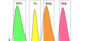
3 Fluorochrome Options for Gallios QDots, Hoechst PI, 7AAD, PerCP, GFPs Exercise 2 5 Excitation/Emission of Common Fluorochromes 525nm 575nm 620nm 675nm 700nm 770nm FITC PE ECD PC5 PECy5.5 PC7 488nm 647nm 675nm 700nm 700nm 750nm 750nm APC Alexa 700 APCAlexa 700 APC-H7 APCAlexa nm Pacific Blue 450nm Pacific Krome Orange Orange Sensitive to light 6 3
4 Measuring Brightness Quantitation of Brightness over Background Current method Signal:Noise MFI(pos) / MFI(neg), Alternative Method Stain Index (Sensitivity) MFI(pos) MFI(neg)/ 2 x SD Background Takes into account the spread of the background (negative population) vs positive population More accurate S:N References Maecker et.al Cytometry 62A: (2004) Hulspas et.al Cytometry 76B : (2009) Measuring Brightness Quantitation of Brightness over Background Signal to Noise Ratio: MFI(pos) / MFI(neg), /0.37 = 1136 Staining Index: (MFI(pos) MFI(neg))/2*SD(neg), ( )/2*0.23 = 913 Information provided courtesy of Dr Michael Kapinsky, Beckman Coulter 4


5 Compensation 9 Compensation Exercise
 Texas Red PE (488) Cy5 PE (488) Cy7 PerCP (488) Cy5.5 APC (635) Alexa700 Commercial Product ECD PC5 PC7 PerCPCy5.")

6 Tandem Dye Technology Schematic of Energy Transfer Mechanism 488 nm LASER Donor Molecule Excitation PE Acceptor Molecule Emitted wavelength Cy5 Donor Molecule Acceptor Molecule PE (488) Texas Red PE (488) Cy5 PE (488) Cy7 PerCP (488) Cy5.5 APC (635) Alexa700 Commercial Product ECD PC5 PC7 PerCPCy nm APC Alexa nm 630 nm 675 nm 760 nm 700nm 11 Tandem Dye Technology Better sensitivity over synthetic dyes Not equivalent from manufacturer to manufacturer Issues Variation in energy transfer Impacts compensation/instrument setup Photostability Stability Binding Specificity Cy5 can bind nonspecifically to monocytes Lot to lot variability 12 6


7 Same Tandem Different Vendor 13 Conjugate Choices High density Ag/ Dim dye Fluorochrome choice dependent on antigen density Bright dye - low density antigens Dimmer dye - high density antigens Low density Ag/ Bright dye Critical for multi-color reagents Proper compensation Detection of dim/rare events Dye conjugate interactions do they work together? Non-specific binding Steric Hindrance Fluorescent quenching 14 7
, Synthetic Dyes (Cyan and Alexa Dyes)best Tandem dyes can be degraded by cellular enzymes Alexa Fluor 488 better than")
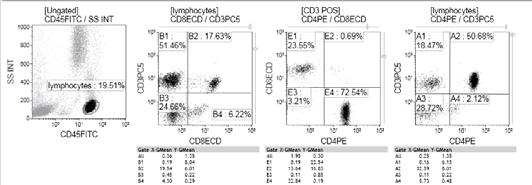
8 PBPs, Tandems, or Synthetic Dyes? Intracellular Antigen Detection Cytoplasmic antigens: Phycobiliproteins (PE, APC), Synthetic Dyes (Cyan and Alexa Dyes)best Tandem dyes can be degraded by cellular enzymes Alexa Fluor 488 better than FITC lower background Nuclear antigens: Phycobiliproteins or tandem dyes: protein size may hinder binding Close proximity can lead to energy transfer between dyes 15 Compensation Normal Human Lymphocytes 16 8
9 Compensation Normal Human Lymphocytes 17 Understanding Colour Compensation Back to Basics Set-up for Compensation Neg controls to set PMT HV Isotype matched and /or blank sample Place negative cells in the first decade Single positive controls for each fluorochrome / CD8 of each flurochrom Use biological sample or antibody capture beads such as VERSACOMP Verifier tube to check settings Set up a single protocol with histograms for all possible fluorochrome combinations Run each tube through protocol, making adjustments to compensation as required Final tube should be a verifier which checks that the settings are correct 18 9

10 19 Compensation Matrix 20 10
 U N T O U C H A B L E S I L E N T Silent = No overspill into")

11 Compensation Adjustment 21 Spillover Guide for the Gallios U N T O U C H A B L E Untouchable = No overspill from other dyes (clean row) U N T O U C H A B L E S I L E N T Silent = No overspill into other channels (clean column) Classification is specific for each combination of antibodies and conjugated dyes on a given hardware configuration Information provided courtesy of Dr Michael Kapinsky, Beckman Coulter 11


12 Multi-Color Combinations: What Can Go Wrong? Charge Size Proximity Concentration Conjugate interactions can make the difference between right and wrong results Decreased Mean Fluorescence or decreased % POS 23 Non-Specific Binding of Dyes Root Cause Low affinity binding of conjugate to irrelevant populations usually FcR Specific to cyanine dyes Other large molecule conjugates also display problem Monocyte binding of Cyanine dye Blocking antibody - unlabelled Same species as test Ab Bind to FcR Use Ig fraction rather than serum 30ug Ig fraction per 10 6 cells in 100 ul
13 Optimising Moabs Perform titration curves for each conjugate by serial dilution assay Plot Fluorescence Intensity vs dose for both positive and negative signal Choose optimal dose Saturation area of curve Highest Signal to Noise Prepare combination for Moab Cocktails Matrix Titre Evaluate for Non-specific binding Steric Hindrance Fluorescence Quenching 25 Dose Titration Dilution Perform titration curves for each conjugate by serial dilution assay Mean Channel Mean Channel MoAb conjugate positive pop negative pop 1:8 1:4 1:2 NEAT Plot Fluorescence intensity vs dose for both positive signal and noise Determine Signal/Noise Ratio Choose optimal conc range (2-3 points) Saturation area of curve Highest S/N Ab conc./test (μg) / Dilution 26 13
14 Moab Cocktail Optimisation Use titration information for Moab cocktail matrix CDx at three doses 1,2,3 CDy at three doses a,b,c Ideal [Ab] ug/test staining 1X10 6 cells in 100ul Determine saturation point Note: each Moab volume dilutes the other! Evaluate performance for major interactions Non-specific binding Steric Hindrance Fluorescence Quenching Verify on multiple samples x N 1:2 1:4 N 1:2 1:4 y 27 Optimal Formulation 20.0 Single CD4 FITC 20.0 CD4 FITC (with CD8 PE) Fluorescencence S/N Positive MFI Negative MFI Fluorescencence S/N Positive MFI Negative MFI Dose (ug/test) Dose (ug/test) Formulation Choice Choose highest Signal/Noise within saturation area of curve Moab mix must demonstrate equivalent performance to single color reagent controls No evidence of any interactions 28 14


15 Specimen Processing Sample presentation Anticoagulants ETDA, Heparin Cell Lines Adherent, Non-adherent Cell Media Phenol Red Clumping Flow Cytometer nightmare! Age of sample EDTA 8 30 hours Gate out dead cells Dye for Viability PI, 7AAD Red Cell Contamination Many RBC Lysis Products Optilyse Immunoprep Ammonium Chloride Ficoll Hyapaque 29 Sample Preparation Pre and Post Wash Remove interfering dyes from Culture medium Post wash can improve signal to noise Loss of Cells Washing Medium Check ph of PBS ph7.2 FCS? B etter than BSA? Check ph after adding buffering serum Autofluorescence Gate autofluorescence population and backgate onto Scatter plot ensure cells of interest are not contained 30 15
16 Sample Preparation Antibody-Antigen binding is a dynamic process Influenced by three parameters Time 1min 15mins Size of fluorochrome, cocktail size, Ag density Temperature Room Temperature. If delay - refrigerate Concentration of Moab Use in excess rather than less Are you using adequate Moab for the worst" case scenario Questionable data?» Use it at manufacturer s recommendation on a normal specimen.» Run Moabs as a SINGLE stain if using in a cocktail 31 Inhouse Cocktails! Do not store pre-mixes for extended periods of time Do NOT dilute / pre-mix Moab in original vial Do a time course study Add protein Dark vials When in doubt run a normal control specimen! 32 16
17 Negative Controls what do they do? Fluorescence due to non-specific binding of MoAb to cell Mainly due to binding to Fc receptors on the cell surface Dependant on IgG subclass IgG1<IgG2a~IgG2b<IgM (Most sticky ) 33 Why Use Negative Controls Set fluorescence PMT HV to obtain best signal to noise ratio Usually in the first decade Set Analysis regions 0.1-2% 34 17

18 Negative Control A non-specific MoAb of the same IgG subclass as the specific MoAb used to assess positive staining Should be at the same protein concentration as the MoAb Should have the same Fluorchrome/Protein ratio as the specific MoAb More common to use the cells that stain negative for the moab of interest internal Control 35 Where do we put the region? Negative 2% Positive R 1 Often needs to be moved with test samples 45% R 1 Subjective. Move the region if necessary! 38% R 1 36 Fluorescence 18


19 Where do we put the region? 37 Low Ag density on cells How do we handle this sample? 30% pos or 100% weak pos? Cells that are negative for an Ag may be more useful than a neg control Negative control Specific stain R1 (Log) Fluorescence 38 19


20 Application Standardisation Standardise the fluorescent light emitting from the Photo Multiplier Tubes Enables data comparison between samples and across instruments Maintain compensation settings 39 Mean Channel Standardisation-Using FlowSet beads PMT HV = 456 V PMT HV = 456 V DAY 24 DAY 0 DAY 24 - Drug XYZ DAY 24 HV to 350 V Decrease PMT HV to 350 V 40 20

21 Standards In Cytometry 41 21
Designing and Implementing a High-Level Multicolor Flow Cytometry Assay. Brent Wood MD PhD Department of Laboratory Medicine University of Washington
 Designing and Implementing a High-Level Multicolor Flow Cytometry Assay Brent Wood MD PhD Department of Laboratory Medicine University of Washington Define Purpose of Assay Most important question What
Flow Cytometry - The Essentials
 Flow Cytometry - The Essentials Pocket Guide to Flow Cytometry: 1. Know your Cytometer 2. Understanding Fluorescence and Fluorophores 3. Gating Process 4. Controls 5. Optimization 6. Panel Building 7.
Anatomy of a flow cytometer
 Anatomy of a flow cytometer Fluidics Optics Electronics Cells in suspension flow in single-file through an illuminated volume where they scatter light and emit fluorescence that is collected, filtered,
A previous ICCS Module entitled Instrument optimization - Adjusting PMT voltages and compensation 1 should be read as a prerequisite to this module.
 Sponsored and reviewed by ICCS Quality and Standards Committee Title: Compensation Tips for Beckman Coulter 10-Color Navios Platform Written by: Salima Janmohamed-Anastasakis Ph.D., Applications Scientist,
AMREP FLOW CYTOMETRY CORE FACILITY. Flow Cytometry Data Analysis Workshop Friday 24 th October
 AMREP FLOW CYTOMETRY CORE FACILITY Flow Cytometry Data Analysis Workshop Friday 24 th October Polychromatic Flow Cytometry Data Analysis: Immunophenotyping Lymphocyte sub-populations and targeting their
Expanding Multicolor Options in Flow Cytometry with Novel Brilliant Violet TM Fluorophores. Carsten Wiethe Scientific Application Manager BioLegend
 Expanding Multicolor Options in Flow Cytometry with Novel Brilliant Violet TM Fluorophores Carsten Wiethe Scientific Application Manager BioLegend Seminar Outline BrilliantViolet TM (BV)fluorophores Instrument
Beyond Colorful Science. A Practical Approach to Setting Up Multicolor Panels
 Beyond Colorful Science A Practical Approach to Setting Up Multicolor Panels ISCT Paris April 2014 Layout: Multicolor Panel Design Identify the pitfalls Have a look at issues with spectral overlap How
Principles of flow cytometry: overview of flow cytometry and its uses for cell analysis and sorting. Shoreline Community College BIOL 288
 Principles of flow cytometry: overview of flow cytometry and its uses for cell analysis and sorting Shoreline Community College BIOL 288 Flow Cytometry What is Flow Cytometry? Measurement of cells or particles
Principles of Multicolor Panel Design BD. BD, the BD Logo and all other trademarks are property of Becton, Dickinson and Company.
 1 Principles of Multicolor Panel Design 2 Common Multicolor Applications Intracellular cytokine staining Regulatory T cells (Tregs) Protein phosphorylation (BD Phosflow) Leukemia and lymphoma phenotyping
Designing and executing a successful flow cytometry experiment
 Designing and executing a successful flow cytometry experiment 1x1 0 5 Orange Fluorescent Protein 10000 1000 100 10 0 1000 2000 3000 4000 Forward Scatter This presentation is based on true events, however,
Ruud Hulspas, Mike Keeney, Ben Hedley and Andrea Illingworth
 Sponsored and reviewed by ICCS Quality and Standards Committee Title: Quality of Reagents Monoclonal Antibodies Written by: Date: March 6, 2018 Ruud Hulspas, Mike Keeney, Ben Hedley and Andrea Illingworth
a Beckman Coulter Life Sciences: White Paper
 a Beckman Coulter Life Sciences: White Paper Long Term Stabilization of Tandem Dyes for Use in High Content, Multi Variant Flow Cytometry Authors: Snehita Sattiraju 1, Tewfik Miloud 2, Neha Girish 1, Murthy
Incorporating New, Bright Fluorochromes into Multicolor Panel Design
 Incorporating New, Bright Fluorochromes into Multicolor Panel Design Maria C. Jaimes, MD Senior Staff Scientist BD Biosciences 23-14684-00 Overview Multicolor flow: successful application prerequisites
Each question may have MULTIPLE correct answers. Select all that are correct.
 Knowledge Assessment Flow Cytometry Workshop, Part 1 April 20, 2015 Each question may have MULTIPLE correct answers. Select all that are correct. 1. Tandem dyes are a. highly stable fluorophores after
Selected Topics in Electrical Engineering: Flow Cytometry Data Analysis
 Selected Topics in Electrical Engineering: Flow Cytometry Data Analysis Bilge Karaçalı, PhD Department of Electrical and Electronics Engineering Izmir Institute of Technology Outline Experimental design
Identification of red and white blood cells from whole blood samples using the Agilent 2100 bioanalyzer. Application Note
 Identification of red and white blood cells from whole blood samples using the Agilent 2100 bioanalyzer Application Note Sylvie Veriac Valérie Perrone Madeleine Avon Abstract Agilent Equipment: 2100 bioanalyzer
Application Note. Assay Portability on the BD FACSVerse System. Summary. Maria Jaimes, Yibing Wang, Catherine McIntyre, and Dev Mittar
 September Assay Portability on the BD FACSVerse System Maria Jaimes, Yibing Wang, Catherine McIntyre, and Dev Mittar Contents Summary Introduction 3 Objective 4 Methods 6 Results Discussion Conclusions
High-throughput automation with the Attune NxT Autosampler: consistent results across all wells and across plates
 APPLICATION NOTE Attune NxT Flow Cytometer with Autosampler High-throughput automation with the Attune NxT Autosampler: consistent results across all wells and across plates Introduction The emerging field
What you need to know before designing a panel
 Design What you need to know before designing a panel For Research Use Only. Not for use in diagnostic or therapeutic procedures. Alexa Fluor is a registered trademark of Life Technologies Corporation.
FACS Core Facility User Group Meeting. 25. April 2018 Janine Bögli
 FACS Core Facility User Group Meeting 25. April 2018 Janine Bögli Overview Basis: Forensic Flow Cytometry by Jennifer Wilshire, PhD, Stemcell Technologies You will suggest the crime in each case 8 Practical
Selecting Reagents for Multicolor Flow Cytometry
 HotLines Platinum Edition f a l l 0 0 6 Selecting Reagents for Multicolor Flow Cytometry By Holden Maecker and Joe Trotter The availability of flow cytometers capable of detecting 6, 8, and more colors
Flow Cytometry SOP: Monocytes from Frozen Cells
 Flow Cytometry SOP: Monocytes from Frozen Cells Purpose This SOP standardizes the procedure for measuring immune cells using flow cytometry in ACTG Immunology Laboratories. Materials 1. 12x75mm flow tubes
Designing and Validating a Multicolor Flow Cytometry Assay. Brent Wood MD PhD Department of Laboratory Medicine University of Washington
 Designing and Validating a Multicolor Flow Cytometry Assay Brent Wood MD PhD Department of Laboratory Medicine University of Washington Specimen Handling Sample Requirements 5 ml Peripheral blood (EDTA,
Application Information Bulletin: DuraClone IM Tubes Compensation setup for high content DuraClone reagents
 Application Information Bulletin: DuraClone IM Tubes Compensation setup for high content DuraClone reagents Compensation setup for high content DuraClone reagents INTRODUCTION High content flow cytometry
Visualization of digital data Interpretation of the visual
 Technical aspects of 8-color flow cytometry in the diagnosis and classification of hematopoietic malignancies Tomas Kalina!"#$%&'()*+,&$'+-./(0 *1 (2#34%-.(56(7&1+3+*&/( 8$#94&/(!:&3"(;&
Spherotech, Inc. 1. SPHERO TM Technical Note STN-8 Rev C
 SPHERO TM Technical Note STN- Rev C. 0070 CALIBRATION AND PERFORMANCE TRACKING OF FLOW CYTOMETERS USING SPHERO TM CALIBRATION PARTICLES Introduction The SPHERO TM Calibration Particles are versatile, stable,
Flow Cytometry Immune Activation SOP
 Flow Cytometry Immune Activation SOP Purpose This SOP standardizes the procedure for measuring immune activation of T cells using flow cytometry in ACTG Immunology Laboratories. Materials 1. 12x75mm flow
Best practices in panel design to optimize the isolation of cells of interest
 Sort Best practices in panel design to optimize the isolation of cells of interest For Research Use Only. Not for use in diagnostic or therapeutic procedures. Alexa Fluor is a registered trademark of Life
Flow Cytometry SOP: 14 color flow for immune activation, senescence, and exhaustion
 Flow Cytometry SOP: 14 color flow for immune activation, senescence, and exhaustion Purpose This SOP standardizes the procedure for measuring immune cells using flow cytometry in ACTG Immunology Laboratories.
Compensation: Fundamental Principles
 Flow Cytometry Seminar Series 2017 : Fundamental Principles Spillover correction in multicolor flow cytometry 28.02.2017 http://www.cytometry.uzh.ch Contents Fluorescence and its detection Absorption and
Your Research, Revolutionized
 Your Research, Revolutionized Drive Your Research Forward Your research needs are evolving and with the CytoFLEX flow cytometer you ll see just how far your data can take you. CytoFLEX has the advanced
Flowcytometry Dirk Pacholsky
 Flowcytometry Dirk Pacholsky Flowcytometry: Overview 1 2 Overview Flow Cyto Metry Fluid Cell Measurement measuring cell properties of cells in suspension Most Flow Cytometer have Spatially separated Lasers
Your Research, Revolutionized
 Your Research, Revolutionized Drive Your Research Forward Your research needs are evolving and with the CytoFLEX flow cytometer you ll see just how far your data can take you. CytoFLEX has the advanced
11/19/2013. Janine Zankl FACS Core Facility 13. November Cellular Parameters. Cellular Parameters. Monocytes. Granulocytes.
 DEPARTEMENT BIOZENTRUM Janine Zankl FACS Core Facility 13. November 2013 Cellular Parameters Granulocytes Monocytes Basophils Neutrophils Lymphocytes Eosinophils Cellular Parameters 1 What Is Flow Cytometry?
Using Fluorescence Spillover to Advantage
 Using Fluorescence Spillover to Advantage Boston June 9, 2011 Compensation: Why and When is it Necessary? Clare Rogers, Marketing and Applications Accuri Cytometers, Inc. Ann Arbor/St.Ives 1 June 11 Summary
Application Information Bulletin: Set-Up of the CytoFLEX Set-Up of the CytoFLEX* for Extracellular Vesicle Measurement
 Application Information Bulletin: Set-Up of the CytoFLEX Set-Up of the CytoFLEX* for Extracellular Vesicle Measurement Andreas Spittler, MD, Associate Professor for Pathophysiology, Medical University
SANTA CRUZ BIOTECHNOLOGY, INC.
 TECHNICAL SERVICE GUIDE: Western Blotting 2. What size bands were expected and what size bands were detected? 3. Was the blot blank or was a dark background or non-specific bands seen? 4. Did this same
How to run Alpha assay: How to setup an Alpha assay Make your own assay!
 How to run Alpha assay: How to setup an Alpha assay Make your own assay! 1 2009 PerkinElmer AlphaLISA kits - recommendations before starting the assay Samples: Phenol red and hemoglobin: choose AlphaLISA
BD Multicolor CompBeads
 4/2015 23-9955-01 IVD BD Multicolor CompBeads 100 compensation setups Catalog No. 644204 BD, BD Logo and all other trademarks are property of Becton, Dickinson and Company. 2015 BD Becton, Dickinson and
Novel Developments in Cytometry. Martin Adelmann Marketing Manager Cellular Analysis & Life Science Beckman Coulter EMEAI
 Novel Developments in Cytometry Martin Adelmann Marketing Manager Cellular Analysis & Life Science Beckman Coulter EMEAI 1 Innovation in Flow Cytometry Cellios EPICS 1V 2 lasers, 3 colors EPICS Profile
a Beckman Coulter Life Sciences: White Paper
 a Beckman Coulter Life Sciences: White Paper DuraClone improves standardization of compensation workflow for high content multicolor applications by flow cytometry Authors: Neha Girish 2, Sudharsan Sathyamurthy
EdU Flow Cytometry Kit. User Manual
 User Manual Ordering information: (for detailed kit content see Table 2) EdU Flow Cytometry Kits for 50 assays: Product number EdU Used fluorescent dye BCK-FC488-50 10 mg 6-FAM Azide BCK-FC555-50 10 mg
Detecting human circulating endothelial cells using the Attune Acoustic Focusing Cytometer
 APPLICATION NOTE Attune Acoustic Focusing Cytometer Detecting human circulating endothelial cells using the Attune Acoustic Focusing Cytometer Circulating endothelial cells (CECs) are mature cells shed
Calibración de equipos de citometría para empleo de paneles EuroFlow
 Calibración de equipos de citometría para empleo de paneles EuroFlow Departamento de Medicina, Centro de Investigación del Cáncer y Servicio de Citometría. Universidad de Salamanca, Salamanca, España.
INTRODUCTION TO FLOW CYTOMETRY
 DEPARTEMENT BIOZENTRUM INTRODUCTION TO FLOW CYTOMETRY F ACS C ore F acility Janine Zankl FACS Core Facility 3. Dezember 2015, 4pm Cellular Parameters Granulocytes Monocytes Basophils Lymphocytes Neutrophils
EdU Click FC ROTI kit for Flow Cytometry
 USER MANUAL EdU Click FC EdU Click FC Introduction and product description: The detection of cell proliferation is of utmost importance for assessing cell health, determining genotoxicity or evaluating
Titration of Fluorochrome-Conjugated Antibodies for Labeling Cell Surface Markers on Live Cells
 Titration of Fluorochrome-Conjugated Antibodies for Labeling Cell Surface Markers on Live Cells Ruud Hulspas 1 UNIT 6.29 1 Cytonome/ST, Boston, Massachusetts ABSTRACT Nonspecific antibody binding is best
SUPPORTING INFORMATION
 Electronic Supplementary Material (ESI) for Dalton Transactions. This journal is The Royal Society of Chemistry 2015 Terbium-Based Time-Gated Förster Resonance Energy Transfer Imaging for Evaluating Protein-Protein
determine optimum instrument settings for their own instruments and establish their own daily values.
 PC7 (770/488) SETUP KIT 6607121 PN 4299504-C FLOW CYTOMETER ALIGNMENT VERIFICATION FLUOROSPHERES FLOW CYTOMETER DETECTOR STANDARDIZATION FLUOROSPHERES INTENDED USE For Research Use Only. Not for use in
TECHNICAL BULLETIN. QUANTUM SIMPLY CELLULAR KIT Product Numbers QSC-20 AND QSC-100 Storage Temperature 2-8 C. Do Not Freeze
 QUANTUM SIMPLY CELLULAR KIT Product Numbers QSC-20 AND QSC-100 Storage Temperature 2-8 C. Do Not Freeze TECHNICAL BULLETIN Product Description The Quantum Simply Cellular Kit provides a convenient method
Flow Cytometry Support Reagents
 Excite and inspire Flow Cytometry Support Reagents Introduction Miltenyi Biotec is a leading supplier of flow cytometry products, offering one of the broadest ranges of antibodies, kits, assays, and support
Challenges for Flow Cytometry in Regulated Bioanalysis
 Challenges for Flow Cytometry in Regulated Bioanalysis Minesh Patel Merck Millipore Discovery & Development Solutions. Oxford, UK. Overview Flow cytometry principles Current uses and regulatory environments
Qdot nanocrystal. wide range of biological investigations, Qdot nanocrystals are powerful complements
 Feature nanocrystal conjugates for flow cytometry Take the easy route to multicolor flow cytometry. With applications across a wide range of biological investigations, nanocrystals are powerful complements
DURACLONE IM ACCELERATE YOUR PACE IN IMMUNE SYSTEM RESEARCH. For Reseach Use Only - Not for use in Diagnostic procedures
 DURACLONE IM ACCELERATE YOUR PACE IN IMMUNE SYSTEM RESEARCH Your clinical research trial companion For Reseach Use Only - Not for use in Diagnostic procedures ACCELERATE YOUR PACE IN IMMUNE SYSTEM RESEARCH
Introduction to Flow Cytometry. -- BD FACSCanto II TM. Daisy Kuo Application Specialist BDBiosciences
 Introduction to Flow Cytometry -- BD FACSCanto II TM Daisy Kuo Application Specialist E-mail: daisy_kuo@bd.com BDBiosciences Outline Basic Concept of Flow Cytometry FACSCanto II System Introduction Application
ACTG Laboratory Technologist Committee Revised Version 2.0 ACTG Lab Man CD38 Quantitation 07 May 2004
 1. Principle Flow Cytometric quantitation of CD38 expression on CD8+ T lymphocytes The purpose of this assay is to quantitate the surface expression of the CD38 molecule on CD8 positive T lymphocytes.
BASICS OF FLOW CYTOMETRY
 BASICS OF FLOW CYTOMETRY AUTHOR: Ana Isabel Vieira APPROVAL: Henrique Veiga Fernandes Ana Sílvia Gonçalves SOP.UCF.002 03-09-2015 Pag. 1/9 Overview Flow: Fluid Cyto: Cell Metry: Measurement Flow cytometry
ACTG Laboratory Technology Committee Version 1.0 ACTG Lab Man Dye Dilution (CFSE) Proliferation 12 April 2004
 Proliferation 12 April 2004") LYMPHOCYTE PROLIFERATION USING SUCCINIMIDYL ESTER OF CARBOXYFLIORESCEIN DIACETATE 1. PRINCIPLE: The succinimidyl ester of carboxyfluorsecein diacetate [5(6)]- CFSE is the best reagent currently available
LYMPHOCYTE PROLIFERATION USING SUCCINIMIDYL ESTER OF CARBOXYFLIORESCEIN DIACETATE 1. PRINCIPLE: The succinimidyl ester of carboxyfluorsecein diacetate [5(6)]- CFSE is the best reagent currently available
Flexible, Intuitive and Affordable
 Data Sheet guava easycyte Flow Cytometry Systems Flexible, Intuitive and Affordable The guava flow cytometry systems are easy to use and deliver complete and comprehensive cell analysis right on your benchtop.
Flow Cytometry. Flow Cytometry Basics Guide
 Flow Cytometry Flow Cytometry Basics Guide Table of Contents Chapter 1 Chapter 2 Chapter 3 Chapter 4 Chapter 5 Principles of the Flow Cytometer Fluidics System.... 3 Optics and Detection.... 4 Signal and
Boundary-breaking acoustic focusing cytometry
 Boundary-breaking acoustic focusing cytometry Introducing the Attune NxT Acoustic Focusing Cytometer a high-performance system that s flexible enough for any lab One of the main projects in my laboratory
More on fluorescence
 More on fluorescence Last class Fluorescence Absorption emission Jablonski diagrams This class More on fluorescence Common fluorophores Jablonski diagrams to spectra Properties of fluorophores Excitation
FLOW CYTOMETRY. CyAn ADP. Analyzer
 FLOW CYTOMETRY CyAn ADP Analyzer Experience the Power of the CyAn ADP and its optimal performance The Power of Detection The Power of Speed The Power of Ease The CyAn ADP Analyzer is the next step in Advanced
Reagent Titration for the ICS Assay
 Purpose This standard operating procedure (SOP) describes how to titrate new lots of fluorochromeconjugated antibody reagents and the cell viability marker for use in the intracellular cytokine staining
Goat Anti Rabbit IgG Antibodies
 Goat Anti Rabbit IgG Antibodies Table 1. Contents and storage information. Material Amount Concentration Storage Upon Receipt Stability Whole antibodies 0.5 ml F(ab ) 2 fragments 250 µl 2 mg/ml in 0.1
Introduction to. BD FACSAria TM Cell Sorter. Flow = Fluid Cyto = Cell Metry = Measurement
 What is Flow Cytometry? Introduction to BD FACSAria TM Cell Sorter Flow = Fluid Cyto = Cell Metry = Measurement BD Biosciences Application Specialist 產品應用專員 Daisy Kuo 郭正佼 A variety of measurements are
A guide to selecting control, diluent and blocking reagents
 Specializing in Secondary Antibodies and Conjugates A guide to selecting control, diluent and blocking reagents Optimize your experimental protocols with Jackson ImmunoResearch Secondary antibodies and
A guide to selecting control, diluent and blocking reagents
 Specializing in Secondary Antibodies and Conjugates A guide to selecting control, diluent and blocking reagents Optimize your experimental protocols with Jackson ImmunoResearch Secondary antibodies and
InhibiScreen Kinase Inhibitor Assay
 InhibiScreen Kinase Inhibitor Assay Flow Cytometric assessment of efficacy of PI3Kγ, PI3Kδ, BTK and SYK pathway inhibitors using the measurement of basophil activation in human EDTA and Heparin Whole Blood.
Minimum Information about a Flow Cytometry Experiment (MIFlowCyt) Annotation
 Annotation") Minimum Information about a Flow Cytometry Experiment (MIFlowCyt) Annotation 1. Experiment Overview 1.1 Purpose The purpose of these sets of experiments is to develop a methodology of identifying and quantifying
Minimum Information about a Flow Cytometry Experiment (MIFlowCyt) Annotation 1. Experiment Overview 1.1 Purpose The purpose of these sets of experiments is to develop a methodology of identifying and quantifying
Introduction to Flow Cytometry. -- BD FACSCanto II TM. Daisy Kuo Assistant Product Manager BDBiosciences
 Introduction to Flow Cytometry -- BD FACSCanto II TM Daisy Kuo Assistant Product Manager E-mail: daisy_kuo@bd.com BDBiosciences Outline Basic Concept of Flow Cytometry FACSCanto II System Introduction
Lab 2. Isolation of mononuclear cells from peripheral blood and separation into subpopulations.
 Lab 2 Isolation of mononuclear cells from peripheral blood and separation into subpopulations. Supervisors: Ulrika Andreasson ulrika.andreasson@immun.lth.se tel: 222 9264 Niclas Olsson niclas.olsson@immun.lth.se
a Beckman Coulter Life Sciences: White Paper
 a Beckman Coulter Life Sciences: White Paper CytoFLEX Instrument Evaluation Using Biological Specimens Authors: James Tung 1, Dan Condello 3, Albert Donnenberg 4, Erika Duggan 3, Jesus Lemus 1, John Nolan
The NIAID Flow Cytometry Advisory Committee; the Guidelines Subcommittee
 January, 1999 From: To: Subject: The NIAID Flow Cytometry Advisory Committee; the Guidelines Subcommittee NIAID DAIDS Flow Cytometry Laboratories Comparison study information for labs wishing to switch
Flow Cytometric Devices Draft Guidance for Industry and Food and Drug Administration Staff DRAFT GUIDANCE
 1 2 3 4 5 6 7 8 9 10 11 12 13 14 15 16 17 18 19 20 21 22 23 24 25 26 27 28 36 Flow Cytometric Devices Draft Guidance for Industry and Food and Drug Administration Staff DRAFT GUIDANCE This guidance document
Welcome to the CORES Flow Cytometry Sorter Facility
 Welcome to the CORES Flow Cytometry Sorter Facility The FACSAria I and FACSARIA III are twin laser high speed sorters capable of analyzing cells based on 7-8 distinct fluorescent properties (besides FSC
Immunophenotyping UNIT
 Immunophenotyping UNIT 6.2 There are four basic methods for staining cells with antibodies for immunophenotyping by flow cytometry. The first method (see Basic Protocol 1) is an indirect one that employs
ab Glucose Uptake Assay Kit (Cell-based)
") ab204702 Glucose Uptake Assay Kit (Cell-based) Instructions for Use For measuring glucose uptake via flow cytometry and fluorescent microscopy. This product is for research use only and is not intended
ab204702 Glucose Uptake Assay Kit (Cell-based) Instructions for Use For measuring glucose uptake via flow cytometry and fluorescent microscopy. This product is for research use only and is not intended
Attune NxT Acoustic Focusing Cytometer The next generation in acoustic cytometry
 Attune NxT Acoustic Focusing Cytometer The next generation in acoustic cytometry Maybelline Giam Field Application Scientist The world leader in serving science Attune NxT Flow Cytometer Attune NxT Acoustic
Flow Cytometry For New PhDs
 Flow Cytometry For New PhDs 2012 Simon Monard SCRM and CIR smonard@staffmail.ed.ac.uk Derek Davies CRUK derek.davies@cancer.org.uk Monday 20th and Tues 21st Feb 2012 Program Day 1: Day 2: Basics of flow
Product Information. Before you begin. Component A 1 vial of 30 ul vial of 300 ul each Glycerol. Tris
 Glowing Products for Science Mix-n-Stain Antibody Labeling Kits Size: 1 labeling per kit Storage: -20 o C Stability: Stable for at least 1 year from date of receipt when stored as recommended. Components:
FLUORESCENT PEPTIDES. Outstanding Performance and Wide Application Range
 FLUORESCENT PEPTIDES Peptides and amino acids labeled with and Tide Quencher TM We offer peptides and amino acids tagged with fluorescent dyes. They meet highest demands in fluorescence intensity and photo-stability,
Phagocytosis Assay Kit (IgG PE)
") Phagocytosis Assay Kit (IgG PE) Item No. 600540 www.caymanchem.com Customer Service 800.364.9897 Technical Support 888.526.5351 1180 E. Ellsworth Rd Ann Arbor, MI USA TABLE OF CONTENTS GENERAL INFORMATION
Phagocytosis Assay Kit (IgG PE) Item No. 600540 www.caymanchem.com Customer Service 800.364.9897 Technical Support 888.526.5351 1180 E. Ellsworth Rd Ann Arbor, MI USA TABLE OF CONTENTS GENERAL INFORMATION
7-amino actinomycin D (7ADD) was added to all samples 10 minutes prior to analysis on the flow cytometer in order to gate 7AAD viable cells.
 was added to all samples 10 minutes prior to analysis on the flow cytometer in order to gate 7AAD viable cells.") Antibody staining for Ho uptake analyses For HSC staining, 10 7 BM cells from Ho perfused mice were stained with biotinylated lineage antibodies (CD3, CD5, B220, CD11b, Gr-1, CD41, Ter119), anti Sca-1-PECY7,
Antibody staining for Ho uptake analyses For HSC staining, 10 7 BM cells from Ho perfused mice were stained with biotinylated lineage antibodies (CD3, CD5, B220, CD11b, Gr-1, CD41, Ter119), anti Sca-1-PECY7,
THE BASICS OF IMMUNOHISTOCHEMISTRY
 THE BASICS OF IMMUNOHISTOCHEMISTRY Introduction Immunohistochemistry (IHC) identifies specific tissue components by means of a specific antigen/antibody reaction tagged with a visible label. IHC makes
J. Philip McCoy, Jr., Ph.D., H.C.L.D. J. Philip McCoy, Jr., PhD
 J. Philip McCoy, Jr., Ph.D., H.C.L.D. Email: mccoyjp@mail.nih.gov 1 Introduction 2 How It All Works A very brief description of how a flow cytometer works. Please read it it will help you design your experiments.
CLEARLLAB LS LYMPHOID SCREEN REAGENT
 CLEARLLAB LS LYMPHOID SCREEN REAGENT CE MARKED ANTIBODY COMBINATION FOR LEUKEMIA / LYMPHOMA ANALYSIS Because Your Patient is Her Everything BECAUSE YOUR PATIENT IS HER EVERYTHING ClearLLab LS Lymphoid
Spherotech, Inc. 1. SPHERO TM Technical Note STN-9 Rev D
 SPHERO TM Technical Note STN-9 Rev D. 00 MEASURING MOLECULES OF EQUIVALENT FLUOROCHROME (MEF) USING SPHERO TM RAINBOW AND ULTRA RAINBOW CALIBRATION PARTICLES Introduction The Molecules of Equivalent Fluorochrome
BD Human Pluripotent Stem Cell Transcription Factor Analysis Kit
 BD Human Pluripotent Stem Cell Transcription Factor Analysis Kit Instruction Manual Catalog No. 560589 ii BD Human Pluripotent Stem Cell Transcription Factor Analysis Kit 2009, Becton, Dickinson and Company.
Navios EX FLOW CYTOMETER POWERFUL, DEPENDABLE FLOW CYTOMETRY
 Navios EX FLOW CYTOMETER POWERFUL, DEPENDABLE FLOW CYTOMETRY BECAUSE EVERY EVENT MATTERS The Navios EX flow cytometer offers a solution for advanced cytometry applications with optimized workflows for
phab Amine and Thiol Reactive Dyes for Antibody Internalization Studies Nidhi Nath, Ph.D. Group Leader, Protein Analysis Promega Corporation
 phab Amine and Thiol Reactive Dyes for Antibody Internalization Studies Nidhi Nath, Ph.D. Group Leader, Protein Analysis 1 Outline 1. phab Dyes 2. Protocols for conjugating phab Dyes to antibodies 3. Applications:
Quality Control in polychromatic Flow Cytometry - EuroFlow interlaboratory standardization
 IV. International Symposium Flow Cytometry/Molecular Biology HOSPITAL ISRAELITA ALBERT EINSTEIN São Paulo 2012 Quality Control in polychromatic Flow Cytometry - EuroFlow interlaboratory standardization
Challenges in receptor occupancy determination assays by flow cytometry in drug development
 Challenges in receptor occupancy determination assays by flow cytometry in drug development DATE 17 November 2016 PRESENTED BY Martine Broekema, Ph.D. Associated Director of Bioanalytical Sciences Large
Secondary Detection Probes & Kits
 Secondary Detection Probes & Kits Secondary Antibody Conjugates Biotin/Streptavidin Conjugates Enzyme Labeled Conjugates Our Mission AAT Bioquest is committed to constantly meet or exceed its customer
SOPVII-7. Panel X: NK-characterization
 Created by judith.eckl Page 1 of 8 09/06/2011 SOPVII-7 Panel X: NK-characterization Date: Author: Petra Prinz, Judith Eckl Experimenter: Date: 08/06/2011 Experiment description: Version: 1.0 Start: End:
Selecting Fluorochrome Conjugates for Maximum Sensitivity
 2004 Wiley-Liss, Inc. Cytometry Part A 62A:169 173 (2004) Technical Primer Selecting Fluorochrome Conjugates for Maximum Sensitivity Holden T. Maecker,* Tom Frey, Laurel E. Nomura, and Joe Trotter BD Biosciences,
Quality Control in Flow. Dr David Westerman Head of Haematopathology Peter MacCallum Cancer Centre
 Quality Control in Flow Dr David Westerman Head of Haematopathology Peter MacCallum Cancer Centre Aims Quality Assurance Quality Control Literature In house competencies SHOT DATA 1996-2009 Ref: SHOT Annual
Antibody Titrations. Institut für HIV Forschung SOP #06-03 (Nov BS) Background. Reagents
 Background. Reagents") Institut für HIV Forschung SOP #06-03 (Nov 2017 - BS) Antibody Titrations Reagents Reagent Vendor Catalogue # Stock Conc. FACS Tubes BD Falcon 352054 - Anti-CD28/CD49d Antibody BD Fastimmune 347690 - GolgiPlug
Institut für HIV Forschung SOP #06-03 (Nov 2017 - BS) Antibody Titrations Reagents Reagent Vendor Catalogue # Stock Conc. FACS Tubes BD Falcon 352054 - Anti-CD28/CD49d Antibody BD Fastimmune 347690 - GolgiPlug
Detection of antibody-stained cell surface and intracellular protein targets with the Agilent 2100 bioanalyzer. Application
 Detection of antibody-stained cell surface and intracellular protein targets with the Agilent 2100 bioanalyzer Application Gerd Luedke and Tobias Preckel Abstract This Application Note describes how the
CytoPainter Golgi Staining Kit Green Fluorescence
 ab139483 CytoPainter Golgi Staining Kit Green Fluorescence Instructions for Use Designed for the detection of Golgi bodies by microscopy This product is for research use only and is not intended for diagnostic