SmartLab. Automated multipurpose X-ray diffractometer. Rigaku s flagship X-ray diffractometer
|
|
|
- Lucinda Fletcher
- 5 years ago
- Views:
Transcription


1 SmartLab Automated multipurpose X-ray diffractometer Rigaku s flagship X-ray diffractometer

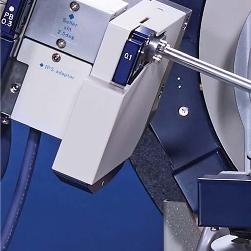
2 Leading With Innovation Next generation Rigaku SmartLab intelligent multipurpose X -ray diffractometer A highly versatile automated X -ray diffraction (XRD) system, the newest SmartLab diffractometer offers continued refinement of the ease-of-use features that enabled the original SmartLab diffractometer to receive the coveted R&D 100 Award, such as automatic alignment, component recognition, Cross Beam Optics and a 2D detector. SmartLab began as the flagship model from Rigaku in 2006 and new leading-edge, advanced technologies have been continuously introduced over the years. This newest addition to the SmartLab series of high-resolution X-ray diffraction analyzers is engineered to provide the best performance in all X -ray diffraction or scattering applications by offering not only breakthrough hardware, but also advanced User Guidance functionality within the new SmartLab Studio II software, to establish a new industry standard for multipurpose X -ray diffractometers. Key features and benefits of the new SmartLab include: Highest flux X-ray source: PhotonMax HyPix-3000 high energy resolution 2D detector New CBO family, with fully automated beam switchable CBO-Auto and high-resolution micro area CBO-μ Various operando measurements with the new SmartLab Studio II Multi-year component warranty contributes to low cost of ownership 1

3 2


4 High performance with an extended warranty PhotonMax The new generation rotating anode, PhotonMax, reduces the X-ray generator s environmental footprint, although it still produces 9 kw of high-power X-rays. The flux generated by the PhotonMax is about 5 times greater than a conventional sealed X-ray tube loaded at 1.8 kw. This enables you to see the fine details of your sample within a reasonable measurement time. The PhotonMax has a newly designed anode that provides more than 3 times longer lifetime than its predecessor. This maximizes the uptime of the instrument and minimizes the cost of ownership while reducing the environmental footprint. 3


5 A new mechanical seal has been introduced to substantially improve the lifetime of the target. 3-year or 10,000-hour warranty 4


 Efficiencies Cr, Co, Cu: ~99% Mo: ~38% Energy resolution 40% better than")

6 Leading-edge hybrid pixel array detector HyPix-3000 * Multidimensional semiconductor detector 5-year warranty Supports 0D, 1D and 2D measurement modes Excellent energy resolution to suppress XRF Keeps background noise to an absolute minimum Wide dynamic range Shutterless measurement Maintenance free Active area 2,984 mm 2 ( mm) Pixel size 100 μm 100 μm Number of pixels = 298,375 pixels Global count rate > (> cps/pixel) Efficiencies Cr, Co, Cu: ~99% Mo: ~38% Energy resolution 40% better than previous type Fully compatible with 5-axis goniometer design Hybrid pixel array detector (HPAD) design Shutterless high-speed in-situ measurement *This product was jointly developed by Department of Measurement and Electronics, AGH University of Science and Technology (Poland) and Rigaku Corporation. 5


 XRF suppression mode (new HyPix-3000)")
7 Achieve excellent energy resolution X-ray fluorescence background emitted by samples makes it difficult to detect minute peaks or scattering signals from amorphous components. It may also impede the correct calculation of the intensity of any detected peaks. Unnecessary X-ray fluorescence can be removed using a monochrometer placed between the sample and the detector. For two-dimensional (2D) measurement intended to detect Debye rings, however, no optical system can be inserted between the sample and the detector. For the removal of X-ray fluorescence during 2D measurement, a mode in which the energy resolution of the detector can be used to disable counting of unnecessary X-ray fluorescence is available. The excellent energy resolution greatly contributes to background suppression. 2D diffraction image measured in standard mode 2D diffraction image measured in XRF suppression mode Standard mode XRF suppression mode (previous HyPix-3000) XRF suppression mode (new HyPix-3000) Standard mode XRF suppression mode (previous HyPix-3000) XRF suppression mode (new HyPix-3000) 1D diffraction pattern obtained by standard mode, XRF suppression mode (previous HyPix-3000) and XRF suppression mode (new HyPix-3000) A graph of the same data as in the left figure normalized by the background noise intensities HyPix-3000 functions not only as a 2D detector but also as a 0/1-D detector. All applications can be handled with this single detector, eliminating the inconvenience of preparing and switching individual detectors for different applications. 6
 CBO-f Converges line beams to a minute point")

 75 μm 2.")
 Pixel size 100 μm 100 μm Global count rate >3.")
8 Optical configurations for various applications CBO (Cross Beam Optics) CBO Divergent beam Parallel beam CBO-E Divergent beam/convergent beam CBO-α Divergent beam (High intensity, low background noise) CBO-f Converges line beams to a minute point of 400 μm. No need to change the X-ray tube focus. CBO-μ Converges line beams to a high-intensity and parallel beam of 100 μm. No need to change the X-ray tube focus. Detectors 1D semiconductor detector D/tex Ultra250/250HE D/tex Ultra250/250HE Active area Spatial resolution Global count rate Efficiencies 384 mm 2 ( mm) 75 μm ( cps/pixel) Cr, Co, Cu: ~99% Mo: ~40%, ~70% (250 HE) Multidimensional semiconductor detector HyPix-400 * HyPix-400 * Active area 369 mm 2 ( mm) Pixel size 100 μm 100 μm Global count rate > cps (> cps/pixel) Efficiencies Cr, Co, Cu: ~99% Mo: ~38% *This product was jointly developed by Department of Measurement and Electronics, AGH University of Science and Technology (Poland) and Rigaku Corporation. 7
 is the standard measurement method for generic powder samples. For samples with specific orientation or large grains (i.e., powder, solid, or films), the transmission method is the optimal approach.")
 / CBO-Auto (Mo) Reflection/transmission ASC-6 Transmission mode High-precision goniometer with optical encoders Reproducibility of")
, 150-300 mm (2D)")
9 CBO-Auto: Fully automatic switch between reflection and transmission optics and geometries Reflection mode The optimal measurement method depends on the type of sample or the application. The Bragg-Brentano focusing (reflection mode) is the standard measurement method for generic powder samples. For samples with specific orientation or large grains (i.e., powder, solid, or films), the transmission method is the optimal approach. SmartLab provides fully automatic switching between the reflection and transmission methods. Ts axis Optics Sample stage CBO-Auto Automatic control CBO-Auto (Cu) / CBO-Auto (Mo) Reflection/transmission ASC-6 Transmission mode High-precision goniometer with optical encoders Reproducibility of the peak positions Rel. peak position [ ] [times] 10-year warranty Encoder controlled high-precision goniometer Type Vertical goniometer with sample horizontal mount Goniometer radius 300 mm (0D, 1D), mm (2D) Minimum step size Peak position stability after 50 times repeat of 2 - scan or 004 diffraction of silicon single crystal substrate. Distribution is within the range of reference accuracy ± Long-term warranty information. The warranty will be valid if the product breaks down when used in our environment and usage methods. When used under conditions other than our specifications, the warranty will not be applicable even within the warranty period. 8

10 Designed for functionality and safety 9


11 Shutter CLOSE lamp Shutter OPEN lamp X-rays on lamp EMO button Interlock Main key LED light Error lamp Door lock lamp Generator on lamp Power on lamp Safety-friendly enclosure design Leakage X-ray dosage 0.1 μsv or less Safe interlock mechanism mounted even in case of erroneous operation Design based on ergonomics Easy access to sample position Wide door opening improves accessibility to the inside of the device, which allows smooth changing of attachments High visibility design Six wide-angle windows let users check the state of the sample from various angles Indicator for easy confirmation of equipment status An easily recognizable LED lamp system has been adopted 10


 Typical")


12 SmartLab Studio II software suite SmartLab Studio II is an integrated software platform with all functions from measurement to analysis. Selection of sample type, Auto selection of measurement program Optics change guidance/ (Auto optics adjustment) Typical applications Powder Phase identification Quantification Crystallite size and distortion Precise lattice parameter determination Percent crystallinity Indexing Structural determination Precise crystalline structure determination Stress Sin 2 ψ method 2D method Multiple-HKL method Small Angle Scattering (SAXS) Grain size distribution Pore size distribution Long period Micro area measurement Specifications of beam size Collimator optics 50 μm to 1 mm CBO-f 400 μm CBO-μ 100 μm No need to change X-ray tube focus 11


13 Measurement, data processing, analysis Reporting Control/analysis history view Thin Film Film thickness Density Roughness Composition Texture Pole figure Stereographic projection ODF calculation Reverse pole figure Radial distribution PDF PDF calculation Simulation Large samples stage Specifications Movable range Min step width of each axis Max sample weight X-axis: -37~50 mm, Y-axis: -50~50 mm, Z-axis: -20~20 mm mm 20 kg 12

 method.")
 method.")
 PhotonMax CuKα 1 high resolution optical configuration with Johansson optics Johansson monochrometer Auto divergence slit")
14 Advanced powder X-ray diffractometry Leading-edge 2D pattern direct qualitative analysis (2D-ID: 2D pattern phase identification). Seamless execution from qualitative analysis to Rietveld refinement. XRD data docking with DSC or other data. Auto divergence slit Bragg-Brentano focusing optics Selection slit HyPix-3000 (1D mode) SmartLab Studio II Rietveld refinement Automatic input of required initial crystal structure from qualitative analysis result. Fitting with WPPF (Whole Powder Pattern Fitting) method. Crystallite size distribution analysis using FP (Fundamental Parameter) method. Estimation of oxidization state of metal atoms using BVS (Bond valence sum) method. Unknown crystal structure analysis package including from indexing to structure determination is available. CBO(BB) PhotonMax CuKα 1 high resolution optical configuration with Johansson optics Johansson monochrometer Auto divergence slit PhotonMax CBO(BB) Selection slit HyPix-3000 (1D mode) 13 Cement qualitative analysis and quantitative analysis using Rietveld refinement Organic powder precise crystalline structure determination


15 Advantage of qualitative analysis from 2D diffraction patterns For powder X-ray diffractometry, samples are generally crushed into grains small enough to obtain ideal diffraction patterns. However, the crushing process may cause crystal phase transition. In addition, there is often a need to measure bulk or thin film samples without crushing. For powders with large crystal grains (coarse particles) or with preferred orientation, the conventional powder measurement method may produce unreliable peak intensities or no observable peak at all, which has been a barrier to ensure reliable qualitative analysis. The HyPix D detector can be used to obtain 2D patterns for powder diffraction. These 2D patterns include distinctive features implying coarse particles or preferred orientation. These patterns can be processed to enable qualitative analysis in which the 2D pattern information is added to the 1D patterns. Qualitative analysis of a carbide tool using 2D diffraction patterns Coming from crystals with preferred orientation Coming from large particles Coming from large particles WC TiC C SiC Pharmaceutical solutions Compliance to FDA 21 CFR Part11 High-level security Audit trail IQ/OQ/PQ Computer system validation Validation document IQ/OQ/PQ Electronic recording/signature (ER/ES) software Unique XRD-DSC attachment Simultaneous XRD-DSC measurement of tolbutamide (antidiabetic drug) 14
.")


16 Residual stress analysis Wavelength can be selected for the type of metal or ceramics (Cr, Co, Cu, Mo). Supports sin 2 ψ, Multiple HKL and 2D (triaxial stress) methods. Analyses of thin films or materials with shear stress are available. Using micro area optics allows measurement of micro area stress mapping. sin 2 ψ method: Ni plate stress measurements Residual Stress measurement optical configuration Sealed tube χφz attachment platform CBO(PB) Selection slit HyPix-3000 (2D mode) Triaxial stress: Shot peened steel stress measurements Multi HKL method: Ni thin film stress measurements 15


17 Pole figure and ODF analysis In-plane or transmission measurement allows the acquisition of a whole pole figure. The α-β attachment allows γ rocking, alleviating the effect of large particles. With the use of a 2D detector, pole figures of two or more planes can be captured in a single measurement session. For analysis of complicated crystal texture, simulation analysis using not only pole acquisition but also ODF (Orientation Distribution Function) analysis allows calculation of the actual orientation distribution rate. Reverse pole figures can be used to determine the orientation of the lattice plane parallel to the sample surface. Pole figure measurement optical configuration PhotonMax CBO(PB) αβ attachment Selection slit HyPix-3000 (2D mode) SUS430 pole and ODF analyses Reverse pole figures of aluminum foils 16


18 in-situ/operando measurements Temperature-controlled measurement optical configuration Optical configuration with a laminate cell attachment Selection slit CBO-f Selection slit Laminate cell attachment HyPix-3000 (2D mode) HyPix-3000 (2D mode) CBO(PB) CBO-E(CB) PhotonMax Temperature-control attachment PhotonMax HyPix-3000 detector measures 30 range of 2θ by single shot. It can repeat the shot every 0.1 s. minimum. The Reactor X achieves high-speed temperature increase with infrared heating. It can accept many different types of gas because the heater section is separated from the sample chamber. Cu powder in-situ high-speed temperature increase measurement (300 /min) 2θ-temperature-intensity A battery cell attachment for evaluation of charging/ discharging, indispensable for battery material assessment, and a laminated cell attachment are available. Bulk data obtained from in-situ/operando measurement can be processed as a single unit by software. In addition, effective visual 3D diagrams can easily be created by adding a time axis to the angle/intensity axis. Li(Ni,Mn)O 4 operando charge/discharge measurement 2θ-time-intensity Cu 2 O Cu 2 O CuO CuO Discharge C C Cu Cu Charge A B A B 17
 HyPix-3000 (0D mode) CBO(PB) PhotonMax CBO-E(CB) PhotonMax Capillary attachment SAXS can be used to analyze")
 is also available, achieving measurement of minimum grain size of 1000 nm.")

19 Nanomaterials analysis SAXS measurement optical configuration PDF measurement optical configuration Selection slit Selection slit HyPix-3000 (1D mode) HyPix-3000 (0D mode) CBO(PB) PhotonMax CBO-E(CB) PhotonMax Capillary attachment SAXS can be used to analyze the grain size of a material (minimum 100 nm) and its distribution as well as the periodic structure. Furthermore, USAXS (Ultra SAXS) is also available, achieving measurement of minimum grain size of 1000 nm. PDF is an approach to derive real space information by inverse Fourier transformation of reciprocal space data. It can analyze interatomic distances, coordination number and periodicity irrespective of crystallinity of samples. Au nanoparticle grain size and distribution analysis PDF analysis of Al metal 4.6 nm 18


 measurement allows analysis of thin film structure information.")

20 Advanced thin film analysis A goniometer equipped with χ and φ axes as standard specifically designed for thin films is used to support the various measurements necessary for thin film analysis. Capable of obtaining thin film surface information by limiting the incidence angle to a micro angle. The CuK ₁ high-resolution parallel beam optics using Ge crystal monochrometer allows evaluation of single-crystal thin films including epitaxial films. High-resolution rocking curve (HRRC) measurement allows analysis of thin film structure information. Out-of-plane measurement optical configuration Sealed tube CBO(PB) Selection slit HyPix-3000 (0D mode) χφz attachment platform InGaN/GaN MQW thin film thickness analysis using HRRC High-speed wide reciprocal lattice map measurement of ferroelectric thin films using 2D detector 19


 Selection slit χφz attachment")
21 Rigaku s original in-plane axes allow in-plane diffractometry with the X-ray incidence angle accurately controlled. Reciprocal lattice map measurement allows user to learn the orientation relationship between the substrate and the film as well as the crystalline state. The use of reciprocal lattice simulation allows user to easily determine the film condition. The 2D detector HyPix-3000 can be effectively used to obtain a wide reciprocal lattice map in a short time. The map is drawn in real time during measurement, resulting in a shorter analysis time. In-plane measurement optical configuration Sealed tube CBO(PB) Selection slit χφz attachment platform with in-plane arm HyPix-3000 (0D mode) Out-of-plane/in-plane measurement IGZO film thickness and density analysis using XRR 20


 Collimator Vacuum path HyPix-3000 (2D mode) χφz attachment platform Preferred")
22 2D SAXS, WAXS and GI-SAXS Transmission 2D patterns of film materials can be measured. Circular measurement of Debye rings is available. This makes it easy to evaluate the oriented state and the degree of orientation. Transmission WAXS (Wide Angle X-ray Scattering) and SAXS (Small Angle X-ray Scattering) can be switched to obtain information including from long period structure to lattice level. With holders for different sample shapes, the equipment can accept powder, films and even textile samples, and provide mapping measurement within a sample. Measurement from the direction of the film cross section is also available. Transmission SAXS/WAXS measurement optical configuration Sealed tube Selection slit CBO(PB) Collimator Vacuum path HyPix-3000 (2D mode) χφz attachment platform Preferred orientation of Polypropylene observed y transmission 2D WAXS measurements Co-oriented Uniaxial oriented Biaxial oriented These are schematic images showing different preferred orientations on the Polypropylene samples. 21
 allows even clearer profile capturing in the in-plane direction.")

23 The GI-SAXS/WAXS unit can be used for 2D measurement of thin film materials. A newly developed aperture slit (patent pending) allows even clearer profile capturing in the in-plane direction. Information in both the lamination and the in-plane directions can be captured in a single measurement session. The oriented state and crystallinity of organic thin layers can be evaluated in a short time. Reflection SAXS/WAXS measurement optical configuration Sealed tube χφz attachment platform Selection slit CBO(PB) Collimator HyPix-3000 (2D mode) Thin-film phase Phase identification of Pentacene thin film with GI-WAXS In-plane direction Thin-film phase is detected. 22


")


24 Optional attachments and sample holders Sample plate Attachment head Attachment base Non-ambient chambers 4 wafer sample plate Battery cell ATT. head Standard ATT. base DHS 1100* 4 wafer sample plate (9 mm) Capillary spin ATT. head ASC-6 ATT. HTK 1200N* 6 wafer sample plate Standard ATT. head χφ ATT. TTK 600* 8 wafer sample plate RxRy-ATT. head αβ ATT. DSC attachment Height reference sample plate XY 20 mm-att. head β ATT. base Reactor X (infrared heating high temperature attachment) Transmission SAXS sample plate XY 4-inch ATT. head φ ATT. base Multipurpose high temperature attachment *Products of Anton Paar. Many other Anton Paar attachments are available for SmartLab. 23
 10 200 ma Installation requirements Enclosure dimensions")
 Weight (without any options) ~750 kg, ~1,653 lb for standard configuration ~850 kg, ~1,874 lb for standard")
.")
25 Specifications X-ray generation* X-ray generator 3 kw for sealed X-ray tube 9 kw for PhotonMax rotating anode Tube voltage variable range kv kv Tube current variable range 2 50 ma (option 60mA) ma Installation requirements Enclosure dimensions 1,300 x 1,300 x 1,880 mm, 51.2 x 51.2 x 74.0 inch (W x D x H) Weight (without any options) ~750 kg, ~1,653 lb for standard configuration ~850 kg, ~1,874 lb for standard configuration Power supply Three phases AC200 V, 50/60 Hz, 30 A or Three phases AC200 V, 50/60 Hz, 60 A single phase AC V, 50/60 Hz, 40 A Ground resistance 100 Ω *The maximum rated values depend on the type of X-ray tube (target, focus). Please refer to the instruction manual of the X-ray tube for details. Dimensions (unit: mm) Example of installation layout 24


 series, which facilitates optical system")

26 Technologies from Rigaku X-ray generators In 1952, Rigaku was the first company in the world to commercialize a rotating anode X-ray generator. Today, Rigaku s product line ranges in output from 50 to 9000 W, and at our Yamanashi plant and US facilities we develop and produce everything from high-brightness, high-output types, to sealed tube microfocus X-ray generators. In the area of high-voltage generation power supplies, we develop and produce molded types more compact and stable than previous systems. X-ray optics At Rigaku Innovative Technologies, which became part of our group in 2000, we are developing and manufacturing X-ray spectroscopy and focusing components. A key part of high-precision, high-sensitivity X-ray analysis is focusing X-rays and making them monochromatic or parallel using optic elements fabricated with sophisticated technology for artificial multilayer stacked films. At our Osaka factory, we develop and manufacture analyzing crystals for wavelength dispersive X-ray fluorescence spectrometers. The CBO (Cross Beam Optics) series, which facilitates optical system switching, also broadens the possibilities of X-ray analysis. MultiMax-9 MicroMax-007HF Confocal Mirror RX series Multipurpose rotating anode X-ray generator Microfocus rotating anode X-ray generator Multilayer optics Multilayer optics MicroMax-003 CBO series K 1 optics Multipurpose sealed tube X-ray generator FR-X Cross Beam Optic units Ultrahigh-intensity microfocus rotating anode X-ray generator Yamanashi plant Rigaku Innovative Technologies, Inc. 25


27 X-ray detectors Among Rigaku s X-ray detectors, the mainstay systems are direct-detection semiconductor detectors such as the 1-dimensional D/tex Ultra250/250HE series and 2-dimensional HyPix series, which combine high speed, low noise, high resolution and other features. Our Yamanashi factory is equipped with a cleanroom, a semiconductor process line and bonding equipment, and we produce detectors in-house. At Rigaku Innovative Technologies Europe s.r.o., established in 2008, we are also developing devices such as ultra-high-resolution CCDs. We are continuing our detector innovation in pursuit of greater convenience, such as simple switching between 0, 1 and 2 dimensions. HyPix-6000C/6000HE Hybrid pixel array detector HyPix-3000 Hybrid pixel array detector HyPix-400 Hybrid pixel array detector D/teX Ultra250/250HE High-resolution and high-speed 1D silicon strip detector XTOP High-sensitivity X-ray camera HR-XTOP High-sensitivity, high-resolution X-ray camera XRM High-resolution camera Rigaku Yamanashi Plant has obtained the international quality system certificate according to ISO 9001 and ISO and is addressing continual improvement with the PDCA cycle to provide reliable products to customers. 26


28 SmartLab Automated multipurpose X-ray diffractometer Rigaku Corporation and its Global Subsidiaries website: SmartLab_brochure_en_Ver1_ Copyright Rigaku Corporation. All rights reserved.
What if your diffractometer aligned itself?
 Ultima IV Perhaps the greatest challenge facing X-ray diffractometer users today is how to minimize time and effort spent on reconfiguring of the system for different applications. Wade Adams, Ph.D., Director,
Technical articles Micro-area X-ray diffraction measurement by SmartLab μ
 Technical articles Micro-area X-ray diffraction measurement by SmartLab μhr diffractometer system with ultra-high brilliance microfocus X-ray optics and two-dimensional detector HyPix-3000 Yuji Shiramata*
Fundamentals of X-ray diffraction and scattering
 Fundamentals of X-ray diffraction and scattering Don Savage dsavage@wisc.edu 1231 Engineering Research Building (608) 263-0831 X-ray diffraction and X-ray scattering Involves the elastic scattering of
Benchtop XRD diffractometer. MiniFlex. Analysis of materials by X-ray diffraction
 Benchtop XRD diffractometer MiniFlex Analysis of materials by X-ray diffraction More power More flexibility More results The new MiniFlex is available in two models. The MiniFlex 600 is the most powerful
MiniFlex. Analysis of materials by X-ray diffraction. Benchtop XRD diffractometer
 MiniFlex Analysis of materials by X-ray diffraction Benchtop XRD diffractometer More power More flexibility More results The new MiniFlex is available in two models. The MiniFlex 600 is the most powerful
Precision Without Compromise
 D1 EVOLUTION Precision Without Compromise Versatile and user-friendly high resolution and multipurpose X-ray diffractometer for the characterization of advanced materials www.jvsemi.com D1 Overview Introduction
X-RAY DIFFRACTION IN SEMICONDUCTOR INDUSTRY AND RESEARCH
 X-RAY DIFFRACTION IN SEMICONDUCTOR INDUSTRY AND RESEARCH M. Leszczyński High Pressure Research Center UNIPRESS, Sokolowska 29/37, 01 142 Warsaw, Poland, e-mail: mike@unipress.waw.pl ABSTRACT The paper
Lesson 1 Good Diffraction Data
 Lesson 1 Good Diffraction Data Nicola Döbelin RMS Foundation, Bettlach, Switzerland Digital Diffractometers Transmission Geometry Debye-Scherrer Geometry Reflective Geometry Bragg-Brentano Geometry Glass
ATTACHMENTES FOR EXPLORER DIFFRACTOMETER. Monochromators
 Monochromators Secondary flat and curved graphite monochromators suitable for Ag, Cr, Fe, Cu, Co and Mo radiations This attachment is installed in the X-ray detection unit. It is designed to remove continuous
Thermo Scientific ARL EQUINOX 100. X-ray Diffractometers
 Thermo Scientific ARL EQUINOX 100 X-ray Diffractometers High performance in a compact size Thermo Scientific ARL EQUINOX 100 X-ray diffractometer (XRD) is designed to meet structural and phase analysis
Thermo Scientific ARL EQUINOX X-ray Diffractometers
 Thermo Scientific ARL EQUINOX 1000 X-ray Diffractometers High performance in a compact size Thermo Scientific ARL EQUINOX 1000 X-ray diffractometer (XRD) is designed to meet structural and phase analysis
ATTACHMENTES FOR APD 2000 PRO POWDER X-RAY DIFFRACTOMETER. Monochromators
 Monochromators Secondary graphite monochromator Johansson Ka 1 monochromator Parabolic monochromator Secondary flat and curved graphite monochromators suitable for Ag, Cr, Fe, Cu, Co and Mo radiations
Lesson 3 Sample Preparation
 Lesson 3 Sample Preparation Nicola Döbelin RMS Foundation, Bettlach, Switzerland January 14 16, 2015, Bern, Switzerland Repetition: Bragg-Brentano Diffractometer Typical Configuration (with Kβ filter)
LECTURE 7. Dr. Teresa D. Golden University of North Texas Department of Chemistry
 LECTURE 7 Dr. Teresa D. Golden University of North Texas Department of Chemistry Diffraction Methods Powder Method For powders, the crystal is reduced to a very fine powder or microscopic grains. The sample,
Instrument Configuration for Powder Diffraction
 Instrument Configuration for Powder Diffraction Advanced X-ray Workshop S.N. Bose National Centre for Basic Sciences, 14-15/12/2011 Innovation with Integrity Overview What is the application? What are
Europe. Benchtop X-Ray Diffractometer.
 Europe Benchtop X-Ray Diffractometer www.gnr.it benchtop x-ray diffractometer Europe, High Performance in a compact configuration GNR is a worldwide market leader supplying advanced X-Ray (XRD, XRF) and
This lecture is part of the Basic XRD Course.
 This lecture is part of the Basic XRD Course. Basic XRD Course 1 A perfect polycrystalline sample should contain a large number of crystallites. Ideally, we should always be able to find a set of crystallites
Technical Specification for Laboratory X-ray diffraction system for measurements of Crystallographic Texture and Residual Stress
 INDIAN INSTITUTE OF TECHNOLOGY BOMBAY MATERIALS MANAGEMENT DIVISION Direct : (+91-22) 2576 8800 (DR) / 8803 (Local) / 8804 (Import) / 8805 (Enquiry & Bill tracking), 8802(Progress) Email : drmm@iitb.ac.in,
Earth & Planetary Science Applications of X-Ray Diffraction: Advances Available for Research with our New Systems
 Earth & Planetary Science Applications of X-Ray Diffraction: Advances Available for Research with our New Systems James R. Connolly Dept. of Earth & Planetary Sciences University of New Mexico 401/501
OPTIMIZING XRD DATA. By: Matthew Rayner
 OPTIMIZING XRD DATA By: Matthew Rayner 1 XRD Applications PANalytical classifies XRD applications in 4 groups 1. Powders 2. Nanomaterials 3. Solid objects 4. Thin films Many day-to-day samples cross these
Identification of Crystal Structure and Lattice Parameter. for Metal Powders Using X-ray Diffraction. Eman Mousa Alhajji
 Identification of Crystal Structure and Lattice Parameter for Metal Powders Using X-ray Diffraction Eman Mousa Alhajji North Carolina State University Department of Materials Science and Engineering MSE
Thermo Scientific X-ray product range. For chemical and phase analysis of solids, liquids and powders
 X-ray product range For chemical and phase analysis of solids, liquids and powders Metals, cement, mining, petrochemicals, environment, electronics, geology, glass, polymers, forensics, materials science,
Dedication in X-ray powder diffraction
 X PERT 3 POWDER Dedication in X-ray powder diffraction The Analytical X-ray Company x-ray diffraction Let materials work for you Advancing materials research From geological exploration, through processing
Single crystal X-ray diffraction. Zsolt Kovács
 Single crystal X-ray diffraction Zsolt Kovács based on the Hungarian version of the Laue lab description which was written by Levente Balogh, Jenő Gubicza and Lehel Zsoldos INTRODUCTION X-ray diffraction
Bruker AXS D8 FABLINE. X-Ray Metrology Solutions. think forward
 Bruker AXS D8 FABLINE X-Ray Metrology Solutions think forward XRD & µxrf D8 FABLINE Metrology for Semiconductor Manufacturing The functional units of semiconductor and compound semiconductor devices shrink
JSM-7800F Field Emission Scanning Electron Microscope
 JSM-7800F catalogue JSM-7800F Field Emission Scanning Electron Microscope We provide high performance The Ultimate Research Tool for Multi-Disciplinary Research Institutions Extreme resolution The super
X-ray diffraction
 2.2.3.- X-ray diffraction 2.2.3.1.- Origins and fundamentals of the technique The first experimental evidence concerning x-ray diffraction was given by Max von Laue who in 1912 demonstrated that x-rays
High Resolution X-ray Diffraction
 High Resolution X-ray Diffraction Nina Heinig with data from Dr. Zhihao Donovan Chen, Panalytical and slides from Colorado State University Outline Watlab s new tool: Panalytical MRD system Techniques:
Introduction to Powder Diffraction/Practical Data Collection
 Durham University Chemistry Department Introduction to Powder Diffraction/Practical Data Collection Dr Ivana Evans Durham, January 2007 Durham Outline Information in a powder pattern What is diffraction
AN INNOVATED LABORATORY XAFS APPARATUS
 Copyright (c)jcpds-international Centre for Diffraction Data 2002, Advances in X-ray Analysis, Volume 45. 397 AN INNOVATED LABORATORY XAFS APPARATUS TAGUCHI Takeyoshi XRD Division, Rigaku Corporation HARADA
Background Statement for SEMI Draft Document 5945 New Standard: Test Method for Determining Orientation of A Sapphire Single Crystal
 Background Statement for SEMI Draft Document 5945 New Standard: Test Method for Determining Orientation of A Sapphire Single Crystal Notice: This background statement is not part of the balloted item.
Observation in the GB (Gentle Beam) Capabilities
 Capabilities") A field-emission cathode in the electron gun of a scanning electron microscope provides narrower probing beams at low as well as high electron energy, resulting in both improved spatial resolution and
A field-emission cathode in the electron gun of a scanning electron microscope provides narrower probing beams at low as well as high electron energy, resulting in both improved spatial resolution and
Characterization of Materials Using X-Ray Diffraction Powder Diffraction
 Praktikum III, Fall Term 09 Experiment P1/P2; 23.10.2009 Characterization of Materials Using X-Ray Diffraction Powder Diffraction Authors: Michael Schwarzenberger (michschw@student.ethz.ch) Philippe Knüsel
Structural Biology. Single crystal X-ray diffraction and SAXS. Systems designed for structural biology
 Structural Biology Single crystal X-ray diffraction and SAXS Systems designed for structural biology Rigaku Oxford Diffraction THE POWER OF SYNERGY Combining the single crystal groups from Rigaku and Oxford
Wavelength Dispersive XRF Spectrometer
 ISO9001 ISO14001 for Wafers, Media Disks, and Large Samples Wavelength Dispersive XRF Spectrometer ADVANCED XRF For Large and Irregularly Shaped Samples Is there any solution to analyze a large sample
Central Purchase unit National Institute of Technology Srinagar Tel: / / / Fax:
 Central Purchase unit National Institute of Technology Srinagar-190006 Tel:- 0194-2424792/2429423/2424809/2424797 Fax:- 0194-2420475 *************************************************************** No.
Diffraction Basics. The qualitative basics:
 The qualitative basics: Diffraction Basics Coherent scattering around atomic scattering centers occurs when x-rays interact with material In materials with a crystalline structure, x-rays scattered in
Non-ambient X-ray Diffraction and Nanostructure Analysis. Sample Stages Overview
 Non-ambient X-ray Diffraction and Nanostructure Analysis Sample Stages Overview Setting the Pace in Non-ambient XRD Non-ambient X-ray diffraction (XRD) has become an indispensable technique to understand
The Empyrean Tube. Advanced eco-friendly design, powerful performance
 The Empyrean Tube Advanced eco-friendly design, powerful performance The Empyrean Tube The industrial benchmark, redefined PANalytical s Empyrean Tubes set the standard, both for X-ray diffraction (XRD)
Specimen Preparation Technique for a Microstructure Analysis Using the Focused Ion Beam Process
 Specimen Preparation Technique for a Microstructure Analysis Using the Focused Ion Beam Process by Kozue Yabusaki * and Hirokazu Sasaki * In recent years the FIB technique has been widely used for specimen
Fundamentals of Crystalline State and Crystal Lattice p. 1 Crystalline State p. 2 Crystal Lattice and Unit Cell p. 4 Shape of the Unit Cell p.
 Fundamentals of Crystalline State and Crystal Lattice p. 1 Crystalline State p. 2 Crystal Lattice and Unit Cell p. 4 Shape of the Unit Cell p. 7 Crystallographic Planes, Directions, and Indices p. 8 Crystallographic
Lesson 1 X-rays & Diffraction
 Lesson 1 X-rays & Diffraction Nicola Döbelin RMS Foundation, Bettlach, Switzerland February 11 14, 2013, Riga, Latvia Electromagnetic Spectrum X rays: Wavelength λ: 0.01 10 nm Energy: 100 ev 100 kev Interatomic
Fundamentals of Crystalline State p. 1 Introduction p. 1 Crystalline state p. 2 Crystal lattice and crystal structure p. 4 Shape of the unit cell p.
 Preface p. xvii Fundamentals of Crystalline State p. 1 Introduction p. 1 Crystalline state p. 2 Crystal lattice and crystal structure p. 4 Shape of the unit cell p. 6 Content of the unit cell p. 7 Asymmetric
Copyright JCPDS - International Centre for Diffraction Data 2004, Advances in X-ray Analysis, Volume
 Copyright JCPDS - International Centre for Diffraction Data 2004, Advances in X-ray Analysis, Volume 47. 240 SIMULTANEOUS MEASUREMENTS OF X-RAY DIFFRACTION (XRD) AND DIFFERENTIAL SCANNING CALORIMETRY (DSC)
NEBRASKA NANOSCALE FACILITY CHARACTERIZATION FACILITIES
 NEBRASKA NANOSCALE FACILITY CHARACTERIZATION FACILITIES Jeff Shield Department of Mechanical & Materials Engineering Nebraska Center for Materials and Nanoscience National Nanotechnology Coordinated Infrastructure
SYSTEMATIC ERRORS IN LINEAR PSD BASED HTXRD SYSTEMS
 Copyright(c)JCPDS-International Centre for Diffraction Data 2,Advances in X-ray Analysis,Vol.43 267 SYSTEMATIC ERRORS IN LINEAR PSD BASED HTXRD SYSTEMS E.A. Payzant and W.S. Harrison, III * Metals and
ARL X TRA Powder X-ray Diffraction System. Uncompromised Intensity and Resolution
 e l e m e n t a l a n a l y s i s ARL X TRA Powder X-ray Diffraction System Uncompromised Intensity and Resolution Analyze Detect Measure Control ARL X TRA Powder X-ray Diffraction System ARL X TRA High
Thermo Scientific ARL X TRA Powder X-ray Diffraction System Uncompromised Intensity and Resolution
 e l e m e n t a l a n a l y s i s Thermo Scientific ARL X TRA Powder X-ray Diffraction System Uncompromised Intensity and Resolution Part of Thermo Fisher Scientific ARL X TRA Powder X-ray Diffraction
Travaux Pratiques de Matériaux de Construction
 Travaux Pratiques de Matériaux de Construction Section Matériaux 6 ème semestre 2009 Etude de Matériaux Cimentaire Par Diffraction des Rayons X Responsable: Silke Ruffing E-Mail: silke.ruffing@epfl.ch
Thin Film Scattering: Epitaxial Layers
 Thin Film Scattering: Epitaxial Layers 6th Annual SSRL Workshop on Synchrotron X-ray Scattering Techniques in Materials and Environmental Sciences: Theory and Application May 29-31, 2012 Thin films. Epitaxial
Smithsonian Museum Conservation Institute
 Smithsonian Museum Conservation Institute XRD Analysis of the Corrosion Products from a Tlingit Copper Rattle MCI#6241 Object: Tlingit Stikine Rattle Owner/Custodian: National Museum of the American Indian
Diffraction: Powder Method
 Diffraction: Powder Method Diffraction Methods Diffraction can occur whenever Bragg s law λ = d sin θ is satisfied. With monochromatic x-rays and arbitrary setting of a single crystal in a beam generally
Strain. Two types of stresses: Usually:
 Stress and Texture Strain Two types of stresses: microstresses vary from one grain to another on a microscopic scale. macrostresses stress is uniform over large distances. Usually: macrostrain is uniform
Characterization of Surfaces and Thin Films Using a High Performance Grazing Incidence X-ray Diffractometer
 Copyright(c)JCPDS-International Centre for Diffraction Data 2000,Advances in X-ray Analysis,Vol.43 177 Characterization of Surfaces and Thin Films Using a High Performance Grazing Incidence X-ray Diffractometer
Aeris. Research edition
 Aeris Research edition It s like making a cup of coffee except it s even easier Watch our video on www.xrdiseasy.com Surprisingly intuitive Meet the Research edition of Aeris PANalytical s easy-to-operate
Materials Lab 1(MT344) X-ray Diffractometer Operation and Data Analysis. Instructor: Dr. Xueyan Wu ( 吴雪艳 )
 X-ray Diffractometer Operation and Data Analysis. Instructor: Dr. Xueyan Wu ( 吴雪艳 )") Materials Lab 1(MT344) X-ray Diffractometer Operation and Data Analysis Instructor: Dr. Xueyan Wu ( 吴雪艳 ) Goals To give students a practical introduction into the use of X-ray diffractometer and data collection.
Materials Lab 1(MT344) X-ray Diffractometer Operation and Data Analysis Instructor: Dr. Xueyan Wu ( 吴雪艳 ) Goals To give students a practical introduction into the use of X-ray diffractometer and data collection.
The object of this experiment is to test the de Broglie relationship for matter waves,
 Experiment #58 Electron Diffraction References Most first year texts discuss optical diffraction from gratings, Bragg s law for x-rays and electrons and the de Broglie relation. There are many appropriate
Chapter 3 Basic Crystallography and Electron Diffraction from Crystals. Lecture 9. Chapter 3 CHEM Fall, L. Ma
 Chapter 3 Basic Crystallography and Electron Diffraction from Crystals Lecture 9 Outline The geometry of electron diffraction Crystallography Kinetic Theory of Electron diffraction Diffraction from crystals
Travaux Pratiques de Matériaux de Construction. Etude de Matériaux Cimentaires par Diffraction des Rayons X sur Poudre
 Travaux Pratiques de Matériaux de Construction Section Matériaux 6 ème semestre 2015 Etude de Matériaux Cimentaires par Diffraction des Rayons X sur Poudre Study Cementitious Materials by X-ray diffraction
Physics 6180: Graduate Physics Laboratory. Experiment CM5: X-ray diffraction and crystal structures
 Physics 6180: Graduate Physics Laboratory Experiment CM5: X-ray diffraction and crystal structures References: Preston and Dietz, Expt. 10 pp. 180-197 Eisberg and Resnick, Quantum Physics, Sec. 9 Kittel,
A. KISHI AND H. TORAYA
 THE RIGAKU JOURNAL VOL. 21 / NO. 1 / 2004, 25 30 SIMULTANEOUS MEASUREMENTS OF X-RAY DIFFRACTION (XRD) AND DIFFERENTIAL SCANNING CALORIMETRY (DSC) DATA UNDER CONTROLLED HUMIDITY CONDITION: INSTRUMENTATION
CHARACTERIZATION OF X-RAY DIFFRACTION SYSTEM WITH A MICROFOCUS X-RAY SOURCE AND A POLYCAPILLARY
 Copyright(c)JCPDS-International Centre for Diffraction Data 2001,Advances in X-ray Analysis,Vol.44 278 CHARACTERIZATION OF X-RAY DIFFRACTION SYSTEM WITH A MICROFOCUS X-RAY SOURCE AND A POLYCAPILLARY OPTIC
Epitaxy and Roughness Study of Glancing Angle Deposited Nanoarrays. Hamid Alouach and G. J. Mankey
 Epitaxy and Roughness Study of Glancing Angle Deposited Nanoarrays Hamid Alouach and G. J. Mankey Introduction Objective: Approach: Characterization: Fabrication of nanoscale magnetic wires for spin transport
CHARACTERISATION OF CRYSTALLINE AND PARTIALLY CRYSTALLINE SOLIDS BY X-RAY POWDER DIFFRACTION (XRPD)
") 2.9.33. Characterisation of crystalline solids by XRPD EUROPEAN PHARMACOPOEIA 6.0 with its standard deviation. The mean values for x 10 and x 90 must not deviate by more than 5 per cent from the certified
2.9.33. Characterisation of crystalline solids by XRPD EUROPEAN PHARMACOPOEIA 6.0 with its standard deviation. The mean values for x 10 and x 90 must not deviate by more than 5 per cent from the certified
Study of amorphous, extraordinary absorbing, high-surface area magnesium carbonate using a laboratory diffractometer
 X-ray XRD SAXS Study of amorphous, extraordinary absorbing, high-surface area magnesium carbonate using a laboratory diffractometer PDF Olga Narygina 1, Marco Sommariva 1, Sara Frykstrand 2, Johan Forsgren
D2 PHASER. 2nd Generation. Innovation with Integrity XRD. Diffraction Solutions
 D2 PHASER 2nd Generation Diffraction Solutions Innovation with Integrity XRD Compact all-in-one benchtop design Innovative high-end goniometer design Integrated PC / monitor DIFFRAC.SUITE software Leading
F. J. Cadieu*, I. Vander, Y. Rong, and R. W. Zuneska, Physics Department, Queens College of CUNY, Flushing, NY
 Copyright JCPDS-International Centre for Diffraction Data 2012 ISSN 1097-0002 1 X-Ray Measurements of Nanometer Thick Ta x O 1-x and Hf x O 1-x Films on Silicon Substrates for Thickness and Composition
Spatially resolved crystal domain identification: Implementing Laue-mapping technique on the M4 TORNADO spectrometer
 Spatially resolved crystal domain identification: Implementing Laue-mapping technique on the M4 TORNADO spectrometer Bruker Nano Analytics, Berlin, Germany Webinar, July 14 th, 2016 Innovation with Integrity
Stress Mitigation of X-ray Beamline Monochromators using a Topography Test Unit
 128 Stress Mitigation of X-ray Beamline Monochromators using a Topography Test Unit J. Maj 1, G. Waldschmidt 1 and A. Macrander 1, I. Koshelev 2, R. Huang 2, L. Maj 3, A. Maj 4 1 Argonne National Laboratory,
Physical structure of matter. Monochromatization of molybdenum X-rays X-ray Physics. What you need:
 X-ray Physics Physical structure of matter Monochromatization of molybdenum X-rays What you can learn about Bremsstrahlung Characteristic radiation Energy levels Absorption Absorption edges Interference
Di rect beam J' / o 20, " - l To tally reftected. 20, X Scan / "-
 THE RIGAKU JOURNAL VOl. 8 / NO. 1 / 1991 Technical Note THIN FILM X-RAY DIFFRACTOMETRY H. ARAKI Rigaku Corporation. Tokvo. Japan 1. Introduction X-ray diffraction methods have been very popular in recent
Thin Film Scattering: Epitaxial Layers
 Thin Film Scattering: Epitaxial Layers Arturas Vailionis First Annual SSRL Workshop on Synchrotron X-ray Scattering Techniques in Materials and Environmental Sciences: Theory and Application Tuesday, May
Bragg diffraction using a 100ps 17.5 kev x-ray backlighter and the Bragg Diffraction Imager
 LLNL-CONF-436071 Bragg diffraction using a 100ps 17.5 kev x-ray backlighter and the Bragg Diffraction Imager B. R. Maddox, H. Park, J. Hawreliak, A. Comley, A. Elsholz, R. Van Maren, B. A. Remington, J.
An Introduction to X-Ray Powder Diffraction. credits to: Scott A Speakman, Patrick McArdle Edited by Di Cicco 2014
 An Introduction to X-Ray Powder Diffraction credits to: Scott A Speakman, Patrick McArdle Edited by Di Cicco 2014 LATTICE ARRAYS AND BRAVAIS LATTICES Crystalline materials differ from amorphous materials
Practical X-Ray Diffraction
 Typical Example Practical X-Ray Diffraction White powder sample of NaCl,KCl,KNO 3 (trace of H 2 O) Département de chimie Université Laval Prof. Josée BRISSON Dr. Wenhua BI 2014-03-20 Powder X-Ray Diffraction
X-ray Diffraction (XRD)
") هب انم خدا X-ray Diffraction (XRD) 1.0 What is X-ray Diffraction 2.0 Basics of Crystallography 3.0 Production of X-rays 4.0 Applications of XRD 5.0 Instrumental Sources of Error 6.0 Conclusions Bragg s
هب انم خدا X-ray Diffraction (XRD) 1.0 What is X-ray Diffraction 2.0 Basics of Crystallography 3.0 Production of X-rays 4.0 Applications of XRD 5.0 Instrumental Sources of Error 6.0 Conclusions Bragg s
Transmission Electron Microscopy (TEM) Prof.Dr.Figen KAYA
 Prof.Dr.Figen KAYA") Transmission Electron Microscopy (TEM) Prof.Dr.Figen KAYA Transmission Electron Microscope A transmission electron microscope, similar to a transmission light microscope, has the following components along
Transmission Electron Microscopy (TEM) Prof.Dr.Figen KAYA Transmission Electron Microscope A transmission electron microscope, similar to a transmission light microscope, has the following components along
CHAPTER 7 MICRO STRUCTURAL PROPERTIES OF CONCRETE WITH MANUFACTURED SAND
 99 CHAPTER 7 MICRO STRUCTURAL PROPERTIES OF CONCRETE WITH MANUFACTURED SAND 7.1 GENERAL Characterizing the mineralogy of the samples can be done in several ways. The SEM identifies the morphology of the
Non-ambient solutions. State-of-the-art in situ X-ray analysis made easy
 Non-ambient solutions State-of-the-art in situ X-ray analysis made easy PANalytical s approach to the implementation of non-ambient attachments is characterized by: Ease of use Versatility Delivering the
Advanced Methods for Materials Research. Materials Structure Investigations Materials Properties Investigations
 Advanced Methods for Materials Research Materials Structure Investigations Materials Properties Investigations Advanced Methods for Materials Research 1. The structure and property of sample and methods
SmartLab: Advanced measurements with simple operation
 Introduction SmartLab: Advanced measurements with simple operation Powered by Rigaku s proprietary Guidance software system, SmartLab incorporates intelligent, applicationspecific protocols in all measurement
Towards the Epitaxial Growth of Silver on Germanium by Galvanic Displacement
 Electronic Supplementary Material (ESI) for CrystEngComm. This journal is The Royal Society of Chemistry 2014 Towards the Epitaxial Growth of Silver on Germanium by Galvanic Displacement Sayed Youssef
RIX3100. Fully Automated Sequential X-ray Spectrometer System. Product Information
 The Rigaku Journal Vol. 14/ number 1/ 1997 Product Information Fully Automated Sequential X-ray Spectrometer System RIX3100 1. Introduction Wavelength dispersive X-ray fluorescence (WDXRF) analysis methods
Lesson 1 Rietveld Refinement and Profex / BGMN
 Lesson 1 Rietveld Refinement and Profex / BGMN Nicola Döbelin RMS Foundation, Bettlach, Switzerland June 13 15, 2018, Bettlach, CH Diffraction Pattern 1000 Diffraction Angle 800 Absolute Intensity Intensity
X-RAY DIFFRACTION CHARACTERIZATION OF MULTILAYER EPITAXIAL THIN FILMS DEPOSITED ON (0001) SAPPHIRE
 SAPPHIRE") The Rigaku Journal Vol. 13/No. 1/ 1996 CONTRIBUTED PAPERS X-RAY DIFFRACTION CHARACTERIZATION OF MULTILAYER EPITAXIAL THIN FILMS DEPOSITED ON (0001) SAPPHIRE THOMAS N. BLANTON AND LIANG-SUN HUNG Imaging
The Rigaku Journal Vol. 13/No. 1/ 1996 CONTRIBUTED PAPERS X-RAY DIFFRACTION CHARACTERIZATION OF MULTILAYER EPITAXIAL THIN FILMS DEPOSITED ON (0001) SAPPHIRE THOMAS N. BLANTON AND LIANG-SUN HUNG Imaging
RAPID QUANTITATIVE MEASUREMENT SYSTEM FOR RETAINED AUSTENITE (Multi-PSPC System)
") The Rigaku Journal Vol. 3/ No. 2/1986 Product Information RAPID QUANTITATIVE MEASUREMENT SYSTEM FOR RETAINED AUSTENITE (Multi-PSPC System) Fig. 1. Rigaku/Rapid Quantitative Measurement System for Retained
The Rigaku Journal Vol. 3/ No. 2/1986 Product Information RAPID QUANTITATIVE MEASUREMENT SYSTEM FOR RETAINED AUSTENITE (Multi-PSPC System) Fig. 1. Rigaku/Rapid Quantitative Measurement System for Retained
S8 TIGER Series 2. Lab Report XRF 144. Innovation with Integrity. Accurate Quality Control of Tool Steels XRF
 Lab Report XRF 144 S8 TIGER Series 2 Accurate Quality Control of Tool Steels Tool steels are iron-based alloys that are particularly well-suited to be made into tools. Their suitability comes from their
Wavelength-dispersive X-ray fluorescence spectrometer
 New products Wavelength-dispersive X-ray fluorescence spectrometer 1. Introduction The newly-released sequential general-purpose wavelength-dispersive X-ray fluorescence spectrometer, ZSX PrimusIII is
UNIT V -CRYSTAL STRUCTURE
 UNIT V -CRYSTAL STRUCTURE Solids are of two types: Amorphous and crystalline. In amorphous solids, there is no order in the arrangement of their constituent atoms (molecules). Hence no definite structure
Philips Analytical, Lelyweg 1, 7602 EA Almelo, The Netherlands
 Copyright(c)JCPDS-International Centre for Diffraction Data 2001,Advances in X-ray Analysis,Vol.44 284 MICRO-DIFFRACTION WITH MONO-CAPILLARIES M.J. Fransen, J.H.A. Vasterink and J. te Nijenhuis Philips
X-Ray Diffraction by Macromolecules
 N. Kasai M. Kakudo X-Ray Diffraction by Macromolecules With 351 Figures and 56 Tables Kodansha ~Springer ... Contents Preface v Part I Fundamental 1. Essential Properties of X-Rays................. 3 1.1
INGE Engineering Materials. Chapter 3 (cont.)
") Some techniques used: Chapter 3 (cont.) This section will address the question how do we determine the crystal structure of a solid sample? Electron microscopy (by direct and indirect observations) Scanning
Some techniques used: Chapter 3 (cont.) This section will address the question how do we determine the crystal structure of a solid sample? Electron microscopy (by direct and indirect observations) Scanning
LECTURE 8. Dr. Teresa D. Golden University of North Texas Department of Chemistry
 LECTURE 8 Dr. Teresa D. Golden University of North Texas Department of Chemistry Practical applications for lattice parameter measurements: -determine composition (stoichiometry) of the sample -determine
Crystallographic Textures Measurement
 Crystallographic Textures Measurement D. V. Subramanya Sarma Department of Metallurgical and Materials Engineering Indian Institute of Technology Madras E-mail: vsarma@iitm.ac.in Macrotexture through pole
X-Ray Diffraction. Nicola Pinna
 X-Ray Diffraction Nicola Pinna Department of Chemistry, CICECO, University of Aveiro, 3810-193 Aveiro, Portugal. School of Chemical and Biological Engineering, College of Engineering, Seoul National University
EBSD Basics EBSD. Marco Cantoni 021/ Centre Interdisciplinaire de Microscopie Electronique CIME. Phosphor Screen. Pole piece.
 EBSD Marco Cantoni 021/693.48.16 Centre Interdisciplinaire de Microscopie Electronique CIME EBSD Basics Quantitative, general microstructural characterization in the SEM Orientation measurements, phase