A Brief History of XRD 1895: Röntgen discovers X-Rays received the first Nobel prize in physics in 1901
|
|
|
- Silvester Allen
- 6 years ago
- Views:
Transcription
1 X-ray Diffraction


, now International Center for Diffraction")
2 A Brief History of XRD 1895: Röntgen discovers X-Rays received the first Nobel prize in physics in : Laue diffracts X-Rays from single crystal 1914 Nobel prize in Physics 1912: Bragg analyzes crystal structures 1915 Nobel prize in physics 1917: Ewald develops dynamical theory of X-Ray diffraction 1918: Scherrer uses X-Rays to determine crystallite size of nanocrystalline gold 1935: X-Ray powder diffractometer developed by Le Galley 1947: First commercial powder diffractometer 1969: Establishment of the Joint Committee on Powder Diffraction Standards (JCPDS), now International Center for Diffraction Data (ICDD)
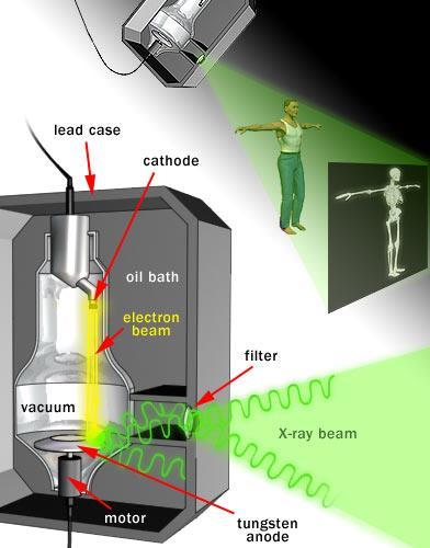
3 Generation of X-rays
4 X-ray Spectrum from an Iron target Short Wavelength Limit Continuous spectrum I CS SWL (nm) AiZV m V Characteristic X-ray Moseley s Law C( Z ) I Bi( V ) K V k n λ SWL
5 Scattering fundamentals Scattering can be broadly defined as the redirection of radiation out of the original direction of propagation, usually due to interactions with molecules and particles Reflection, refraction, diffraction etc. are actually all just forms of scattering Matter is composed of discrete electrical charges (atoms and molecules dipoles) Light is an oscillating EM field excites charges, which radiate EM waves These radiated EM waves are scattered waves, excited by a source external to the scatterer The superposition of incident and scattered EM waves is what is observed
 Forward")
6 Scattering geometry Backward scattering (backscattering) Forward scattering
7 Types of scattering Elastic scattering the wavelength (frequency) of the scattered light is the same as the incident light (Rayleigh and Mie scattering) Inelastic scattering the emitted radiation has a wavelength different from that of the incident radiation (Raman scattering, fluorescence) Quasi-elastic scattering the wavelength (frequency) of the scattered light shifts (e.g., in moving matter due to Doppler effects)


8 How Diffraction occurs Diffraction occurs when objects in a periodic array scatter radiation coherently, producing constructive interference at specific angles X-Rays can diffract from a periodic array of elastic scatterers, such as atoms in a crystal
9 Diffraction of light through an aperture a Intensity
10 Intensity Minima Maxima sinθ λ n a n = 0, 1,.. sin 2n 1 2a n = 1, 2,..
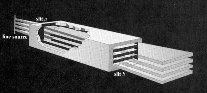
11 Young s Double slit experiment d sinθ = mλ, m = 1,2,3.. Constructive Interference d sinθ = (m+½)λ, m = 1,2,3.. Destructive Interference


12 Interference Phase Difference = 0 Phase Difference = 90 Phase Difference = 180
13 Elastic Scattering When x-rays or electrons interact with matter, the dominant effect is scattering. Considering x-rays and electrons as waves we deal with elastic scattering (rather than as particles, where we deal with inelastic scattering) For elastic scattering, x-rays and electrons are scattered with no loss of energy, and give rise to scattered radiation of the same wavelength
14 Constructive Interference The distance between atoms (d hkl ) are on the same order of size as the wavelength of an x-ray (Cu Ka =1.54Å) Interference phenomena is concentrated in directions related to the crystal lattice The intensity of the diffracted x-rays gives rise to peaks for each set of wave vectors which make up diffraction patterns The positions of the atoms in the material (the crystal lattice of the solid) and the wavelength of the x-rays determine the positions and intensities of the diffracted peaks. Another kind of scattering, incoherent (Compton), is easiest understood in terms of the particle nature of photons: the photon deviates from path and electron takes part of its energy. The scattered photon has lost energy (so has a longer wavelength), and there is no relationship between the phases of the two waves. There is no interference and of little significance here (though it is for XRF) and we will not consider it further.
15 Conditions Required for X-Ray Diffraction Bragg s law - a model to describe the position of observed diffraction peaks 2d sin hkl Constructive interference only occurs when Bragg s law is satisfied for parallel planes of atoms, with a space d hkl between them Each plane of atoms produces a diffraction peak at a specific angle Wavelengths of the excited filament material should be similar in dimension with the interplanar spacing d (most common emission being copper K α Å)
16 Conditions Required for X-Ray Diffraction Miller indices (hkl) define a series of parallel planes in a crystal with interplanar spacing d The interplanar distance is dependent on the Miller indices. When combined with Bragg s law: This equation helps us determine the Miller indices of crystal planes that diffract x-rays For example: {001} is the planar index for (h 2 +k 2 +l 2 )=1 Or {110} is the planar index for (h 2 +k 2 +l 2 )=2 This calculation is not practical because Miller indices for the diffracting planes and the diffraction angles for a known wavelength are presented for all crystalline materials by the International Center for Diffraction Data (ICDD)
17 Diffraction Data is Essential
18 Conditions Required for X-Ray Diffraction Not all diffracted lights from crystal planes that satisfy Bragg s law are detectable. Detection of the diffracted light depends on its intensity X-ray diffraction in a crystal happens in all direction from all atoms within the crystal that satisfy the Bragg s law All these atoms contribute to the intensity of the diffracted light An X-ray can be scattered by an electron in all directions in space The intensity of the scattered light is a function of the angle (2θ) between the incident photon and the scattered: I o is the intensity of the incident photon r is the distance between the electron and the detector K is a constant depending on the properties of the atom The mathematical term represents the effect of the scattering angle on intensity Polarization factor
19 Conditions Required for X-Ray Diffraction Total intensity of the scattered (diffracted) X-rays from an atom at a specific scattering angle is less than the theoretical intensity of X-rays generated by the electrons The reason is absorption (fluorescence) of some of the diffracted X-rays by electrons at suitable positions around the atom Scattered X-rays follow different paths with different path lengths for each diffraction angle Atomic scattering factor determines the intensity of the diffracted X-rays as f= Number of X-rays scattered from an atom/ Number of X-rays scattered from all electrons in the atom
20 Structural photon absorption
21 Conditions Required for X-Ray Diffraction Structural absorption of diffracted X-ray intensity is determined from the Structural factor (F) It is independent of the shape (cubic, hexagonal, etc) or size of the crystal structure In a unit cell within the (hkl) plane and consisting of N atoms, the position of the nth atom is defined as u n, v n, w n Structural factor can be calculated if the atomic scattering factor f n is known
22 Structure Factor Structural absorption of diffracted X-ray intensity is determined from the Structural factor (F) For a plane within a body centered cubic crystal, the structural factor in the (001) and (002) planes are found as: Simple crystal structures like BCC, FCC, SC have the same structural factors in general
23 Systematic Absences N 2 i( hun kvn lwn) Fhkl fne Intensity of the diffracted beam F 2 1 h,k,l : indices of the diffraction plane under consideration u,v,w : co-ordinates of the atoms in the lattice N : number of atoms f n : scattering factor of a particular type of atom Bravais Lattice Reflections possibly present Reflections absent Simple All None Body Centered (h+k+l): Even (h+k+l): Odd Face Centered h, k, and l unmixed i.e. all odd or all even h, k, and l: mixed
24 Permitted Reflections Simple Cubic (100), (110), (111), (200), (210), (211), (220), (300), (221) BCC FCC (110), (200), (211), (220), (310), (222). (111), (200), (220), (311)..
25 The result of structural absorption
26 Diffraction Instrument Modern powder diffractometers typically use the Bragg-Brentano parafocusing geometry The incident angle between the X-Ray source and the sample is w The diffraction angle, between the incident beam and the detector angle, is 2 The Bragg-Brentano geometry constrains w to be always ½ of the detector angle 2 Two main diffractometer models: The tube is fixed, sample and detector rotate The sample is fixed, tube and detector rotate X-ray tube Detector w 2
27 Two Circle Diffractometer For polycrystalline Materials
28 Four Circle Diffractometer For single crystals
29 Geometry and Configuration Theta-Theta Theta-2Theta Source and detector move θ, sample fixed Sample moves θ and detector 2θ, source fixed Incident Beam Part Diffracted Beam Part Source Incident Beam Optics Sample Diffracted Beam Optics Detector
30 Beam Optics A material with an absorption edge between the K- alpha and K-beta wavelengths can be used as a beta filter. This is often the element just below the target material on the periodic table For example, when using Cu radiation, Cu K-alpha = Å Cu K-beta= Å The Ni absorption edge= Å Choice of proper thickness The Ni absorption of Cu raidation is: 50% of Cu K-alpha 99% of Cu K-beta
31
32
33
34
35
36 Mirror Parallel beam Soller slit Source Detector Sample Mirror Para-focusing Source Detector Sample Detector Point focus Source Sample
37 Comparison Parallel beam X-rays are aligned Lower intensity for bulk samples Higher intensity for small samples Instrumental broadening independent of orientation of diffraction vector with specimen normal Suitable for GI-XRD Texture, stress Para-focusing X-rays are diverging Higher intensity Lower intensity Instrumental broadening dependent of orientation of diffraction vector with specimen normal Suitable for Bragg-Brentano Powder diffraction

38 Diffraction Methods 1. Laue method: a single crystal is held stationary in a beam of x- ray white radiation. The crystal diffracts the discrete values of l for which {hkl} planes exist of spacing d hkl and incidence angle θ for each wavelength. Used to determine symmetry of a crystal. 2. Rotating-crystal method: a single crystal is rotated about a fixed axis in a beam of monchromatic x-rays. The variation in θ brings different atomic planes into position for reflection. 3. Powder (Debye-Scherrer-Hull) method: a finely powdered sample is placed in a holder in a monochromatic x-ray beam, with the angle θ gradually changing due synchronous movement of holder and detector. Assuming random orientation of the tiny crystallites, there will be diffraction off of different {hkl} planes at specific angles.
39 Diffraction Methods Method Wavelength Angle Specimen Laue Variable Fixed Single Crystal Rotating Crystal Fixed Variable (in part) Single Crystal Powder Fixed Variable Powder


40 Laue Method Transmission Zone axis Reflection Zone axis crystal crystal Incident beam Film Incident beam Film Uses Single crystal Uses White Radiation Used for determining crystal orientation and quality


41 Rotating Crystal Method Used for determination of unknown crystal structures

42 Powder Method Incident Beam Sample Film Useful for determining lattice parameters with high precision and for identification of phases
43

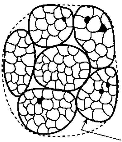
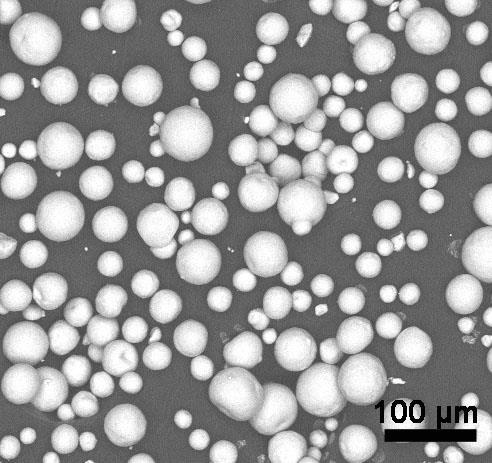
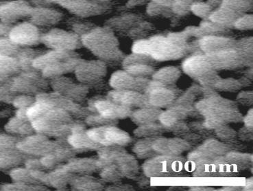
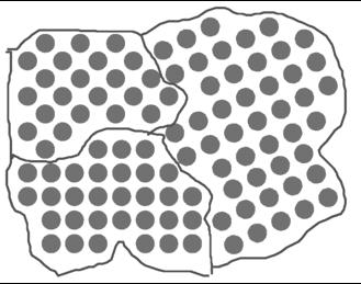
44 What does a powder really mean? Single Oriented Random

45 Limited crystals vs. many crystals
46 X-Ray Powder Diffraction Each crystallite in the powder is a small single crystal with a random orientation There is roughly equal statistically relevant number of crystallites for every set of randomly oriented planes that will diffract the incident beam Samples can be powder, sintered pellets, coatings on substrates, engine block The resultant XRD pattern is a unique fingerprint of the polycrystalline material which contains a lot of information The position, intensity, width, and shape of the observed diffraction peaks tells us about the crystal structure and, in some cases, microstructure of the sample
47
48
49
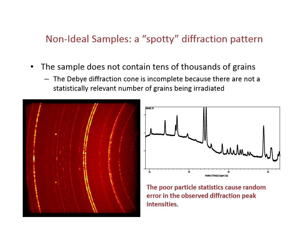
50
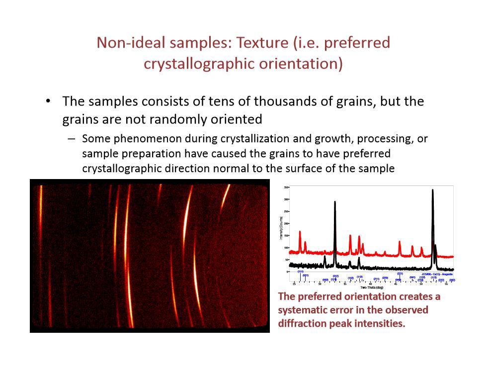
51
52 Diffraction from a variety of materials (From Elements of X-ray Diffraction, B.D. Cullity, Addison Wesley)
53
54 What can be done with XRD? Identify phase composition Measure unit cell lattice parameters Estimate crystallite size, microstrain Measure residual stress Measure texture and/or epitaxy Evaluate thin film quality Measure multilayer thin film thickness, roughness, and density Determine orientation of single crystals Solve or refine crystal structures Analyze ordered meso- and nanostructures


55 Diffraction Pattern Diffraction peak positions and intensity contain information about the crystal structure of a material Each diffraction peak is produced by a family of atomic planes. The position of the diffraction peak indicates the length d hkl between those atomic planes this ultimately correlates to the bond distances between atoms The intensity of the diffraction peak is determined by what atoms are present on the diffracting plane, their scattering factors, and the structure factor Intensity (a.u.) (200) (220) (111) (deg.) XRD pattern of NaCl
56 Diffraction of a Single Crystal A single crystal in a Bragg-Brentano diffractometer produces one family of peaks in the diffraction pattern 2
57 Diffraction of a Polycrystalline Sample A polycrystalline sample contains thousands of crystallites All diffraction peaks should be observed There will be a small percentage of crystallites that are properly oriented to diffract for every set of planes 2 2 2
58 Diffraction Peaks The absolute peak intensity is measured as photons counted or count rate The absolute intensity can vary due to instrumental and experimental parameters When normalized to the most intense peak, relative intensities are obtained which should be instrument independent Peak areas are much more reliable than peak heights as a measure of intensity The intensity of diffracted photon tells us about the atoms that are on those planes
59 Stick pattern from JCPDS
 Bulk electrodeposited nanocrystalline")
60 Actual Pattern Lattice parameter, phase diagrams Texture, Strain (micro and residual) Size, microstructure (twins and dislocations) Bulk electrodeposited nanocrystalline nickel
61 Intensity(Counts) (deg.) Qualitative Phase Identification Mostly used for analysis of processes and quality control When You found something in the laboratory and want to know what it is or You tried to synthesize a material and want to know if you really did or You want to check if your raw materials are pure Example: You have some monoclinic Y 2 O 3 in the lab for a long time and want to see if it degraded in time > cubic Y 2 O > monoclinic Y 2 O 3
62 (deg.) Qualitative Phase Identification can be used to study reactions ex situ Comparing and identifying the XRD patterns of a material before and after a phase changing reaction Example: You heated the monoclinic Y 2 O 3 to 1000 C and want to see the level of degradation Heat treatment converted all of the monoclinic Y 2 O 3 into cubic Y 2 O 3 original, as-made after annealing Intensity(Counts)
63 Qualitative Phase Identification can also be used to study reactions in situ A phase changing reaction such as heat treatment is monitored directly by the attached X-Ray diffractometer Example: You heat the monoclinic Y 2 O 3 to 1000 C and want to see the rate of degradation original, as-made after annealing Intensity(Counts) (deg.) time
64 Intensity(Counts) Qualitative Phase Identification can also be used to discern between isostructural compounds and polymorphs Differences in electron density can be mapped because X-Rays scatter proportionally to Z 2 Example: the cubic phases of CaTiO 3 and SrTiO 3 have identical crystal structures, with the cation replaced by Ca or Sr respectively CaTiO3 cubic perovskite SrTiO3 cubic perovskite (deg.)
65 Intensity (a.u.) Analysis of Single Phase 2 ( ) d (Å) (I/I 1 )* I 1 : Intensity of the strongest peak
66 Procedure Note first three strongest peaks at d 1, d 2, and d 3 In the present case: d 1 : 2.82; d 2 : 1.99 and d 3 : 1.63 Å Search JCPDS manual to find the d group belonging to the strongest line: between Å There are 17 substances with approximately similar d 2 but only 4 have d 1 : 2.82 Å Out of these, only NaCl has d 3 : 1.63 Å It is NaCl Specimen and Intensities Substance File Number x (ErSe) 2 Q x NaCl x (NH 4 ) 2 WO 2 Cl x (BePd)2C Caution: It could be much more tricky if the sample is oriented or textured or your goniometer is not calibrated
67 Presence of Multiple phases More Complex Several permutations combinations possible e.g. d 1 ; d 2 ; and d 3, the first three strongest lines show several alternatives Then take any of the two lines together and match It turns out that 1 st and 3 rd strongest lies belong to Cu and then all other peaks for Cu can be separated out Pattern for Cu d (Å) I/I d (Å) I/I * * * * * * *
68 Presence of Multiple phases Now separate the remaining lines and normalize the intensities Look for first three lines and it turns out that the phase is Cu 2 O If more phases, harder to solve Pattern of Cu 2 O d (Å) I/I d (Å) Remaining Lines Observed I/I 1 Normalized
69 Diffraction Data is Essential
 Quantitative Phase Identification In depth analysis for precise monitoring of phase composition When You know which phases are present and want to know how much Example: You mix certain powders to")
70 (deg.) Quantitative Phase Identification In depth analysis for precise monitoring of phase composition When You know which phases are present and want to know how much Example: You mix certain powders to make a red paint pigment then wonder how much of each is present Red Paint Pigment Mixture Intensity(Counts) 28 wt% Hematite, Fe 2 O 3 21 wt% Anatase, TiO 2 51 wt% Rutile, TiO 2
71 Relative Intensity Approach The ratio of peak intensities varies linearly with the ratio of weight fractions I I K is determined from the mass absorption coeffcicients of all phases in the sample Not the most accurate and reliable way K X X Simplification of the ratio of the general intensity equation Constant for an experimental setup Structure factor Absorption factor Multiplicity factor Lorentz polarization factor
72 Relative Intensity Approach K can be determined : by using published relative intensity values (I/Ic) External Standard Method empirically, by building calibration curves Internal Standard Method by simulating the diffraction pattern: whole pattern refinement Reitveld Analysis To equate the concentration of a given phase with the intensity of the peaks in the multiphase pattern, a single peak, least precision a number of peaks, considerable precision all of the peaks ultimate precision (Reitveld analysis) can be utilized The shape of the peaks is decisive in choosing the intensity measurement method When the diffraction peak is sharp and non-overlapped, peak height is taken as the representative value as it is considered proportional to the peak area Background corrected integrated intensities are measured by subtracting the background intensity from the peak height
73 a) Peak height is proportional to peak area. Peak height is measured b) Peak height not proportional to peak area. Combined intensity is calculated c) Peak area is overlapped by other peaks This is a common case. Peak profiles should be adjusted
74 External Standard Method The complexity of the analysis of multiple phases in a mixture is greatly reduced by referring all of the pure phase peak intensities to a single standard W a W s = 1, so P I RIR P I s s where I a is the intensity of the 100% peak of phase α, and I s is the intensity of the 100% peak of a reference phase s, taken by convention to be α-al 2 O 3, corundum, in a 50:50 mixture by weight Generalized External Standard Equation The use of RIR values is much faster and less prone to error than the determination of all of the weight fractions from the starting equation which would require reference to the integrated intensities of the 100% peak of each phase in its pure form
75 Internal Standard Method The proportionality constant K is determinded empirically by building calibration curves from mixtures containing known quantities of internal standard I I K X X MgSiO3:Al2O3 YSZ:Al2O3 K Example Three component mixture consisting of predetermined concentrations of MgSiO 3, YSZ, Al 3 O X (Al2O3) Sample MgSiO 3 wt% YSZ wt% Al 3 O 2 wt%
76 Determining Crystallinity using Standard Additions Adding a predetermined amount of well crystallized standard to a sample containing amorphous phases Calculate the observed amount of all phases using any of the quantitative analysis methods, ignoring the amorphous content Apply a normalization to all calculated amounts based on the initial standard amount Example Fe, Fe 2 O 3 mixture with 17% Si standard the discrepancy due to the amorphous content Intensity(Counts) 22.2wt% Si 73.5wt% Fe 2 O 3 4.3wt% Fe the discrepancy due to the amorphous content Sample Fe wt% Fe 2 O 3 wt% Si wt% Calculated Actual Amorphous (deg.)
77 Determining Crystallinity without Standard Additions If the mass absorption coefficient of an amorphous phase is the same as the crystalline content, the ratio of intensities can be used to determine % crystallinity Area Crystalline Peaks: cts Area Amorphous Hump: cts % Crystalline: 46.5% % Amorphous: 53.5% Intensity(Counts) (deg.)
78 Reitveld Refinement Rietveld realised that the detailed profile of a powder-diffraction pattern contained a lot more information than the extracted intensities of composite peaks and stated that "The method of using the total integrated intensities of the separate groups of overlapping peaks in the least-squares refinement of structures, leads to the loss of all the information contained in the often detailed profile of these composite peaks. By the use of these profile intensities instead of the integrated quantities in the refinement procedure, however, this difficulty is overcome and it allows the extraction of the maximum amount of information contained in the powder diagram." H. M. Rietveld

79 Reitveld Refinement Reitveld found that the detailed profile could be fitted on a point by point basis using the simple Gaussian peak-shape function without any need to extract intensities of composite groups of reflections Refinement implies taking an approximate model of the structure and converting it so that diffraction data calculated from the model structure has a closer resemblance to the measured data Many complex computer algorythms operating on the basis of using a least-squares procedure to refine the initial structure model in order to improve the agreement between the observed diffraction data and that calculated from the model In other words Reitveld refinement is a broad numerical technique used to make the XRD pattern as true and error-free as possible
80 Intensity(Counts) Lattice Parameter Determination Changes in the interplanar spacing of a material such as substitution of an atom can be detected by XRD The position of the diffraction peaks are a product of the space between planes of atoms The change in peak intensity due to substitution of atoms with similar Z is subtle, however peak angles which indicate the interplanar space change Example Substitution of Zr in YSZ with Y 10% Y in ZrO 2 50% Y in ZrO (deg.)
81 Crystallite Size Broadening Ideally a peak in the Bragg diffraction pattern is a line without a width. In reality the peaks have some width that originate from the instrument and the size of the crystals
82 Crystallite Size Broadening A diffraction peak is produced by a plane with B that satisfies Bragg s law However X-ray beam in parafocusing geometry is not perfectly parallel and is diffracted at angles between 1 and 2. So X-rays scattered at angles between 1 and 2 do not interfere with each other perfectly constructive or destructively In this case X-rays scattered from plane 0 and 1 have angles 1 that correspond to Bragg s number n less than an integer These X-rays will interfere with X-rays scattered from plane m that scatters rays with an angle that correspond to n/2 (out of phase - destructive interference) For very fine crystals the plane m is not present so that X-rays diffracted at 1 will be detected as peak broadening
83 Kristal Boyutu Peak genişliği ile kristal boyutu ters orantılıdır. Peak genişliği (B) arttıkça kristal boyutu küçülür. Krital Boyut hesabı (t) :
84 Crystallite Size Broadening No crystal is perfect due to its finite size The deviation from perfect crystallinity leads to a broadening of the diffraction peaks which is negligible above a certain size (~0.1-1 micron) Peak broadening occurs for powder samples with crystallites around 100 nm in diameter L B K 2 cos K= Sherrer Equation for samples with fine crysrallites Peak width is measured at the half of the maximum peak intensity (FWHM) Various methods to determine the crystallite size of an unknown fine powder mixed with a coarse standard powder B 2 = B U 2 BS 2 Warren s equation the simplest form
85 Crystallite Size Broadening Example - Sherrer analysis of the main peak for a heat treated polycrystalline La 2 Zr 2 O 7 to estimate the crystallite size Calcined at 900 C for 1 hr: ~10 nm average crystallite size Sintered at 1000 C for 96 hrs: ~30-40 nm average size Sintered at 1500 C for 2 hrs: >100 nm average crystallite size Figure adapted from ``Ion Transport Membranes for H 2 Separation:Y-doping of La 2 Zr 2 O 7`` by Carneim et al.
86 Factors That Contribute to Crystallite Size Broadening Instrumental Broadening Crystallite Size Microstrain Faulting Dislocations Antiphase Domain Boundaries Grain Surface Relaxation Solid Solution Inhomogeneity Temperature Factors Non ideal optics Wavelength Dispersion Sample Transparency Axial Divergence Flat Sample Effect Detector resolution The peak profile is a combination of the profiles from all of these contributions Peak broadening analysis is most accurate when the broadening due to crystallite size effects is at least twice the contribution due to instrumental broadening

87 Microstrain Determination Inhomogeneous strain is the second main source of specimen broadening a compressive stress would make the d spacings smaller and a tensile stress would make the d spacings larger which would cause only peak shifting if the strain is inhomogeneous then different crystallites will be strained by different amounts and the shifts in 2θ will be variable resulting in peak broadening B(s) = C tanθ, C=4-5
88 Microstrains
89 Seperating the size and microstrain contributions to peak broadening Williamson and Hall simplified the contributions of size and strain to a basic sum or sum of squares B 2 K Lcos B B(s) = C tanθ K 2 B s C tan Lcos total B B K L total cos C sin By plotting a Williamson-Hall graph, where B(total)cosθ and sinθ are the axes, the strain component is obtained from the slope (C) and the size component from (L) the intercept
90 Seperating the size and microstrain contributions to peak broadening Williamson-Hall method lacks precision due to its simplifications but it can be a useful method if used to observe trends in the relative sense. Example - a study of many powder patterns of the same chemical compound, but synthesized under different conditions
91 Thin Film Specimen Grazing angle (very small, ~1-5 ) B Film or Coating B Substrate Smaller volume i.e. less intensity of the scattered beam from the film Grazing angle Useful only for polycrystalline specimens
92
93
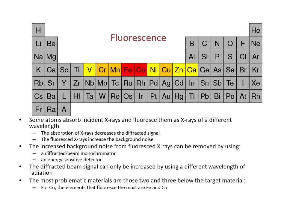
94 Data collection and analysis procedure Choose 2θ range Step size and time per step Hardware: slit size, filter, sample alignment Fast scan followed with a slower scan Look for fluorescence Collected data: Background subtraction, K α2 stripping Normalize data for comparison I/I max
95 Thank you for your interest! The following sources provide more information on X-Ray Diffraction: Elements of X-Ray Diffraction (The Bible) by B. D. Cullity Fundamentals of Powder Diffraction and Structural Characterization of Materials by V. K. Pecharsky et al. Principles and Applications of Powder Diffraction by A. Clearfield et al.
A Brief History of XRD 1895: Röntgen discovers X-Rays received the first Nobel prize in physics in 1901
 X-ray Diffraction A Brief History of XRD 1895: Röntgen discovers X-Rays received the first Nobel prize in physics in 1901 1912: Laue diffracts X-Rays from single crystal 1914 Nobel prize in Physics 1912:
Fundamentals of X-ray diffraction and scattering
 Fundamentals of X-ray diffraction and scattering Don Savage dsavage@wisc.edu 1231 Engineering Research Building (608) 263-0831 X-ray diffraction and X-ray scattering Involves the elastic scattering of
LECTURE 7. Dr. Teresa D. Golden University of North Texas Department of Chemistry
 LECTURE 7 Dr. Teresa D. Golden University of North Texas Department of Chemistry Diffraction Methods Powder Method For powders, the crystal is reduced to a very fine powder or microscopic grains. The sample,
Identification of Crystal Structure and Lattice Parameter. for Metal Powders Using X-ray Diffraction. Eman Mousa Alhajji
 Identification of Crystal Structure and Lattice Parameter for Metal Powders Using X-ray Diffraction Eman Mousa Alhajji North Carolina State University Department of Materials Science and Engineering MSE
X-Ray Diffraction. Nicola Pinna
 X-Ray Diffraction Nicola Pinna Department of Chemistry, CICECO, University of Aveiro, 3810-193 Aveiro, Portugal. School of Chemical and Biological Engineering, College of Engineering, Seoul National University
Introduction to Powder Diffraction/Practical Data Collection
 Durham University Chemistry Department Introduction to Powder Diffraction/Practical Data Collection Dr Ivana Evans Durham, January 2007 Durham Outline Information in a powder pattern What is diffraction
Diffraction Basics. The qualitative basics:
 The qualitative basics: Diffraction Basics Coherent scattering around atomic scattering centers occurs when x-rays interact with material In materials with a crystalline structure, x-rays scattered in
Instrument Configuration for Powder Diffraction
 Instrument Configuration for Powder Diffraction Advanced X-ray Workshop S.N. Bose National Centre for Basic Sciences, 14-15/12/2011 Innovation with Integrity Overview What is the application? What are
Lesson 1 Good Diffraction Data
 Lesson 1 Good Diffraction Data Nicola Döbelin RMS Foundation, Bettlach, Switzerland Digital Diffractometers Transmission Geometry Debye-Scherrer Geometry Reflective Geometry Bragg-Brentano Geometry Glass
Thin Film Scattering: Epitaxial Layers
 Thin Film Scattering: Epitaxial Layers 6th Annual SSRL Workshop on Synchrotron X-ray Scattering Techniques in Materials and Environmental Sciences: Theory and Application May 29-31, 2012 Thin films. Epitaxial
Single crystal X-ray diffraction. Zsolt Kovács
 Single crystal X-ray diffraction Zsolt Kovács based on the Hungarian version of the Laue lab description which was written by Levente Balogh, Jenő Gubicza and Lehel Zsoldos INTRODUCTION X-ray diffraction
9/29/2014 8:52 PM. Chapter 3. The structure of crystalline solids. Dr. Mohammad Abuhaiba, PE
 1 Chapter 3 The structure of crystalline solids 2 Home Work Assignments HW 1 2, 7, 12, 17, 22, 29, 34, 39, 44, 48, 53, 58, 63 Due Sunday 12/10/2014 Quiz # 1 will be held on Monday 13/10/2014 at 11:00 am
Diffraction: Powder Method
 Diffraction: Powder Method Diffraction Methods Diffraction can occur whenever Bragg s law λ = d sin θ is satisfied. With monochromatic x-rays and arbitrary setting of a single crystal in a beam generally
This lecture is part of the Basic XRD Course.
 This lecture is part of the Basic XRD Course. Basic XRD Course 1 A perfect polycrystalline sample should contain a large number of crystallites. Ideally, we should always be able to find a set of crystallites
It is instructive however for you to do a simple structure by hand. Rocksalt Structure. Quite common in nature. KCl, NaCl, MgO
 Today the structure determinations etc are all computer -assisted It is instructive however for you to do a simple structure by hand Rocksalt Structure Quite common in nature KCl, NaCl, MgO 9-1 Typical
9/28/2013 9:26 PM. Chapter 3. The structure of crystalline solids. Dr. Mohammad Abuhaiba, PE
 Chapter 3 The structure of crystalline solids 1 2 Why study the structure of crystalline solids? Properties of some materials are directly related to their crystal structure. Significant property differences
X-ray diffraction
 2.2.3.- X-ray diffraction 2.2.3.1.- Origins and fundamentals of the technique The first experimental evidence concerning x-ray diffraction was given by Max von Laue who in 1912 demonstrated that x-rays
X-ray Diffraction (XRD)
") هب انم خدا X-ray Diffraction (XRD) 1.0 What is X-ray Diffraction 2.0 Basics of Crystallography 3.0 Production of X-rays 4.0 Applications of XRD 5.0 Instrumental Sources of Error 6.0 Conclusions Bragg s
هب انم خدا X-ray Diffraction (XRD) 1.0 What is X-ray Diffraction 2.0 Basics of Crystallography 3.0 Production of X-rays 4.0 Applications of XRD 5.0 Instrumental Sources of Error 6.0 Conclusions Bragg s
Thin Film Scattering: Epitaxial Layers
 Thin Film Scattering: Epitaxial Layers Arturas Vailionis First Annual SSRL Workshop on Synchrotron X-ray Scattering Techniques in Materials and Environmental Sciences: Theory and Application Tuesday, May
Materials Lab 1(MT344) X-ray Diffractometer Operation and Data Analysis. Instructor: Dr. Xueyan Wu ( 吴雪艳 )
 X-ray Diffractometer Operation and Data Analysis. Instructor: Dr. Xueyan Wu ( 吴雪艳 )") Materials Lab 1(MT344) X-ray Diffractometer Operation and Data Analysis Instructor: Dr. Xueyan Wu ( 吴雪艳 ) Goals To give students a practical introduction into the use of X-ray diffractometer and data collection.
Materials Lab 1(MT344) X-ray Diffractometer Operation and Data Analysis Instructor: Dr. Xueyan Wu ( 吴雪艳 ) Goals To give students a practical introduction into the use of X-ray diffractometer and data collection.
An Introduction to X-Ray Powder Diffraction. credits to: Scott A Speakman, Patrick McArdle Edited by Di Cicco 2014
 An Introduction to X-Ray Powder Diffraction credits to: Scott A Speakman, Patrick McArdle Edited by Di Cicco 2014 LATTICE ARRAYS AND BRAVAIS LATTICES Crystalline materials differ from amorphous materials
X-RAY DIFFRACTION. X- Ray Sources Diffraction: Bragg s Law Crystal Structure Determination
 X-RAY DIFFRACTION X- Ray Sources Diffraction: Bragg s Law Crystal Structure Determination Part of MATERIALS SCIENCE & ENGINEERING A Learner s Guide AN INTRODUCTORY E-BOOK Anandh Subramaniam & Kantesh Balani
Atomic Densities. Linear Density Number of atoms per length whose centers lie on the direction vector for a specific crystallographic direction.
 Atomic Densities Linear Density Number of atoms per length whose centers lie on the direction vector for a specific crystallographic direction. Planar Density Number of atoms per unit area that are centered
Spreadsheet Applications for Materials Science
 Spreadsheet Applications for Materials Science Introduction to X-ray Powder Diffraction Introduction X-ray powder diffraction is a powerful analytical technique that is widely used in many fields of science
9/16/ :30 PM. Chapter 3. The structure of crystalline solids. Mohammad Suliman Abuhaiba, Ph.D., PE
 Chapter 3 The structure of crystalline solids 1 Mohammad Suliman Abuhaiba, Ph.D., PE 2 Home Work Assignments HW 1 2, 7, 12, 17, 22, 29, 34, 39, 44, 48, 53, 58, 63 Due Sunday 17/9/2015 3 Why study the structure
X-RAY DIFFRACTIO N B. E. WARREN
 X-RAY DIFFRACTIO N B. E. WARREN Chapter 1 X-Ray Scattering by Atom s 1.1 Classical scattering by a free electron 1 1.2 Polarization by scattering 4 1.3 Scattering from several centers, complex representation
Atomic Densities. Linear Density. Planar Density. Linear Density. Outline: Planar Density
 Atomic Densities Outline: Atomic Densities - Linear Density - Planar Density Single- vs poly- crystalline materials X-ray Diffraction Example Polymorphism and Allotropy Linear Density Number of atoms per
Chapter 3 Basic Crystallography and Electron Diffraction from Crystals. Lecture 9. Chapter 3 CHEM Fall, L. Ma
 Chapter 3 Basic Crystallography and Electron Diffraction from Crystals Lecture 9 Outline The geometry of electron diffraction Crystallography Kinetic Theory of Electron diffraction Diffraction from crystals
X-RAY DIFFRACTION IN SEMICONDUCTOR INDUSTRY AND RESEARCH
 X-RAY DIFFRACTION IN SEMICONDUCTOR INDUSTRY AND RESEARCH M. Leszczyński High Pressure Research Center UNIPRESS, Sokolowska 29/37, 01 142 Warsaw, Poland, e-mail: mike@unipress.waw.pl ABSTRACT The paper
Strain. Two types of stresses: Usually:
 Stress and Texture Strain Two types of stresses: microstresses vary from one grain to another on a microscopic scale. macrostresses stress is uniform over large distances. Usually: macrostrain is uniform
Lesson 3 Sample Preparation
 Lesson 3 Sample Preparation Nicola Döbelin RMS Foundation, Bettlach, Switzerland January 14 16, 2015, Bern, Switzerland Repetition: Bragg-Brentano Diffractometer Typical Configuration (with Kβ filter)
X ray diffraction in materials science
 X ray diffraction in materials science Goals: Use XRD spectra to determine the orientation of single crystals and preferred orientations in a thin film. Understand how grain size and strain affect the
Basic X-ray Powder Diffraction (XRPD)
") Basic X-ray Powder Diffraction (XRPD) Solid-State, Material Science Crystalline (Scattering : diffraction) Non-crystalline (Scattering) Analytical Tool Qualitative and Quantitative Analysis Quantitative
Basic X-ray Powder Diffraction (XRPD) Solid-State, Material Science Crystalline (Scattering : diffraction) Non-crystalline (Scattering) Analytical Tool Qualitative and Quantitative Analysis Quantitative
Travaux Pratiques de Matériaux de Construction
 Travaux Pratiques de Matériaux de Construction Section Matériaux 6 ème semestre 2009 Etude de Matériaux Cimentaire Par Diffraction des Rayons X Responsable: Silke Ruffing E-Mail: silke.ruffing@epfl.ch
Lesson 1 X-rays & Diffraction
 Lesson 1 X-rays & Diffraction Nicola Döbelin RMS Foundation, Bettlach, Switzerland February 11 14, 2013, Riga, Latvia Electromagnetic Spectrum X rays: Wavelength λ: 0.01 10 nm Energy: 100 ev 100 kev Interatomic
High Resolution X-ray Diffraction
 High Resolution X-ray Diffraction Nina Heinig with data from Dr. Zhihao Donovan Chen, Panalytical and slides from Colorado State University Outline Watlab s new tool: Panalytical MRD system Techniques:
Workshop RIETVELD REFINEMENT OF DIFFRACTION PATTERNS Program Monday June 1st, Introduction to Rietveld refinement S.
 Workshop RIETVELD REFINEMENT OF DIFFRACTION PATTERNS Program Monday June 1st, 2009 9.00 13.00 Introduction to Rietveld refinement S.Enzo Università di Sassari X-ray diffraction for bulk samples and thin
Travaux Pratiques de Matériaux de Construction. Etude de Matériaux Cimentaires par Diffraction des Rayons X sur Poudre
 Travaux Pratiques de Matériaux de Construction Section Matériaux 6 ème semestre 2015 Etude de Matériaux Cimentaires par Diffraction des Rayons X sur Poudre Study Cementitious Materials by X-ray diffraction
Advanced Methods for Materials Research. Materials Structure Investigations Materials Properties Investigations
 Advanced Methods for Materials Research Materials Structure Investigations Materials Properties Investigations Advanced Methods for Materials Research 1. The structure and property of sample and methods
X-Ray Diffraction Analysis
 162402 Instrumental Methods of Analysis Unit III X-Ray Diffraction Analysis Dr. M. Subramanian Associate Professor Department of Chemical Engineering Sri Sivasubramaniya Nadar College of Engineering Kalavakkam
Experiment 2b X-Ray Diffraction* Optical Diffraction Experiments
 * Experiment 2b X-Ray Diffraction* Adapted from Teaching General Chemistry: A Materials Science Companion by A. B. Ellis et al.: ACS, Washington, DC (1993). Introduction Inorganic chemists, physicists,
The object of this experiment is to test the de Broglie relationship for matter waves,
 Experiment #58 Electron Diffraction References Most first year texts discuss optical diffraction from gratings, Bragg s law for x-rays and electrons and the de Broglie relation. There are many appropriate
Lecture C4b Microscopic to Macroscopic, Part 4: X-Ray Diffraction and Crystal Packing
 Lecture C4b Microscopic to Macroscopic, Part 4: X-Ray Diffraction and Crystal Packing X-ray Diffraction Max von Laue won the 1914 Nobel Prize for his discovery of the diffraction of x-rays by crystals.
Practical X-Ray Diffraction
 Typical Example Practical X-Ray Diffraction White powder sample of NaCl,KCl,KNO 3 (trace of H 2 O) Département de chimie Université Laval Prof. Josée BRISSON Dr. Wenhua BI 2014-03-20 Powder X-Ray Diffraction
Physics 6180: Graduate Physics Laboratory. Experiment CM5: X-ray diffraction and crystal structures
 Physics 6180: Graduate Physics Laboratory Experiment CM5: X-ray diffraction and crystal structures References: Preston and Dietz, Expt. 10 pp. 180-197 Eisberg and Resnick, Quantum Physics, Sec. 9 Kittel,
X-ray diffraction. Talián Csaba Gábor University of Pécs, Medical School Department of Biophysics
 X-ray diffraction Talián Csaba Gábor University of Pécs, Medical School Department of Biophysics 2012.10.11. Outline of the lecture X-ray radiation Interference, diffraction Crystal structure X-ray diffraction
A - Transformation of anatase into rutile
 Exercise-Course-XRD.doc 1/12 04/06/2012 A - Transformation of anatase into rutile Anatase and rutile are two distinct phases of titanium dioxide TiO 2. The stable phase is rutile. 1. Structural study Anatase:
X-Ray Analytical Methods
 X-Ray Analytical Methods X-rays were discovered by W.C. Röentgen in 1895, and led to three major uses: X-ray radiography is used for creating images of light-opaque materials relies on the relationship
Carbon nanostructures. (http://www.mf.mpg.de/de/abteilungen/schuetz/index.php?lang=en&content=researchtopics&type=specific&name=h2storage)
") Carbon nanostructures (http://www.mf.mpg.de/de/abteilungen/schuetz/index.php?lang=en&content=researchtopics&type=specific&name=h2storage) 1 Crystal Structures Crystalline Material: atoms arrange into a
Carbon nanostructures (http://www.mf.mpg.de/de/abteilungen/schuetz/index.php?lang=en&content=researchtopics&type=specific&name=h2storage) 1 Crystal Structures Crystalline Material: atoms arrange into a
Electron Microscopy. Dynamical scattering
 Electron Microscopy 4. TEM Basics: interactions, basic modes, sample preparation, Diffraction: elastic scattering theory, reciprocal space, diffraction pattern, Laue zones Diffraction phenomena Image formation:
INGE Engineering Materials. Chapter 3 (cont.)
") Some techniques used: Chapter 3 (cont.) This section will address the question how do we determine the crystal structure of a solid sample? Electron microscopy (by direct and indirect observations) Scanning
Some techniques used: Chapter 3 (cont.) This section will address the question how do we determine the crystal structure of a solid sample? Electron microscopy (by direct and indirect observations) Scanning
X-ray Powder Diffraction in Catalysis
 X-ray Powder Diffraction in Catalysis 0/63 Introduction Introduction: scope of this lecture This lecture is designed as a practically oriented guide to powder XRD in catalysis, not as an introduction into
X-Ray Diffraction by Macromolecules
 N. Kasai M. Kakudo X-Ray Diffraction by Macromolecules With 351 Figures and 56 Tables Kodansha ~Springer ... Contents Preface v Part I Fundamental 1. Essential Properties of X-Rays................. 3 1.1
Example: Compute the wavelength of a 1 [kg] block moving at 1000 [m/s].
![Example: Compute the wavelength of a 1 [kg] block moving at 1000 [m/s].](/thumbs/95/123245062.jpg "Example: Compute the wavelength of a 1 [kg] block moving at 1000 [m/s].") Example: Calculate the energy required to excite the hydrogen electron from level n = 1 to level n = 2. Also calculate the wavelength of light that must be absorbed by a hydrogen atom in its ground state
Example: Calculate the energy required to excite the hydrogen electron from level n = 1 to level n = 2. Also calculate the wavelength of light that must be absorbed by a hydrogen atom in its ground state
OPTIMIZING XRD DATA. By: Matthew Rayner
 OPTIMIZING XRD DATA By: Matthew Rayner 1 XRD Applications PANalytical classifies XRD applications in 4 groups 1. Powders 2. Nanomaterials 3. Solid objects 4. Thin films Many day-to-day samples cross these
Fundamentals of Crystalline State and Crystal Lattice p. 1 Crystalline State p. 2 Crystal Lattice and Unit Cell p. 4 Shape of the Unit Cell p.
 Fundamentals of Crystalline State and Crystal Lattice p. 1 Crystalline State p. 2 Crystal Lattice and Unit Cell p. 4 Shape of the Unit Cell p. 7 Crystallographic Planes, Directions, and Indices p. 8 Crystallographic
UNIT V -CRYSTAL STRUCTURE
 UNIT V -CRYSTAL STRUCTURE Solids are of two types: Amorphous and crystalline. In amorphous solids, there is no order in the arrangement of their constituent atoms (molecules). Hence no definite structure
Characterization of Materials Using X-Ray Diffraction Powder Diffraction
 Praktikum III, Fall Term 09 Experiment P1/P2; 23.10.2009 Characterization of Materials Using X-Ray Diffraction Powder Diffraction Authors: Michael Schwarzenberger (michschw@student.ethz.ch) Philippe Knüsel
MICROSTRUCTURAL CHARACTERIZATION OF NANOCRYSTALLINE POWDERS AND THIN FILMS BY X-RAY POWDER DIFFRACTION
 MICROSTRUCTURAL CHARACTERIZATION OF NANOCRYSTALLINE POWDERS AND THIN FILMS BY X-RAY POWDER DIFFRACTION Zdeněk MATĚJ a, Lea NICHTOVÁ a, Radomír KUŽEL a a Faculty of Mathematics and Physics, Charles University
CHARACTERISATION OF CRYSTALLINE AND PARTIALLY CRYSTALLINE SOLIDS BY X-RAY POWDER DIFFRACTION (XRPD)
") 2.9.33. Characterisation of crystalline solids by XRPD EUROPEAN PHARMACOPOEIA 6.0 with its standard deviation. The mean values for x 10 and x 90 must not deviate by more than 5 per cent from the certified
2.9.33. Characterisation of crystalline solids by XRPD EUROPEAN PHARMACOPOEIA 6.0 with its standard deviation. The mean values for x 10 and x 90 must not deviate by more than 5 per cent from the certified
Earth & Planetary Science Applications of X-Ray Diffraction: Advances Available for Research with our New Systems
 Earth & Planetary Science Applications of X-Ray Diffraction: Advances Available for Research with our New Systems James R. Connolly Dept. of Earth & Planetary Sciences University of New Mexico 401/501
Microstructural Characterization of Materials
 Microstructural Characterization of Materials 2nd Edition DAVID BRANDON AND WAYNE D. KAPLAN Technion, Israel Institute of Technology, Israel John Wiley & Sons, Ltd Contents Preface to the Second Edition
Fundamentals of Crystalline State p. 1 Introduction p. 1 Crystalline state p. 2 Crystal lattice and crystal structure p. 4 Shape of the unit cell p.
 Preface p. xvii Fundamentals of Crystalline State p. 1 Introduction p. 1 Crystalline state p. 2 Crystal lattice and crystal structure p. 4 Shape of the unit cell p. 6 Content of the unit cell p. 7 Asymmetric
Uses of Powder Diffraction. Diffraction
 Powder X-ray X Diffraction Brendan J. Kennedy School of Chemistry The University of Sydney Uses of Powder Diffraction Qualitative Analysis Identification of single-phase materials Identification of multiple
LECTURE 8. Dr. Teresa D. Golden University of North Texas Department of Chemistry
 LECTURE 8 Dr. Teresa D. Golden University of North Texas Department of Chemistry Practical applications for lattice parameter measurements: -determine composition (stoichiometry) of the sample -determine
Structure of silica glasses (Chapter 12)
") Questions and Problems 97 Glass Ceramics (Structure) heat-treated so as to become crystalline in nature. The following concept map notes this relationship: Structure of noncrystalline solids (Chapter 3)
Questions and Problems 97 Glass Ceramics (Structure) heat-treated so as to become crystalline in nature. The following concept map notes this relationship: Structure of noncrystalline solids (Chapter 3)
Powder X-ray Diffraction
 Powder X-ray Diffraction The construction of a simple powder diffractometer was first described by Hull in 1917 1 which was shortly after the discovery of X-rays by Wilhelm Conrad Röntgen in1895 2. Diffractometer
What if your diffractometer aligned itself?
 Ultima IV Perhaps the greatest challenge facing X-ray diffractometer users today is how to minimize time and effort spent on reconfiguring of the system for different applications. Wade Adams, Ph.D., Director,
3. Anisotropic blurring by dislocations
 Dynamical Simulation of EBSD Patterns of Imperfect Crystals 1 G. Nolze 1, A. Winkelmann 2 1 Federal Institute for Materials Research and Testing (BAM), Berlin, Germany 2 Max-Planck- Institute of Microstructure
TEM and Electron Diffraction Keith Leonard, PhD (1999) U. Cincinnati
 U. Cincinnati") TEM and Electron Diffraction Keith Leonard, PhD (1999) U. Cincinnati Electron Microscopes: Electron microscopes, such as the scanning electron microscope (SEM) and transmission electron microscope (TEM)
TEM and Electron Diffraction Keith Leonard, PhD (1999) U. Cincinnati Electron Microscopes: Electron microscopes, such as the scanning electron microscope (SEM) and transmission electron microscope (TEM)
Basics of XRD part IV
 Basics of XRD part IV Dr. Peter G. Weidler Institute of Functional Interfaces IFG 1 10/31/17 KIT The Research University in the Helmholtz Association Name of Institute, Faculty, Department www.kit.edu
VOLUME FRACTION ANALYSIS
 VOLUME FRCTON NLYSS Qualitative (dentification) Lattice parameter versus composition curve (single phase) JCPDS Files: 1) Locate d1 group; 2)Closest match to d2 3) d1, d2, d3 (-wise) 4) Compare all lines
Dr. Teresa D. Golden University of North Texas Department of Chemistry
 Dr. Teresa D. Golden University of North Texas Department of Chemistry Advance X-Ray Diffraction Lecture: TTh 8:00 a.m. 9:20 p.m. CHEM 253 and CHEM 271 Instructor: Dr. Teresa D. Golden Office hours: 3:00-5:00
11.3 The analysis of electron diffraction patterns
 11.3 The analysis of electron diffraction patterns 277 diameter) Ewald reflecting sphere, the extension of the reciprocal lattice nodes and the slight buckling of the thin foil specimens all of which serve
AP 5301/8301 Instrumental Methods of Analysis and Laboratory Lecture 5 X ray diffraction
 1 AP 5301/8301 Instrumental Methods of Analysis and Laboratory Lecture 5 X ray diffraction Prof YU Kin Man E-mail: kinmanyu@cityu.edu.hk Tel: 3442-7813 Office: P6422 Lecture 5: Outline Review on crystallography
IF YOUR ONLY SINGLE CRYSTAL IS NOT REALLY SINGLE
 THE RIGAKU JOURNAL VOL. 12 / NO.1 / 1995 IF YOUR ONLY SINGLE CRYSTAL IS NOT REALLY SINGLE L. W. FINGER Geophysical Laboratory and Center for High-Pressure Research, 5251 Broad Branch Road, N.W. Washington,
X-RAY POWDER DIFFRACTION XRD
 X-RAY POWDER DIFFRACTION XRD for the analyst Getting acquainted with the principles Martin Ermrich nλ = 2d sin θ Detlef Opper The Analytical X-ray Company X-RAY POWDER DIFFRACTION XRD for the analyst Getting
Powder X-ray Diffraction. Brendan J. Kennedy School of Chemistry The University of Sydney
 Powder X-ray Diffraction Brendan J. Kennedy School of Chemistry The University of Sydney State of the Art on Earth1912 Bragg s X-ray tube Laue X-ray Diffractometer State of the Art on Mars 2012 Prototype
An Investigation of Non-Crystalline Materials Using X-ray Powder Diffraction. PPXRD 12 Beijing May 2013 Simon Bates: Triclinic Labs
 An Investigation of Non-Crystalline Materials Using X-ray Powder Diffraction PPXRD 12 Beijing May 2013 Simon Bates: Triclinic Labs 1 This document was presented at PPXRD - Pharmaceutical Powder X-ray Diffraction
X-rays were discovered by the German physicist
 Calculating Crystal Structure and Lattice Parameters Using X-ray Diffraction Robert Welch Abstract Certain materials, such as Molybdenum and NaCl, have repeating crystal structures with lattice parameters
DIFFRACTION METHODS IN MATERIAL SCIENCE. PD Dr. Nikolay Zotov Lecture 7
 DIFFRACTION METHODS IN MATERIAL SCIENCE PD Dr. Nikolay Zotov Email: zotov@imw.uni-stuttgart.de Lecture 7 OUTLINE OF THE COURSE 0. Introduction 1. Classification of Materials 2. Defects in Solids 3+4. Basics
DIFFRACTION METHODS IN MATERIAL SCIENCE. PD Dr. Nikolay Zotov Tel Room 3N16.
 DIFFRACTION METHODS IN MATERIAL SCIENCE PD Dr. Nikolay Zotov Tel. 0711 689 3325 Email: zotov@imw.uni-stuttgart.de Room 3N16 Lecture 5 OUTLINE OF THE COURSE 0. Introduction 1. Classification of Materials
ATTACHMENTES FOR APD 2000 PRO POWDER X-RAY DIFFRACTOMETER. Monochromators
 Monochromators Secondary graphite monochromator Johansson Ka 1 monochromator Parabolic monochromator Secondary flat and curved graphite monochromators suitable for Ag, Cr, Fe, Cu, Co and Mo radiations
Lecture C4a Microscopic to Macroscopic, Part 4: X-Ray Diffraction and Crystal Packing
 Lecture C4a Microscopic to Macroscopic, Part 4: X-Ray Diffraction and Crystal Packing X-ray Diffraction Max von Laue won the 1914 Nobel Prize for his discovery of the diffraction of x-rays by crystals.
ATTACHMENTES FOR EXPLORER DIFFRACTOMETER. Monochromators
 Monochromators Secondary flat and curved graphite monochromators suitable for Ag, Cr, Fe, Cu, Co and Mo radiations This attachment is installed in the X-ray detection unit. It is designed to remove continuous
Metallic crystal structures The atomic bonding is metallic and thus non-directional in nature
 Chapter 3 The structure of crystalline solids Hw: 4, 6, 10, 14, 18, 21, 26, 31, 35, 39, 42, 43, 46, 48, 49, 51, 56, 61 Due Wensday 14/10/2009 Quiz1 on Wensday 14/10/2009 Why study the structure of crystalline
Technical articles Micro-area X-ray diffraction measurement by SmartLab μ
 Technical articles Micro-area X-ray diffraction measurement by SmartLab μhr diffractometer system with ultra-high brilliance microfocus X-ray optics and two-dimensional detector HyPix-3000 Yuji Shiramata*
Basics of XRD part I. 1 KIT 10/31/17. Name of Institute, Faculty, Department. The Research University in the Helmholtz Association
 Basics of XRD part I Dr. Peter G. Weidler Institute of Functional Interfaces IFG 1 KIT 10/31/17 The Research University in the Helmholtz Association Name of Institute, Faculty, Department www.kit.edu Overview
Smithsonian Museum Conservation Institute
 Smithsonian Museum Conservation Institute XRD Analysis of the Corrosion Products from a Tlingit Copper Rattle MCI#6241 Object: Tlingit Stikine Rattle Owner/Custodian: National Museum of the American Indian
Informations: microstructure
 Informations: microstructure Crystallite sizes, anisotropic, distribution Microstrain (III kind), distribution, dislocation or point defects density Antiphase domains (intermetallics ) Stacking and deformation
3.091 Introduction to Solid State Chemistry. Lecture Notes No. 5 X-RAYS AND X-RAY DIFFRACTION
 3.091 Introduction to Solid State Chemistry Lecture Notes No. 5 X-RAYS AND X-RAY DIFFRACTION * * * * * * * * * * * * * * * * * * * * * * * * * * * * * * * * * * * * * * * * * * * * * * * * * * Sources
Lesson 1 Rietveld Refinement and Profex / BGMN
 Lesson 1 Rietveld Refinement and Profex / BGMN Nicola Döbelin RMS Foundation, Bettlach, Switzerland June 13 15, 2018, Bettlach, CH Diffraction Pattern 1000 Diffraction Angle 800 Absolute Intensity Intensity
Thin Film Characterizations Using XRD The Cases of VO2 and NbTiN
 Thin Film Characterizations Using XRD The Cases of VO2 and NbTiN A thesis submitted in partial fulfillment of the requirement for the degree of Bachelor of Arts / Science in Physics from The College of
Microstructural parameters from Multiple Whole Profile (MWP) or Convolutional Multiple Whole Profile (CMWP) computer programs
 or Convolutional Multiple Whole Profile (CMWP) computer programs") Microstructural parameters from Multiple Whole Profile (MWP) or Convolutional Multiple Whole Profile (CMWP) computer programs Iuliana Dragomir-Cernatescu School of Materials Science and Engineering, Georgia
Microstructural parameters from Multiple Whole Profile (MWP) or Convolutional Multiple Whole Profile (CMWP) computer programs Iuliana Dragomir-Cernatescu School of Materials Science and Engineering, Georgia
X-ray Diffraction and Vibrational Spectroscopy of Catalysts for Exhaust Aftertreatment
 Copyright (c)jcpds-international Centre for Diffraction Data 2002, Advances in X-ray Analysis, Volume 45. 139 X-ray Diffraction and Vibrational Spectroscopy of Catalysts for Exhaust Aftertreatment Roger