BioMater Centre. - the equipment and services at the university - Virpi Tiitu Translational research-kuh and UEF opportunities
|
|
|
- Ralph Cain
- 6 years ago
- Views:
Transcription

1 BioMater Centre - the equipment and services at the university - Virpi Tiitu Translational research-kuh and UEF opportunities
2 Basic Role of BioMater Centre "BioMater Centre acts as an independent department to provide services and equipment for researchers in different faculties." Operational focus areas in the Centre: to develop methods and modernize the equipment for biomaterials research to develop and activate education, instrumentation and research for the specific fields of research to utilize the equipment and know-how for the industrial needs One of the goals is to support development of education and research of technical and natural sciences

3 Status of BioMater in UEF Supports many of the strong research areas in UEF: Molecular medicine, drug development, biotechnology Environmental health and climate change Optics and new materials Forestry and environment BioMater acts as an interface to local high tech companies (analytical services, etc.) national companies and research centers and even to international companies and research networks in demanding projects


4 BioMater Centre in the University of Eastern Finland The goverment of the University of Eastern Finland Rector, academic rector Philosophical Faculty Faculty of Science and Forestry Faculty of Health Sciences Faculty of Social Sciences and Business studies BioMater - SMARC - InFotonics SIB-labs 2011
 Electron microscopy (TEM, SEM) Spectroscopy (FTIRi, FTIR, Raman) Sample preparation (EM samples, hard tissues) (Bio)chemistry Structural analysis: microct,")
5 Laboratories in the BioMater Material testing: Centre Instron, Lloyd, hip joint simulator (destructive) Impedance, roughness, etc. (non-destructive) Microscopy: Optical microscopy (incl. confocal microscopy) Electron microscopy (TEM, SEM) Spectroscopy (FTIRi, FTIR, Raman) Sample preparation (EM samples, hard tissues) (Bio)chemistry Structural analysis: microct, ultrasound


,")
 Normal tr.")
6 Microscopy: Optical microscope 1 Nikon Microphot FXA Location S3151 Versatile optics Objectives: 1x - 100x Transmitted light; BF, DF Epifluorescence DIC (differential interference contrast microscopy), Phase contrast Color camera Examples: DIC (toluidine blue) Normal tr.light (epididymis) Fluorescence (pollen)


")
7 Microscopy: Optical microscope2 Zeiss AxioImager M2 Location S3136 Fully motorized (not yet in full operation) Objectives: 2.5x - 40x Transmitted light Polarization Color camera Examples: Bone (2 stains) Cartilage (2.5x) Plant root (40x)


8 Extra options: Zeiss AxioImager Extended focus - Each section in focus Topography using Reflected light


9 Microscopy: Confocal microscope Nikon TE-300 with UltraVIEW confocal unit Location S3151 Inverted microscope Objectives: 4x - 100x Confocal unit: Spinning disc 3-laser excitation 488 nm, 568 nm, 647 nm Z-stack, time-lapse imaging Examples: Pollen Cell culture (2 stains) Cartilage
 imaging the specimen (structure) In vivo imaging Light microscopy Electronmicroscopy Resolution -")
10 Microscopy: Transmission electron microcope TEM is a method, where high energy electrons are used for (high resolution) imaging the specimen (structure) In vivo imaging Light microscopy Electronmicroscopy Resolution - Resolution +

 Magnification: 50x")
11 JEM-2100F, Jeol Location S3144 Operation voltage up to 200 kv (possible also 80 kv) Magnification: 50x 1,5miljx


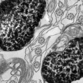
12 Examples: Bacteria: Golgi in a cell: Microvillae: Epithelial cells: Tails of fish sperm:


13 Modern TEM: Various applications Negative Staining Correlative Microscopy Immunogold labeling Cryo-TEM Tomography Elemental analysis


14 Pilot samples studied during autumn 2010 Tissues ImmunoAu labeled tissue Rat brain nanogold particles for labeling Artery in rat brain


15 Case-studies 1 The diabetes-prone NZO/HI stain (Pancreatic immunopathology) Electron micrographs of islet sections. A, Detail of an islet of a 52-week-old female. Fully granulated beta cells surround one pancreatic polypeptide cell (PP). B, Detail of an islet of an age-matched chronically diabetic male. Most beta cells are degranulated.

16 Case study 2: Viral nanoparticles as tools for intravital vascular imaging Intravital fluorescence imaging of chick CAM vasculature and subcellular localization of CPMV.
17 NANOTEM project 3-year projects to develope know-how and modernize equipment in the field of electron microscopy Organized by BioMater Centre Funding: State provincial office of Eastern Finland Also in the future: Tekes? Tekes-RDF Company-related development (2011 -)? Education: NANOTEM Lecture Series Practical training Instrumentation: Modern TEM Elemental analysis Tomography, etc. State provincial office-esf Educational project ( ) State provincial office-rdf Instrumentation ( )

18 Microscopy: Scanning electron microscope ESEM TMP XL30, Fei Company Location S3140 Operation voltage up to 30 kv Magnification 20x max x


19 Examples: ESEM TMP XL30 Pollen: Cultured cells: Bacteria: Red cells:


20 Microscopy: Sample preparation 1 EM sample preparation (Virpi Miettinen) Location S3137 and S3141 Samples for TEM and SEM need fixation dehydration embedding cutting staining fixation dehydration drying coating

")


21 Microscopy: Sample preparation2 Hard tissue laboratory (Ritva Sormunen) Location S3142 For demanding samples consisting metals and soft tissues, or large samples, for example dehydration - embedding - sawing with diamond saw - grinding - staining Examples: hip implant stent in a vein cadaver femoral bone


22 Microscopy: Spectroscopy "Imaging FTIR", PerkinElmer Spotlight Location S3145 Embedded or cryo-sections, special IR window needed "Raman spectrometer", Bruker Optics Senterra 200LX Location S3145 Tissue sections or blocks Optical image Spectral imaging Examples; collagen distribution in cartilage, typing of tumours, homogeneity of tablets, polymorphism


23 Structural Analysis The equipment include for example MicroCT, ultrasonic microscope Examples; - composition of bone, cartilage and tablets using ultrasonic microscope - structure of wood, bone and tablets using MicroCT
24 Contact information For more information, please visit Location: Snellmania Building 3 rd floor, corridor C
25 SIB-labs Three different units: Smarc-material research center (Joensuu), InFotonics Center (Joensuu) and Biomater centre (Kuopio) SIB-labs Research areas/interests: Material techniques Optics Biomaterials Focused Research services Customise research Equipment (maintenance and development) Participate research programs and networks
26 Quite big organization: over 200 persons SIB-labs started beginning of the year
27 Thank you Virpi Tiitu:
Introduction to histology and its methods of study
 Introduction to histology and its methods of study Li shulei lishulei@tom.com Department of Histology & Embryology 1 What is histology Definition Cell: smallest units functions in the human body Tissue
Monday: Y42 G53 Tuesday: Y42 G53 Wednesday: Y42 J11
 Locations: Irchel building 42, Level H and F Locations: Irchel building 42, Level H and F Self-study sessions: Monday: Y42 G53 Tuesday: Y42 G53 Wednesday: Y42 J11 1 Center for Microscopy and Image Analysis
Imaging of endocrine organs
 Imaging of endocrine organs Helen Christian Department of Physiology, Anatomy & Genetics St Anne s College, University of Oxford Diabetesforum, Stockholm 2017 Islets of Langerhan Pituitary gland Renin
Introduction to Histology
 Introduction to Histology The name "Histology" is derived from the Greek word for a tissue "Histos", and "-logos" = the study of It is tightly bounded to molecular biology, genetics, immunology and other
Morphological Investigations - Different Microscopic Techniques (Semicrystalline Polymers)
") Morphological Investigations - Different Microscopic Techniques (Semicrystalline Polymers) Method SEM TEM AFM Typical Sample Preparation Evaporation Surface Etching Ultramicrotomy Selective Staining no
Morphological Investigations - Different Microscopic Techniques (Semicrystalline Polymers) Method SEM TEM AFM Typical Sample Preparation Evaporation Surface Etching Ultramicrotomy Selective Staining no
CENTER FOR BRAIN EXPERIMENT
 CENTER FOR BRAIN EXPERIMENT Section of Brain Structure Associate Professor: ARII, Tatsuo, PhD 1967 Graduated from Tohoku University, Faculty of Science. Completed the doctoral course in Engineering, Nagoya
EMS MICROSCOPY ACADEMY PHARMACEUTICAL MICROSCOPY WORKSHOP
 Examples of the Microscopy of Pharmaceuticals PLM image of anthroquinone cooled after thermal microscopy. Crossed polars with lambda waveplate. Uniaxial interference pattern used to determine optical crystallographic
Correlative Microscopy - From living cells to 3D electron microscopy A comprehensive workflow approach
 Correlative Microscopy - From living cells to 3D electron microscopy A comprehensive workflow approach Maria Marosvoelgyi, Liesbeth Hekking, Ben Lich, Matthias Geisbauer and Alex de Marco (FEI) Maja Guenthert
NINT Supporting Innovative Ideas through Nanotechnology
 NINT Supporting Innovative Ideas through Nanotechnology (Capabilities and Expertise) Dr. Marianna Kulka, PhD Group Leader, NINT National Research Council Marianna.kulka@nrc.ca National Institute for Nanotechnology
EMS MICROSCOPY ACADEMY BIOLOGICAL TEM WORKSHOP: A COMPLETE PICTURE
 Examples of the endless possibilities in the field of Microscopy Bone Marrow: Transmission electron microscope image of a thin section cut through an area of bone marrow area near the cartilage/bone interface
Introduction to Electron Microscopy Andres Kaech
 Center for Microscopy and Image Analysis Introduction to Electron Microscopy Andres Kaech The types of electron microscopes Transmission electron microscope (TEM) Scanning electron microscope (SEM) 1 The
Cell Structure and Function
 Cell Structure and Function Dead White Men Who Discovered (and were made of) Cells: Anton Van Leeuwenhoek Robert Hooke Where the Magic Happened Schleiden Cell Theory All plants are made of cells Schwann
1st Faculty of Medicine, Charles University in Prague Center for Advanced Preclinical Imaging (CAPI)
") ADVANTAGES Optical Imaging OI Optical Imaging is based on the detection of weak light by a highly sensitive and high resolution CCD camera DISADVANTAGES High sensitivity Limited penetration depth Easy
ADVANTAGES Optical Imaging OI Optical Imaging is based on the detection of weak light by a highly sensitive and high resolution CCD camera DISADVANTAGES High sensitivity Limited penetration depth Easy
Supplementary Figure 1. CryoTEM images of the barcoded nanoparticles (a) and
 and") a b Supplementary Figure 1. CryoTEM images of the barcoded nanoparticles (a) and size measurements of the particles (b). Liposomes were loaded with DNA barcodes and were imaged using cryo-tem and measured
a b Supplementary Figure 1. CryoTEM images of the barcoded nanoparticles (a) and size measurements of the particles (b). Liposomes were loaded with DNA barcodes and were imaged using cryo-tem and measured
Microscopy...Seeing the Unseen
 Technical Workshops Series 2013 Three Day Intensive Workshop on Venture Center Microscopy...Seeing the Unseen Organized by Venture Center Learn Organized by For whom When Principles and applications of
Practical 2P8 Transmission Electron Microscopy
 Practical 2P8 Transmission Electron Microscopy Originators: Dr. N.P. Young and Prof. J. M. Titchmarsh What you should learn from this practical Science This practical ties-in with the lecture course on
Supplementary Information Electrospray Deposition-Induced Ambient Phase Transition in Copper Sulphide Nanostructures
 Electronic Supplementary Material (ESI) for Journal of Materials Chemistry A. This journal is The Royal Society of Chemistry 2019 Supplementary Information Electrospray Deposition-Induced Ambient Phase
The principles and practice of electron microscopy
 The principles and practice of electron microscopy Second Edition Ian M. Watt CAMBRIDGE UNIVERSITY PRESS Contents Preface tofirstedition page ix Preface to second edition xi 1 Microscopy with light and
Prof. Steven S. Saliterman
 Department of Biomedical Engineering, University of Minnesota http://saliterman.umn.edu/ Prof. Angela Panoskaltsis-Mortari s BMEn 5361, 3D Bioprinting Tissue engineering Bioprinting Design considerations
Methods of Culturing Microorganisms. Chapter 3. Five Basic Techniques of Culturing Bacteria. Topics
 Chapter 3 Topics Methods of Culturing Microorganisms Microscope (History, Types, Definitions) Staining (Gram s) Methods of Culturing Microorganisms Five basic techniques of culturing Media Microbial growth
BIOLOGICAL SAMPLE PREPARATION FOR TEM OBSERVATION. TEM Seminar Nov 16, 2017 Astari Dwiranti, Ph.D
 BIOLOGICAL SAMPLE PREPARATION FOR TEM OBSERVATION TEM Seminar Nov 16, 2017 Astari Dwiranti, Ph.D Why do we need EM for biological samples? (O'Connor and Adams, 2010) Why do we need EM for biological samples?
Supporting information
 Supporting information One-pot facile synthesis of concentrated Si nanoparticles solution Hua Sun a, Satoshi Miyazaki a, Hironori Tamamitsu a, and Ken-ichi Saitow* a,b a Department of chemistry, Graduate
future s in the making Vibration Control Solutions AVOS
 Vibration Control Solutions AVOS Ostec Corporate Group produces and offers hi-tech innovative scientific and analytical equipment. Our mission is to be a company that finds, selects, protects and develops
Special Techniques 1. Mark Scott FILM Facility
 Special Techniques 1 Mark Scott FILM Facility SPECIAL TECHNIQUES Multi-photon microscopy Second Harmonic Generation FRAP FRET FLIM In-vivo imaging TWO-PHOTON MICROSCOPY Alternative to confocal and deconvolution
SUPPLEMENTARY INFORMATION
 SUPPLEMENTARY INFORMATION Nanogap Engineerable Raman-Active Nanodumbbells for Single-Molecule Detection Dong-Kwon Lim 1,, Ki-Seok Jeon 2,, Hyung Min Kim 2, Jwa-Min Nam 1, *, and Yung Doug Suh 2, * 1 Department
CORE FACILITY USER FEES FEES FLOW CYTOMETRY CORE FACILITY
 CORE FACILITY USER FEES This document is for researchers to use for grant applications in 2018. In December 2018 fees will be increased by the CPI rate for WIMR facilities (current rate as at September
AFM-Raman Characterization of Pharmaceutical Tablets
 AFM-Raman Characterization of Pharmaceutical Tablets Compound Distribution Studies in Pharmaceutical Tablets by Integrated AFM-Raman Instrument 1,2 1 Sergey Shashkov and Pavel Dorozhkin, 1 NT-MDT Co.,
EMS MICROSCOPY ACADEMY PHARMACEUTICAL MICROSCOPY WORKSHOP: TECHNIQUES
 Examples of the Microscopy of Pharmaceuticals Carbamazepine Form 3 twinned crystals by optical microscopy. Carbamazepine Form 3 twinned crystals by SEM. Optical microscopy and SEM allow for unique views
Heteroepitaxy of Monolayer MoS 2 and WS 2
 Supporting Information Seed Crystal Homogeneity Controls Lateral and Vertical Heteroepitaxy of Monolayer MoS 2 and WS 2 Youngdong Yoo, Zachary P. Degregorio, James E. Johns* Department of Chemistry, University
Introduction to Histology
 Introduction to Histology Histology The term "Histology" is derived from the Greek word for a tissue "Histos", and "-logos" = the study of Histology : Is the study of tissues and how they are arranged
E! 6143 MiniTEM Development of benchtop equipment for automated characterization of viruses and other biological nanoparticles
 E! 6143 MiniTEM Development of benchtop equipment for automated characterization of viruses and other biological nanoparticles 3 years from 7.2011 to 6.2014 Brno - Palackého tř. headquarters, R&D, special
REMEDI. Regenerative Medicine Institute (REMEDI) NUI Galway, Ireland GENERAL PRESENTATION. Director: Prof. Frank Barry
 NUI Galway, Ireland GENERAL PRESENTATION. Director: Prof. Frank Barry") Regenerative Medicine Institute (REMEDI) NUI Galway, Ireland Director: Prof. Frank Barry GENERAL PRESENTATION Contact person in NEWGEN: Dr. Jessica Hayes Working Group Involvement: Member of Working Group
Regenerative Medicine Institute (REMEDI) NUI Galway, Ireland Director: Prof. Frank Barry GENERAL PRESENTATION Contact person in NEWGEN: Dr. Jessica Hayes Working Group Involvement: Member of Working Group
A combined method for correlative 3D imaging of biological samples from macro to nano scale
 7 8 9 0 7 8 9 A combined method for correlative D imaging of biological samples from macro to nano scale Manuela Kellner,, *, Marko Heidrich, *, Raoul-Amadeus Lorbeer, Georgios C. Antonopoulos, Lars Knudsen,,
T H E J O U R N A L O F C E L L B I O L O G Y
 Supplemental material Prashar et al., http://www.jcb.org/cgi/content/full/jcb.201304095/dc1 T H E J O U R N A L O F C E L L B I O L O G Y Figure S1. FBT phagocytosis in cells expressing PM-GFP. (A) T-PC
University of Eastern Finland (UEF) Main Research Lines
 Main Research Lines") University of Eastern Finland (UEF) Main Research Lines Asla Pitkänen, MD, PhD Epilepsy Research Laboratory A.I.Virtanen Institute for Molecular Sciences University of Eastern Finland Kuopio, Finland E-mail:
University of Eastern Finland (UEF) Main Research Lines Asla Pitkänen, MD, PhD Epilepsy Research Laboratory A.I.Virtanen Institute for Molecular Sciences University of Eastern Finland Kuopio, Finland E-mail:
*These authors contributed equally to this work
 CUSTOMIZED PATTERNED SUBSTRATES FOR HIGHLY VERSATILE CORRELATIVE LIGHT- SCANNING ELECTRON MICROSCOPY Lorena Benedetti 1,2 *, Elisa Sogne 1,3,4 *, Simona Rodighiero 1, Davide Marchesi 1, Paolo Milani 1,3,
COPYRIGHTED MATERIAL. Tissue Preparation and Microscopy. General Concepts. Chemical Fixation CHAPTER 1
 CHAPTER 1 Tissue Preparation and Microscopy General Concepts I. Biological tissues must undergo a series of treatments to be observed with light and electron microscopes. The process begins by stabilization
A comparative study of cellular uptake and cytotoxicity of multi-walled carbon
 A comparative study of cellular uptake and cytotoxicity of multi-walled carbon nanotube, graphene oxide, and nanodiamond Xiaoyong Zhang,* a,b Wenbing Hu, a Jing Li, a Lei tao, b and Yen wei b Preparation
SCIENTIFIC ANALYSIS AND ENDORSEMENT OF NITTA CASINGS COLLAGEN GEL
 SCIENTIFIC ANALYSIS AND ENDORSEMENT OF NITTA CASINGS COLLAGEN GEL 12/26/2017 For Nitta Casings TABLE OF CONTENTS About Dr. Silvia Minardi 2 Background on Collagen.. 3 Analysis of NITTA GEL. 3 Endorsement
From Forest To Product:
 From Forest To Product: New Solutions For Rapid, Comprehensive Wood And Fibre Analyses Co-authors: Kathy Woo, Ho Fan Jang, Shannon Huntley, James Drummond, Val Lawrence (Paprican) Francides Gomes, University
MEDICAL PHYSICS (MED PHYS)
") Medical Physics (MED PHYS) 1 MEDICAL PHYSICS (MED PHYS) MED PHYS/PHYSICS 265 INTRODUCTION TO MEDICAL PHYSICS Primarily for premeds and other students in the medical and biological sciences. Applications
Medical Physics (MED PHYS) 1 MEDICAL PHYSICS (MED PHYS) MED PHYS/PHYSICS 265 INTRODUCTION TO MEDICAL PHYSICS Primarily for premeds and other students in the medical and biological sciences. Applications
A Brief History of Light Microscopy And How It Transformed Biomedical Research
 A Brief History of Light Microscopy And How It Transformed Biomedical Research Suewei Lin Office: Interdisciplinary Research Building 8A08 Email: sueweilin@gate.sinica.edu.tw TEL: 2789-9315 Microscope
Advanced SEM: ESEM and Cryo-SEM
 Advanced SEM: ESEM and Cryo-SEM Peter Harris www.reading.ac.uk/emlab Electron Microscopy Scanning electron microscope Transmission electron microscope Electron gun Condenser lens Objective lens Specimen
Supporting Information
 Supporting Information Photoinitiated Growth of Sub-7 nm Silver Nanowires within a Chemically Active Organic Nanotubular Template D. M. Eisele 1, H. v. Berlepsch 2, C. Böttcher 2, K. J. Stevenson 3, D.
Practical 2P8 Transmission Electron Microscopy
 Practical 2P8 Transmission Electron Microscopy Originators: Dr. M. L. Jenkins and Prof. J. M. Titchmarsh What you should learn from this practical Science This practical ties-in with the lecture course
The Orthopaedic Research Laboratory (ORL) performs research in the field of orthopaedics.
 performs research in the field of orthopaedics.") The Orthopaedic Research Laboratory (ORL) performs research in the field of orthopaedics. The biomechanical section focusses on bone, soft tissues and on pre-clinical testing of implants. We are specialized
The Orthopaedic Research Laboratory (ORL) performs research in the field of orthopaedics. The biomechanical section focusses on bone, soft tissues and on pre-clinical testing of implants. We are specialized
Confocal Microscopy Analyzes Cells
 Choosing Filters for Fluorescence A Laurin Publication Photonic Solutions for Biotechnology and Medicine November 2002 Confocal Microscopy Analyzes Cells Reprinted from the November 2002 issue of Biophotonics
Chapter 10: Classification of Microorganisms
 Chapter 10: Classification of Microorganisms 1. The Taxonomic Hierarchy 2. Methods of Identification 1. The Taxonomic Hierarchy Phylogenetic Tree of the 3 Domains Taxonomic Hierarchy 8 successive taxa
Resolution of Microscopes Visible light is nm Dry lens(0.5na), green(530nm light)=0.65µm=650nm for oil lens (1.4NA) UV light (300nm) = 0.13µm f
, green(530nm light)=0.65µm=650nm for oil lens (1.4NA) UV light (300nm) = 0.13µm f") Microscopes and Microscopy MCB 380 Good information sources: Alberts-Molecular Biology of the Cell http://micro.magnet.fsu.edu/primer/ http://www.microscopyu.com/ Approaches to Problems in Cell Biology
Microscopes and Microscopy MCB 380 Good information sources: Alberts-Molecular Biology of the Cell http://micro.magnet.fsu.edu/primer/ http://www.microscopyu.com/ Approaches to Problems in Cell Biology
EMS MICROSCOPY ACADEMY BIOLOGICAL SEM WORKSHOP: A COMPLETE PICTURE
 Examples of the endless possibilities in the field of Microscopy: SEM Mouse Lung Tissue: Red blood cells in the lung. Mouse Lung Tissue: Tracchea area. Red blood cells inside blood vessel in the lung.
PREPARATION AND CHARACTERISATION OF CHITOSAN/NANO HYDROXYAPATITE COMPOSITES ABSTRACT INTRODUCTION
 PREPARATION AND CHARACTERISATION OF CHITOSAN/NANO HYDROXYAPATITE COMPOSITES Ismail Zainol, Fadzil Ayad Zakaria, Mohd Razif Saliman and Mohd Affandi Derman Advanced Materials Research Centre (AMREC), SIRIM
Herpes simplex virus type-1 attachment inhibition. by functionalized graphene oxide
 Supporting information Herpes simplex virus type-1 attachment inhibition by functionalized graphene oxide Matias Sametband a, Inna Kalt b, Aharon Gedanken a*, Ronit Sarid b a Department of Chemistry, Kanabar
Analytical Tools for Reliable Detection and Characterization of Protein Particles. Alla Polozova Analytical Biochemistry, MedImmune
 Analytical Tools for Reliable Detection and Characterization of Protein Particles Alla Polozova Analytical Biochemistry, MedImmune Outline What to expect: Types of particles which might be present in protein
HYPERSPECTRAL MICROSCOPE PLATFORM FOR HIGHLY MULTIPLEX BIOLOGICAL IMAGING. Marc Verhaegen
 HYPERSPECTRAL MICROSCOPE PLATFORM FOR HIGHLY MULTIPLEX BIOLOGICAL IMAGING Marc Verhaegen CMCS, MONTREAL, MAY 11 th, 2017 OVERVIEW Hyperspectral Imaging Multiplex Biological Imaging Multiplex Single Particle
Microstructure Analysis by Means of the Orthogonallyarranged
 Hitachi Review Vol. 65 (2016), No. 7 201 Special Contributions Microstructure Analysis by Means of the Orthogonallyarranged FIB-SEM Toru Hara, Dr. Eng. OVERVIEW: Serial sectioning using a combined FIB
The new LSM 700 from Carl Zeiss
 The new LSM 00 from Carl Zeiss Olaf Selchow, Bernhard Goetze To cite this version: Olaf Selchow, Bernhard Goetze. The new LSM 00 from Carl Zeiss. Biotechnology Journal, Wiley- VCH Verlag, 0, (), pp.. .
SUPPLEMENTARY INFORMATION
 Biosynthesis of Luminescent Quantum Dots in an Earthworm S.R. Stürzenbaum, a# M. Hoeckner, a# A. Panneerselvam, b J. Levitt, b J.-S. Bouillard, b S. Taniguchi, b L.-A. Dailey, d R. Ahmad Khanbeigi, d E.
Ilya Turchin. Institute of Applied Physics of the RAS, Nizhny Novgorod, Russia.
 Fluorescence 3D imaging of small animals Ilya Turchin Institute of Applied Physics of the RAS, Nizhny Novgorod, Russia ilya@ufp.appl.sci-nnov.ru http://www.bioimaging.ru German-Russian Forum Biotechnology
High-resolution phase contrast imaging, aberration corrections, and the specifics of nanocrystals
 High-resolution phase contrast imaging, aberration corrections, and the specifics of nanocrystals Pages 450-459, in Methods and applications in crystallographic computing, S.R. Hall and T. Ashida, Proc.
Skills and excellence formation on basis of Laboratory of Plasma Physics & Atomic Spectroscopy Institute of Spectroscopy (ISAN) of Russian Academy of
 of Russian Academy of") 1968 Skills and excellence formation on basis of Laboratory of Plasma Physics & Atomic Spectroscopy Institute of Spectroscopy (ISAN) of Russian Academy of Science 2005 Development of Jet 1 Demo EUV Source
1968 Skills and excellence formation on basis of Laboratory of Plasma Physics & Atomic Spectroscopy Institute of Spectroscopy (ISAN) of Russian Academy of Science 2005 Development of Jet 1 Demo EUV Source
Bioengineering Research Map
 Bioengineering Research Map Lee Makowski, Professor and Chair l.makowski@northeastern.edu Mark Niedre, Associate Professor and Associate Chair For Research m.niedre@northeastern.edu Chloe Tolman, Academic
Cell viability in TE constructs
 Cell viability in TE constructs Module 3, Lecture 4 20.109 Spring 2009 Lecture 3 review What engineering principles may be useful food-for-thought in BE? What difference in live and dead cells is exploited
Supplementary Information
 Supplementary Information Nanofibrous Spongy Microspheres to Distinctly Release mirna and Growth Factors to Enrich Regulatory T Cells and Rescue Periodontal Bone Loss Zhongning Liu, Xin Chen, Zhanpeng
Supporting Online Material for
 www.sciencemag.org/cgi/content/full/327/5969/1126/dc1 Supporting Online Material for A Nodule-Specific Protein Secretory Pathway Required for Nitrogen- Fixing Symbiosis Dong Wang, Joel Griffitts, Colby
CHAPTER 3 MATERIALS AND EXPERIMENTAL METHODS
 45 CHAPTER 3 MATERIALS AND EXPERIMENTAL METHODS This chapter is divided into six different sections. The section one explains the selection of materials and process flow in this research work. Section
JSM-7800F Field Emission Scanning Electron Microscope
 JSM-7800F catalogue JSM-7800F Field Emission Scanning Electron Microscope We provide high performance The Ultimate Research Tool for Multi-Disciplinary Research Institutions Extreme resolution The super
Dr Claire MacDonald Business Development Manager CMAC National Facility
 Dr Claire MacDonald Business Development Manager CMAC National Facility This presentation will cover CMAC Overall Vision CMAC National Facility Capabilities and Expertise API Process Development Secondary
KILLING THROMBUS WITH
 KILLING THROMBUS WITH D. Dash Department of Biochemistry Institute of Medical Sciences Banaras Hindu University Thrombus has two components: (1) Protein Component composed of Insoluble Fibrin Clot (2)
Ionscope SICM. About Ionscope. Scanning Ion Conductance Microscopy. Ionscope A brand of OpenIOLabs Limited
 SICM About is a brand of OpenIOLabs Ltd, headquartered in Cambridge UK, is the worldleader in (SICM), a rapidly emerging Scanning Probe Microscopy (SPM) technique which allows nanoscale topographical mapping
Supplementary Figure legends. Supplementary Methods
 Supplementary Methods Transmission electron microscopy. For transmission electron microscopy (TEM), the cell populations were rinsed with 0.1 Sorensen s buffer (ph 7.5), fixed in 2.5% glutaraldehyde for
Imperial College London Key Research Themes for NBIT and FP7
 Imperial College London Key Research Themes for NBIT and FP7 Professor Richard I Kitney Imperial College Research Systems and Software Solutions Advanced Information Systems (eg CISs) Systems Analysis
Supporting Information. Glutathione-stabilized fluorescent gold nanoclusters vary in their
 Supporting Information Glutathione-stabilized fluorescent gold nanoclusters vary in their influences on the proliferation of pseudorabies virus and porcine reproductive and respiratory syndrome virus Yanli
Ongoing R&D projects in COST member countries Finland
 Ongoing R&D projects in COST member countries Finland Finland Participating Group 1, VTT Technical Research Centre of Finland Hannu Viitanen, Elina Sohlberg, Kirsi Immonen, Participating Group 2, Metla
Supporting Information for. Direct Chemical Synthesis of Plasmonic Black Colloidal Gold Superparticles with Broadband Absorption Properties
 Supporting Information for Direct Chemical Synthesis of Plasmonic Black Colloidal Gold Superparticles with Broadband Absorption Properties Nayoung Kwon,, Hwisu Oh,, Reehyang Kim, Arjyabaran Sinha, Jaeyun
Microstructural Characterization of Materials
 Microstructural Characterization of Materials 2nd Edition DAVID BRANDON AND WAYNE D. KAPLAN Technion, Israel Institute of Technology, Israel John Wiley & Sons, Ltd Contents Preface to the Second Edition
Skin Imaging NTU
 Skin Imaging Research @ NTU A*STAR-NHG-NTU Joint Skin Research Workshop, Oct 19, 2013 A/Prof. Wee Ser ewser@ntu.edu.sg Programme Lead (MISA), NITHM Director, VALENS Research Centre, EEE Nanyang Technological
The preparation of both fixed and living material is a new and equally demanding art.
 W hen viewed using a microscope, all parts of the human body - from chromosomes to sperm can be visually arresting. My work is inspired by scientific images of cells from organisms as diverse as algae
Visualizing Cells Molecular Biology of the Cell - Chapter 9
 Visualizing Cells Molecular Biology of the Cell - Chapter 9 Resolution, Detection Magnification Interaction of Light with matter: Absorbtion, Refraction, Reflection, Fluorescence Light Microscopy Absorbtion
Electron microscopy II
 Electron microscopy II Nanomaterials characterization I RNDr. Věra Vodičková, PhD. Interaction ction: electrons solid matter Signal types SE.secondary e - AE Auger s e - BSE back scattered e - X-ray photons,
Carbon Nanotube Material Quality Assessment
 Carbon Nanotube Material Quality Assessment Sivaram Arepalli 1, Edward Sosa 1, Pasha Nikolaev 1, William Holmes 1, Olga Gorelik 1, and Leonard Yowell 2 1 ERC Inc. / NASA - Johnson Space Center, Houston,
Kinematical theory of contrast
 Kinematical theory of contrast Image interpretation in the EM the known distribution of the direct and/or diffracted beam on the lower surface of the crystal The image on the screen of an EM = the enlarged
Confocal Microscopy & Imaging Technology. Yan Wu
 Confocal Microscopy & Imaging Technology Yan Wu Dec. 05, 2014 Cells under the microscope What we use to see the details of the cell? Light and Electron Microscopy - Bright light / fluorescence microscopy
Electronic Supporting Information
 Electronic Supplementary Material (ESI) for RSC Advances. This journal is The Royal Society of Chemistry 2018 Electronic Supporting Information An Au@Ag Nanocube based Plasmonic Nano-sensor for Rapid Detection
Absorption of an electromagnetic wave
 In vivo optical imaging?? Absorption of an electromagnetic wave Tissue absorption spectrum Extinction = Absorption + Scattering Absorption of an electromagnetic wave Scattering of an electromagnetic wave
Supplementary information. Extreme biomimetic approach: Hydrothermal synthesis of β-chitin/zno nanostructured composites
 Journal Name Dynamic Article Links Cite this: DOI: 10.1039/c0xx00000x www.rsc.org/xxxxxx Supplementary information ARTICLE TYPE Extreme biomimetic approach: Hydrothermal synthesis of β-chitin/zno nanostructured
T H E J O U R N A L O F C E L L B I O L O G Y
 T H E J O U R N A L O F C E L L B I O L O G Y Supplemental material Rodríguez-Fraticelli et al., http://www.jcb.org/cgi/content/full/jcb.201203075/dc1 Figure S1. Cell spreading and lumen formation in confined
Waterford Institute of Technology Waterford Institute of Technology, Cork Road, Waterford
 INNOVATION VOUCHERS INITIATIVE Waterford Institute of Technology Waterford Institute of Technology, Cork Road, Waterford DIRECTORY OF SKILLS AND EXPERTISE College Contact Point for Innovation Voucher Enquires:
Biosensis P1TM Polar Lipid Tracing Reagent. Catalogue Number: TR-600-P1. For research use only, not for use in clinical and diagnostic procedures.
 Biosensis P1TM Polar Lipid Tracing Reagent Catalogue Number: TR-600-P1 For research use only, not for use in clinical and diagnostic procedures. Version: TR-600-P1/v2/Sep2018 Biosensis Pty Ltd., 51 West
Assays for gene expression and protein production
 Assays for gene expression and protein production Module 3, Lecture 5! 20.109 Spring 2011! Topics for Lecture 5 Measuring protein levels! Measuring transcript levels! Imaging assays! 2 Module overview:
2004 Debye Lecture 4 C. B. Murray. Quantum Dot Applications: Sun Screen. Solar Cells. Bio-tagging. Solid State Lighting?
 2004 Debye Lecture 4 C. B. Murray Quantum Dot Applications: Sun Screen Solar Cells Bio-tagging Solid State Lighting? Quantum Dot Solar cells Nanocrystal Solar Cells Double-labeling of mitochondria
SUPPLEMENTARY INFORMATION
 a 14 12 Densitometry (AU) 1 8 6 4 2 t b 16 NMHC-IIA GAPDH NMHC-IIB Densitometry (AU) 14 12 1 8 6 4 2 1 nm 1 nm 1 nm 1 nm sirna 1 nm 1 nm Figure S1 S4 Quantification of protein levels. (a) The microtubule
Optical Observation - Hyperspectral Characterization of Nano-scale Materials In-situ
 Optical Observation - Hyperspectral Characterization of Nano-scale Materials In-situ Research at the nanoscale is more effective, when research teams can quickly and easily observe and characterize a wide
Crystallographic Characterization of GaN Nanowires by Raman Spectral Image Mapping
 Crystallographic Characterization of GaN Nanowires by Raman Spectral Image Mapping Heerad Farkhoor, Adam Schwartzberg, Jeffrey Urban August 12, 2009 Abstract Obtaining structural information of nano-structured
SUPPORTING INFORMATION
 Electronic Supplementary Material (ESI) for Nanoscale. This journal is The Royal Society of Chemistry 2015 SUPPORTING INFORMATION Chemical Sporulation and Germination: Cytoprotective Nanocoating of Individual
A NOVEL TECHNOLOGY IN WET SCANNING ELECTRON MICROSCOPY
 A NOVEL TECHNOLOGY IN WET SCANNING ELECTRON MICROSCOPY Amnon Katz, Arnon Bentur, Konstantin Kovler National Building Research Institute, Faculty of Civil and Environmental Engineering, Technion Israel
Building An Ultrafast Photon-Induced Near-field Transmission Electron Microscope
 Building An Ultrafast Photon-Induced Near-field Transmission Electron Microscope Dr. Tom T.A. Lummen École Polytechnique Fédérale de Lausanne -- LUMES Photonic Instruments 2013 September 11, 2013 Zürich,
Scanning Electron Microscope & Surface Analysis. Wageningen EM Centre Marcel Giesbers
 Scanning Electron Microscope & Surface Analysis Wageningen EM Centre Marcel Giesbers Scanning Electron Microscope & Surface Analysis SEM vs Light Microscope and Transmission EM Secondary Electron Imaging
SUPPLEMENTARY INFORMATION
 High Electrochemical Activity of the Oxide Phase in Model Ceria- and Ceria-Ni Composite Anodes William C. Chueh 1,, Yong Hao, WooChul Jung, Sossina M. Haile Materials Science, California Institute of Technology,
Optical microscopy Theoretical background Galina Kubyshkina
 Optical microscopy Theoretical background Galina Kubyshkina Elektromaterial Lendava d.d., Slovenia Crystalline materials presence of a unit (cell), which is periodically repeated in space regular structure
NORDIC LIFE SCIEN SWEDEN S LIFE SCIENCE STRATEGY ANTIBIOTIC RESISTANCE THE LEADING LIFE SCIENCE BUSINESS MAGAZINE 169 SEK / 20 EUR / 25 USD
 NORDIC LIFE SCIEN THE LEADING LIFE SCIENCE BUSINESS MAGAZINE 169 SEK / 20 EUR / 25 USD // UMEÅ // KUOPIO // TROMSØ ANTIBIOTIC RESISTANCE SWEDEN S LIFE SCIENCE STRATEGY ILLUSTRATION AJAK ARCHITECTS ALL