Supplemental figures Supplemental Figure 1: Fluorescence recovery for FRAP experiments depicted in Figure 1.
|
|
|
- Simon Hodge
- 6 years ago
- Views:
Transcription
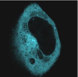
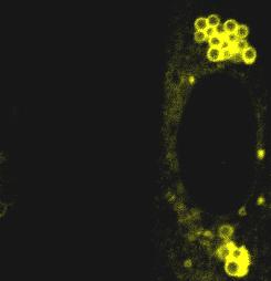
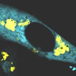
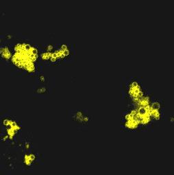
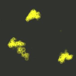
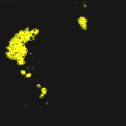
1 Supplemental figures Supplemental Figure 1: Fluorescence recovery for FRAP experiments depicted in Figure 1. Percent of original fluorescence was plotted as a function of time following photobleaching for ADFP-YFP and LSDP5-YFP. Data are means ± SEM from 7 or 8 experiments. Supplemental Figure 2: PAT proteins at the lipid droplet surface recover similarly from 50% photobleaching. FRAP analysis of perilipin-gfp, ADFP-GFP and LDSP5-YFP in CHO- K1 cells incubated overnight with 400 μm oleic acid. Live cells were examined with a confocal microscope using a 40x oil immersion objective. Red boxed regions were bleached at time 0; fluorescence within these regions was monitored at 15 s intervals. Bar: 1 μm. Supplemental Figure 3: Negative and positive controls used for AFRET experiments. (A) Confocal imaging of CHO-K1 cells co-transfected with perilipin-yfp and LSDP5-CFP (top), and CGI-58-CFP and perilipin-yfp (bottom). (B) Measurement of AFRET between perilipin- YFP and LSDP5-CFP, and CGI-58-CFP and perilipin-yfp in basal conditions. Data are means ± SEM from 10 or 12 experiments. (C) of perilipin-yfp and LSDP5-YFP from CHO-K1 cell extracts. Lysates were immunoprecipitated without antibody (None), or with anti-gfp polyclonal antibody, anti-perilipin antibody, or anti-lsdp5 antibody; immunoblots were probed with GFP monoclonal antibody. Experiment is representative of 7 experiments. (D) Co- of perilipin A (untagged) with CGI-58-CFP from CHO-K1 cell extracts in basal (B) conditions. s were performed with control IgG or anti-cgi-58 antibody. Western blot was first incubated with antiperilipin antibody and subsequently with anti-cgi-58 antibody. Experiment is representative of 9 experiments. Supplemental Figure 4: Untagged HSL interacts with untagged perilipin A. CHO cells expressing ectopic Perilipin A (Peri A) cells were transduced with HSL adenovirus. Cells were incubated in basal conditions (B) or stimulated conditions (S). s were performed using a rabbit anti-hsl IgG or pre-immune rabbit IgG (IgG). Immunoprecipitates were analyzed by Western blot probed with an anti-perilipin antibody or an anti-hsl antibody. Experiment is representative of 2 experiments. Supplemental Figure 5: Overexpression of the PAT-1 domain of perilipin prevents binding of phosphorylated HSL to the surfaces of perilipin-coated lipid droplets. (A) CHO cells stably overexpressing untagged perilipin A (Peri A cells) were co-transfected with either 1) HSL-CFP and a construct driving expression of YFP or 2) CFP-HSL and perilipin (1-121)-YFP. Cells were incubated overnight with 400 μm oleic acid. The following day, the cells were incubated with 5 μm triacsin C with no further additions (basal), or stimulated for 15 min with 10 μm forskolin, 1 mm IBMX and 5 μm triacsin C; live cells were examined with confocal microscopy as in Fig 1. Bar, 10 μm. (B) Quantitative analysis for HSL binding to the lipid droplet under PKA-stimulated conditions. Data are means ± SEM from 8 to 13 experiments (*, p <0.05). Supplemental Figure 6: The PAT-1 domains of both LSDP5 and perilipin interact with HSL. (A)Western blot of of HSL with the PAT-1 domain of perilipin (left; 1-121) 1
2 and the PAT-1 domain of LSDP5 (right; LSDP5-YFP 1-123) from CHO-K1 cell lysates. (B) Western blot of of HSL with the full length LSDP5 (top) and the 11-mer repeat sequence (PAT-2 domain) of LSDP5 (right; LSDP5-YFP 11-mer) from CHO-K1 cell lysates. Cells were incubated in basal conditions (B) or stimulated conditions (S). s were performed using a rabbit anti-hsl IgG or mouse anti-rabbit (control) IgG (IgG). Immunoprecipitates were analyzed by Western blot probed with a GFP antibody. Experiment is representative of 2 experiments. Supplemental Figure 7: The phosphorylation of serine 81, 222, 276 of perilipin is required for recruitment of HSL to perilipin-coated lipid droplets. Complete set of representative confocal images for Figure 6, including images for the YFP channel, CFP channel and merged images. Cells were incubated overnight with 400 μm oleic acid. The following day, the cells were incubated with 5 μm triacsin C (basal) or stimulated for 10 min with 10 μm forskolin, 1 mm IBMX and 5 μm triacsin C; live cells were examined with a confocal microscope as in Fig 1. Bar, 10 μm. Supplemental Figure 8: HSL binding to perilipin requires the phosphorylation of serine 81, 222 and 276 of perilipin A. (A) Representative Western blot of s of HSL with mutated forms of perilipin. Plasmids for S81A Perilipin-YFP, S222A perilipin-yfp, S276A perilipin- YFP, perilipin STriA-YFP, and perilipin SQuadA-YFP were transfected into CHO-K1 cells stably expressing. Cells were incubated in basal (B) and stimulated (S) conditions. Cell extracts were used for with control rabbit pre-immune IgG or rabbit anti-mouse HSL IgG, and Western blots were probed using a GFP antibody. Experiment depicted is representative of three experiments. 2
3 Supplemental Figure 1 % Fluorescence recovery ADFP-YFP Time (s) % Fluorescence recovery LSDP5-YFP Time (s) 3
4 Supplemental Figure 2 Pre-bleach Post bleach Peri-GFP ADFP-GFP LSDP5-YFP 4


5 Supplemental Figure 3 A. LSDP5-CFP Merge CGI-58-CFP Merge rcfp-ryfp Pairs CFP/YFP 5 B. C. LSDP5 Peri lipid cytosol CGI-58 Peri D. Negative Control B B None GFP Ab Peri Ab LSDP5 Ab WB: GFP Ab Positive Control IgG B : CGI-58 Ab LSDP5-CFP CGI-58-CFP Peri A WB: CGI-58 Ab and Peri Ab
6 Supplemental Figure 4 WB: HSL Ab and Peri Ab HSL Perilipin A Ab B S B S B S HSL IgG 6


 3 2")
7 A. Supplemental Figure 5 Stimulated Peri A cells HSL-CFP YFP Peri A cells HSL-CFP Peri YFP B. 4 Fold increase in CFP intensity/mm2 at the lipid droplet (normalized to basal conditions) * HSL-CFP/ HSL-CFP/Peri YFP -1 7


 B 8")
8 A. Supplement Figure 6 B B S IgG B S (1-121) B B S IgG B S LSDP5-YFP (1-123) B. LSDP5-YFP B B S IgG B S LSDP5-YFP (11 mer) B B S IgG B S 8
9 Supplement Figure 7A YFP CFP merge a Peri/HSL a Stimulated b Peri S81A/HSL b Stimulated 9
10 Supplement Figure 7B YFP CFP merge c Peri S222A/HSL c Stimulated d Peri S276A/HSL d Stimulated 10


 and m")
 and m")

11 Supplemental Figure 8 and : HSL Ab WB: GFP Ab and m (S81A) and m (S222A) and m (S276A) and m (STriA) and m (SQuadA) B B S S B S B S IgG HSLAb 11
12 Supplemental tables Table 1 Primers used for constructs Construct Name PCR Primer Sequence Digestion Sites HSL-CFP-N1 Forward 5 -GGCAGATCTGATGGATTTACGCACAATGAC-3 Reverse 5 -ATTTAAAGCTTGGTCAGCGGTGCAGCAGG-3 BglII /HindIII Perilipin-YFP-C1 Forward 5 -TAGAGCTCGGATGTCAATGAACAAGGGCC-3 Reverse 5 -TAGGTACCGCAGTCTGCTCAGCTCTTCTTGC-3 SacI /KpnI LDSP5-YFP-C1 Forward 5 -TAGAGCTCAGGAAATGGACCAGAGAGGTGAAG-3 Reverse 5 -TAGGTACCCCTCGATAGTCAGAAGTCCAGCTC-3 SacI /KpnI ADFP-YFP-C1 Forward 5 -TAGGTACCAAAATGGGAGCAGCAGTAGTGGAT-3 Reverse 5 -TAGGTACCAGGAGGGGTTTACTGAGCTTTGAC-3 KpnI /KpnI Tip47-YFP-C1 Forward 5 -TAGAGCTCCCATGTCTAGCAATGGTACAGAT-3 Reverse 5 -TAGGTACCTCCCTACTTCCCTTCAGGGGTTT-3 SacI /KpnI CGI-58-CFP-C1 Forward 5 -CAGTAAGCTTCGAAAGCGATGGCGGCGGAGGA-3 Reverse 5 -CCGTCGGATCCTCAGTCTACTGTGTGGCAGATC-3 HindIII /BamHI 12
13 Table 2 Cell types and antibodies used Protein/protein Interaction Cell line Transfection Immunoprecipitation antibody Western Blot antibody HSL/Perilipin Perilipin-YFP HSL GFP HSL/LDSP5 LDSP5-YFP HSL GFP HSL/ADFP ADFP-YFP HSL GFP HSL/Tip47 Tip47-YFP HSL GFP Perilipin/CGI-58 Perilipin A CGI-58-C CGI-58 Perilipin /CGI-58 Perilipin/LSPD-5 CHO-K1 Perilipin-YFP/ LDSP5-YFP Perilipin/LDSP5 GFP 13
Supplementary Table 1. The Q-PCR primer sequence is summarized in the following table.
 Supplementary Table 1. The Q-PCR primer sequence is summarized in the following table. Name Sequence (5-3 ) Application Flag-u ggactacaaggacgacgatgac Shared upstream primer for all the amplifications of
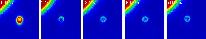
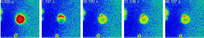
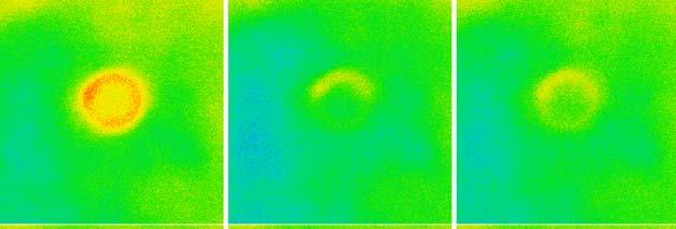
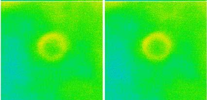
over time using live cell microscopy. The time post infection is indicated in the lower left corner.
 Title of file for HTML: Supplementary Information Description: Supplementary Figures and Supplementary Table Title of file for HTML: Supplementary Movie 1 Description: Fusion of NBs. BSR cells were infected
Figure S1. Figure S2. Figure S3 HB Anti-FSP27 (COOH-terminal peptide) Ab. Anti-GST-FSP27(45-127) Ab.
 Ab. Anti-GST-FSP27(45-127) Ab.") / 36B4 mrna ratio Figure S1 * 2. 1.6 1.2.8 *.4 control TNFα BRL49653 Figure S2 Su bw AT p iw Anti- (COOH-terminal peptide) Ab Blot : Anti-GST-(45-127) Ab β-actin Figure S3 HB2 HW AT BA T Figure S4 A TAG
/ 36B4 mrna ratio Figure S1 * 2. 1.6 1.2.8 *.4 control TNFα BRL49653 Figure S2 Su bw AT p iw Anti- (COOH-terminal peptide) Ab Blot : Anti-GST-(45-127) Ab β-actin Figure S3 HB2 HW AT BA T Figure S4 A TAG
Supplemental Online Material. The mouse embryonic fibroblast cell line #10 derived from β-arrestin1 -/- -β-arrestin2 -/-
 #1074683s 1 Supplemental Online Material Materials and Methods Cell lines and tissue culture The mouse embryonic fibroblast cell line #10 derived from β-arrestin1 -/- -β-arrestin2 -/- knock-out animals
SANTA CRUZ BIOTECHNOLOGY, INC.
 TECHNICAL SERVICE GUIDE: Western Blotting 2. What size bands were expected and what size bands were detected? 3. Was the blot blank or was a dark background or non-specific bands seen? 4. Did this same
SUPPLEMENTARY INFORMATION
 SUPPLEMENTARY INFORMATION Dynamic Phosphorylation of HP1 Regulates Mitotic Progression in Human Cells Supplementary Figures Supplementary Figure 1. NDR1 interacts with HP1. (a) Immunoprecipitation using
Confocal immunofluorescence microscopy
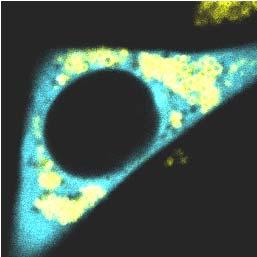
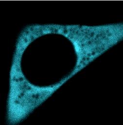
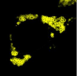
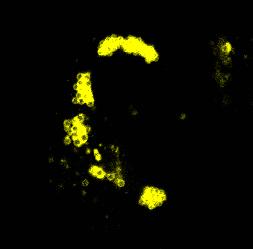
 Confocal immunofluorescence microscopy HL-6 and cells were cultured and cytospun onto glass slides. The cells were double immunofluorescence stained for Mt NPM1 and fibrillarin (nucleolar marker). Briefly,
The microtubule-associated tau protein has intrinsic acetyltransferase activity. Todd J. Cohen, Dave Friedmann, Andrew W. Hwang, Ronen Marmorstein and
 SUPPLEMENTARY INFORMATION: The microtubule-associated tau protein has intrinsic acetyltransferase activity Todd J. Cohen, Dave Friedmann, Andrew W. Hwang, Ronen Marmorstein and Virginia M.Y. Lee Cohen
Supplementary information to accompany: A novel role for the DNA repair gene Rad51 in Netrin-1 signalling
 Supplementary information to accompany: A novel role for the DNA repair gene Rad51 in Netrin-1 signalling Glendining KA 1, Markie D 2, Gardner RJM 4, Franz EA 3, Robertson SP 4, Jasoni CL 1 Supplementary
Supplemental Table 1 Gene Symbol FDR corrected p-value PLOD1 CSRP2 PFKP ADFP ADM C10orf10 GPI LOX PLEKHA2 WIPF1
 Supplemental Table 1 Gene Symbol FDR corrected p-value PLOD1 4.52E-18 PDK1 6.77E-18 CSRP2 4.42E-17 PFKP 1.23E-14 MSH2 3.79E-13 NARF_A 5.56E-13 ADFP 5.56E-13 FAM13A1 1.56E-12 FAM29A_A 1.22E-11 CA9 1.54E-11
Supplemental Information. A Versatile Tool for Live-Cell Imaging. and Super-Resolution Nanoscopy Studies. of HIV-1 Env Distribution and Mobility
 Cell Chemical Biology, Volume 24 Supplemental Information A Versatile Tool for Live-Cell Imaging and Super-Resolution Nanoscopy Studies of HIV-1 Env Distribution and Mobility Volkan Sakin, Janina Hanne,
Short hairpin RNA (shrna) against MMP14. Lentiviral plasmids containing shrna
 against MMP14. Lentiviral plasmids containing shrna") Supplemental Materials and Methods Short hairpin RNA (shrna) against MMP14. Lentiviral plasmids containing shrna (Mission shrna, Sigma) against mouse MMP14 were transfected into HEK293 cells using FuGene6
Supplemental Materials and Methods Short hairpin RNA (shrna) against MMP14. Lentiviral plasmids containing shrna (Mission shrna, Sigma) against mouse MMP14 were transfected into HEK293 cells using FuGene6
mcherry Monoclonal Antibody (16D7) Catalog Number M11217 Product data sheet
 Catalog Number M11217 Product data sheet") Website: thermofisher.com Customer Service (US): 1 800 955 6288 ext. 1 Technical Support (US): 1 800 955 6288 ext. 441 mcherry Monoclonal Antibody (16D7) Catalog Number M11217 Product data sheet Details
Website: thermofisher.com Customer Service (US): 1 800 955 6288 ext. 1 Technical Support (US): 1 800 955 6288 ext. 441 mcherry Monoclonal Antibody (16D7) Catalog Number M11217 Product data sheet Details
Supplementary Figure 1 Activated B cells are subdivided into three groups
 Supplementary Figure 1 Activated B cells are subdivided into three groups according to mitochondrial status (a) Flow cytometric analysis of mitochondrial status monitored by MitoTracker staining or differentiation
A subclass of HSP70s regulate development and abiotic stress responses in Arabidopsis thaliana
 1 2 3 4 5 6 7 8 9 10 11 12 13 14 15 16 17 18 19 20 21 Journal of Plant Research A subclass of HSP70s regulate development and abiotic stress responses in Arabidopsis thaliana Linna Leng 1 Qianqian Liang
supplementary information
 DOI: 1.138/ncb1839 a b Control 1 2 3 Control 1 2 3 Fbw7 Smad3 1 2 3 4 1 2 3 4 c d IGF-1 IGF-1Rβ IGF-1Rβ-P Control / 1 2 3 4 Real-time RT-PCR Relative quantity (IGF-1/ mrna) 2 1 IGF-1 1 2 3 4 Control /
Supplementary material to Alterations in the properties of the cell membrane due to glycosphingolipid accumulation in a model of Gaucher disease
 Supplementary material to Alterations in the properties of the cell membrane due to glycosphingolipid accumulation in a model of Gaucher disease Gyula Batta, Lilla Soltész, Tamás Kovács, Tamás Bozó, Zoltán
Nature Neuroscience: doi: /nn Supplementary Figure 1
 Supplementary Figure 1 PCR-genotyping of the three mouse models used in this study and controls for behavioral experiments after semi-chronic Pten inhibition. a-c. DNA from App/Psen1 (a), Pten tg (b) and
Species predicted to react based on 100% sequence homology: Chicken, Bovine, Dog.
 1 of 5 11/1/2013 10:25 PM Product Pathways - Jak/Stat Pathway Phospho-Stat3 (Tyr705) Antibody #9131 Have you tried your application using our XP monoclonal antibodies? Try products: 9145 PhosphoSitePlus
Immunofluorescence images of different core histones and different histone exchange assay.
 Molecular Cell, Volume 51 Supplemental Information Enhanced Chromatin Dynamics by FACT Promotes Transcriptional Restart after UV-Induced DNA Damage Christoffel Dinant, Giannis Ampatziadis-Michailidis,
Protocol for induction of expression and cell lysate production
 Protocol for induction of expression and cell lysate production AV-04 Doxycyclin induction and cell lysate 1.0 Introduction / Description This method is intended for the treatment of the previously transfected
Supplemental Figure 1. Mutation in NLA Causes Increased Pi Uptake Activity and
 Supplemental Figure 1. Mutation in NLA Causes Increased Pi Uptake Activity and PHT1 Protein Amounts. (A) Shoot morphology of 19-day-old nla mutants under Pi-sufficient conditions. (B) [ 33 P]Pi uptake
Thyroid peroxidase gene expression is induced by lipopolysaccharide involving Nuclear Factor (NF)-κB p65 subunit phosphorylation
-κB p65 subunit phosphorylation") 1 2 3 4 5 SUPPLEMENTAL DATA Thyroid peroxidase gene expression is induced by lipopolysaccharide involving Nuclear Factor (NF)-κB p65 subunit phosphorylation Magalí Nazar, Juan Pablo Nicola, María Laura
1 2 3 4 5 SUPPLEMENTAL DATA Thyroid peroxidase gene expression is induced by lipopolysaccharide involving Nuclear Factor (NF)-κB p65 subunit phosphorylation Magalí Nazar, Juan Pablo Nicola, María Laura
Segments of the obstructed intestinal loops were fixed in 4% paraformaldehyde
 Supplementary text Supplementary materials and methods Histopathological examination Segments of the obstructed intestinal loops were fixed in 4% paraformaldehyde (PFA) and embedded in paraffin wax with
IMMUNOPRECIPITATION TROUBLESHOOTING TIPS
 IMMUNOPRECIPITATION TROUBLESHOOTING TIPS Creative Diagnostics Abstract Immunoprecipitation (IP) is the technique of precipitating a protein antigen out of solution using an antibody that specifically binds
Supplemental Information. Loss of MicroRNA-7 Regulation Leads. to a-synuclein Accumulation and. Dopaminergic Neuronal Loss In Vivo
 YMTHE, Volume 25 Supplemental Information Loss of MicroRNA-7 Regulation Leads to a-synuclein Accumulation and Dopaminergic Neuronal Loss In Vivo Kirsty J. McMillan, Tracey K. Murray, Nora Bengoa-Vergniory,
NTM486-04, NTM174-04,
 Transfection of transformed human trabecular meshwork TM5, and primary human NTM210-05, NTM486-04, NTM174-04, and NTM153-00 cells with Metafectene Easy Adnan Dibas1A,C, Ming Jiang1A,C, Thomas Yorio1A,C.
Immunostaining Protocols
 Immunostaining Protocols Lula L. Hilenski, Ph.D. Director Microscopy in Medicine Core Emory University Variables in standard immunostaining protocol 2-step or indirect immunofluorescence 1. Substrate on
INOS. Colorimetric Cell-Based ELISA Kit. Catalog #: OKAG00807
 INOS Colorimetric Cell-Based ELISA Kit Catalog #: OKAG00807 Please read the provided manual entirely prior to use as suggested experimental protocols may have changed. Research Purposes Only. Not Intended
Kinase Reaction and Alkylation Protocol
 Kinase Reaction and Alkylation Protocol Protocol for the treatment of substrates prior to detection by Thiophosphate Ester antibodies This product is for research use only and is not intended for diagnostic
TECHNICAL BULLETIN. Color. Fig.1. Cell-Based protein phosphorylation procedure
 Cell-Based ELISA Kit for detecting phospho-stat3 (ptyr 705 ) in cultured cell lines adequate for 96 assays (1 96 well plate) Catalog Number RAB0444 Storage Temperature 20 C TECHNICAL BULLETIN Product Description
Custom Antibodies Services. GeneCust Europe. GeneCust Europe
 GeneCust Europe Laboratoire de Biotechnologie du Luxembourg S.A. 2 route de Remich L-5690 Ellange Luxembourg Tél. : +352 27620411 Fax : +352 27620412 Email : info@genecust.com Web : www.genecust.com Custom
Culture media, trypsin, penicillin and streptomycin were from Invitrogen (Breda, the Netherlands).
.") Methods Materials Culture media, trypsin, penicillin and streptomycin were from Invitrogen (Breda, the Netherlands). Bovine fibroblast growth factor (BFGF), thrombin, forskolin, IBMX, H-89, BAPTA-AM and
Methods Materials Culture media, trypsin, penicillin and streptomycin were from Invitrogen (Breda, the Netherlands). Bovine fibroblast growth factor (BFGF), thrombin, forskolin, IBMX, H-89, BAPTA-AM and
MSD Immuno-Dot-Blot Assays. A division of Meso Scale Diagnostics, LLC.
 MSD Immuno-Dot-Blot Assays Example: High Throughput Western Blots Replacements Traditional Western Blots High content Molecular weight and immunoreactivity Labor and protein intensive Inherently low throughput
RayBio Human NF-κB p65 Transcription Factor Activity Assay Kit
 RayBio Human NF-κB p65 Transcription Factor Activity Assay Kit Catalog #: TFEH-p65 User Manual Mar 13, 2017 3607 Parkway Lane, Suite 200 Norcross, GA 30092 Tel: 1-888-494-8555 (Toll Free) or 770-729-2992,
Technical Note Detection of post-immunoprecipitation proteins by Western blot using the Quick Western Kit IRDye 680RD
 Technical Note Detection of post-immunoprecipitation proteins by Western blot using the Quick Western Kit IRDye 680RD Developed for: Aerius, Odyssey Classic, Odyssey CLx and Odyssey Sa Imaging Systems
Figure S2. Response of mouse ES cells to GSK3 inhibition. Mentioned in discussion
 Stem Cell Reports, Volume 1 Supplemental Information Robust Self-Renewal of Rat Embryonic Stem Cells Requires Fine-Tuning of Glycogen Synthase Kinase-3 Inhibition Yaoyao Chen, Kathryn Blair, and Austin
Plasmid DNA transfection of SW480 human colorectal cancer cells with the Biontex K2 Transfection System
 Plasmid DNA transfection of human colorectal cancer cells with the Biontex K2 Transfection System Stephanie Hehlgans and Franz Rödel, Department of Radiotherapy and Oncology, Goethe- University Frankfurt,
IMMUNOPRECIPITATION (IP)
") 1 IMMUNOPRECIPITATION (IP) Overview and Technical Tips 2 CONTENTS 3 7 8 9 12 13 17 18 19 20 Introduction Factors Influencing IP General Protocol Modifications Of IP Protocols Troubleshooting Contact Us
1 IMMUNOPRECIPITATION (IP) Overview and Technical Tips 2 CONTENTS 3 7 8 9 12 13 17 18 19 20 Introduction Factors Influencing IP General Protocol Modifications Of IP Protocols Troubleshooting Contact Us
CERTIFICATE OF ANALYSIS
 5171 Wilfong Road Memphis, TN 38134 Telephone: 901-382-8716 Fax: 901-333-8223 Email: info@meridianlifescience.com www.meridianlifescience.com CERTIFICATE OF ANALYSIS Important Note: Centrifuge before opening
Nature Immunology: doi: /ni Supplementary Figure 1
 Supplementary Figure 1 BALB/c LYVE1-deficient mice exhibited reduced lymphatic trafficking of all DC subsets after oxazolone-induced sensitization. (a) Schematic overview of the mouse skin oxazolone contact
ENCODE RBP Antibody Characterization Guidelines
 ENCODE RBP Antibody Characterization Guidelines Approved on November 18, 2016 Background An integral part of the ENCODE Project is to characterize the antibodies used in the experiments. This document
by Neurobasal medium (supplemented with B27, 0.5mM glutamine, and 100 U/mL
 Supplementary Materials and methods Neuronal cultures and transfection The hippocampus was dissected from E8 rat embryos, dissociated, and neurons plated onto glass coverslips coated with poly-ornithine
1. Cross-linking and cell harvesting
 ChIP is a powerful tool that allows the specific matching of proteins or histone modifications to regions of the genome. Chromatin is isolated and antibodies to the antigen of interest are used to determine
ENCODE DCC Antibody Validation Document
 ENCODE DCC Antibody Validation Document Date of Submission 09/12/12 Name: Trupti Kawli Email: trupti@stanford.edu Lab Snyder Antibody Name: SREBP1 (sc-8984) Target: SREBP1 Company/ Source: Santa Cruz Biotechnology
Supplementary Information
 Supplementary Information Supplementary Figure 1. ZBTB20 expression in the developing DRG. ZBTB20 expression in the developing DRG was detected by immunohistochemistry using anti-zbtb20 antibody 9A10 on
Supplementary Figure 1. Isolation of GFPHigh cells.
 Supplementary Figure 1. Isolation of GFP High cells. (A) Schematic diagram of cell isolation based on Wnt signaling activity. Colorectal cancer (CRC) cell lines were stably transduced with lentivirus encoding
Basic Fluorescence Microscopy and Sample Preparation. Eva Wegel
 Basic Fluorescence Microscopy and Sample Preparation Eva Wegel eva.wegel@bioch.ox.ac.uk Visible Light 390 700 nm visible to the human eye White light is split into its components through a prism Reason:
mcherry Polyclonal Antibody Catalog Number PA Product data sheet
 Website: thermofisher.com Customer Service (US): 1 800 955 6288 ext. 1 Technical Support (US): 1 800 955 6288 ext. 441 mcherry Polyclonal Antibody Catalog Number PA5-34974 Product data sheet Details Size
SensoLyte Anti-alpha-Synuclein Quantitative ELISA Kit (Human/Mouse/Rat) *Colorimetric*
 *Colorimetric*") Catalog # Kit Size SensoLyte Anti-alpha-Synuclein Quantitative ELISA Kit (Human/Mouse/Rat) *Colorimetric* AS-55550 One 96-well strip plate This kit is optimized to detect human/mouse/rat alpha-synuclein
Catalog # Kit Size SensoLyte Anti-alpha-Synuclein Quantitative ELISA Kit (Human/Mouse/Rat) *Colorimetric* AS-55550 One 96-well strip plate This kit is optimized to detect human/mouse/rat alpha-synuclein
Supplemental Data. LMO4 Controls the Balance between Excitatory. and Inhibitory Spinal V2 Interneurons
 Neuron, Volume 61 Supplemental Data LMO4 Controls the Balance between Excitatory and Inhibitory Spinal V2 Interneurons Kaumudi Joshi, Seunghee Lee, Bora Lee, Jae W. Lee, and Soo-Kyung Lee Supplemental
Cycles of vascular plexus formation within the nephrogenic zone of the developing mouse kidney
 1 Supplementary text and data for: 2 3 4 5 Cycles of vascular plexus formation within the nephrogenic zone of the developing mouse kidney Authors: David A. D. Munro 1*, Peter Hohenstein 2, and Jamie A.
This Document Contains:
 This Document Contains: 1. In-Cell Western Protocol II. Cell Seeding and Stimulation Supplemental Protocol III. Complete Assay Example: Detailing the Seeding, Stimulation and Detection of the A431 Cellular
NAME TA SEC Problem Set 4 FRIDAY October 15, Answers to this problem set must be inserted into the box outside
 MIT Biology Department 7.012: Introductory Biology - Fall 2004 Instructors: Professor Eric Lander, Professor Robert A. Weinberg, Dr. Claudette Gardel NAME TA SEC 7.012 Problem Set 4 FRIDAY October 15,
CFTR 2 is a phosphorylation-activated Cl channel expressed at the apical membranes of epithelial cells that are involved in salt secretion or
 THE JOURNAL OF BIOLOGICAL CHEMISTRY VOL. 281, NO. 16, pp. 11312 11321, April 21, 2006 2006 by The American Society for Biochemistry and Molecular Biology, Inc. Printed in the U.S.A. Cysteine String Protein
Discovery and Humanization of Novel High Affinity Neutralizing Monoclonal Antibodies to Human IL-17A
 Discovery and Humanization of Novel High Affinity Neutralizing Monoclonal Antibodies to Human IL-17A Contacts: Marty Simonetti martysimonetti@gmail.com Kirby Alton kirby.alton@abeomecorp.com Rick Shimkets
EGFR (Phospho-Ser695)
") Assay Biotechnology Company www.assaybiotech.com Tel: 1-877-883-7988 Fax: 1-877-610-9758 EGFR (Phospho-Ser695) Colorimetric Cell-Based ELISA Kit Catalog #: OKAG02090 Please read the provided manual entirely
Assay Biotechnology Company www.assaybiotech.com Tel: 1-877-883-7988 Fax: 1-877-610-9758 EGFR (Phospho-Ser695) Colorimetric Cell-Based ELISA Kit Catalog #: OKAG02090 Please read the provided manual entirely
Supplementary Figures Montero et al._supplementary Figure 1
 Montero et al_suppl. Info 1 Supplementary Figures Montero et al._supplementary Figure 1 Montero et al_suppl. Info 2 Supplementary Figure 1. Transcripts arising from the structurally conserved subtelomeres
Figure S1. gfp tola and pal mcherry can complement deletion mutants of tola and pal respectively. (A)When strain LS4522 was grown in the presence of
When strain LS4522 was grown in the presence of") Figure S1. gfp tola and pal mcherry can complement deletion mutants of tola and pal respectively. (A)When strain LS4522 was grown in the presence of xylose, inducing expression of gfp-tola, localization
Figure S1. gfp tola and pal mcherry can complement deletion mutants of tola and pal respectively. (A)When strain LS4522 was grown in the presence of xylose, inducing expression of gfp-tola, localization
Please read manual carefully before starting experiment
 RayBio Cell-Based Phosphorylation ELISA Kit - Preliminary For the semi-quantitative detection of both phosphorylated and pan human, mouse or rat proteins in adherent whole cell lines. User Manual (Revised
How to run Alpha assay: How to setup an Alpha assay Make your own assay!
 How to run Alpha assay: How to setup an Alpha assay Make your own assay! 1 2009 PerkinElmer AlphaLISA kits - recommendations before starting the assay Samples: Phenol red and hemoglobin: choose AlphaLISA
!! PLEASE READ BEFORE USE!!
 In situ Proximity Ligation Assay protocols!! PLEASE READ BEFORE USE!! The test protocol is a guideline, user need to determine their optimal experimental condition for best performance. The following protocol
Nature Immunology: doi: /ni Supplementary Figure 1. Zranb1 gene targeting.
 Supplementary Figure 1 Zranb1 gene targeting. (a) Schematic picture of Zranb1 gene targeting using an FRT-LoxP vector, showing the first 6 exons of Zranb1 gene (exons 7-9 are not shown). Targeted mice
TECHNICAL BULLETIN. MEK Activity Assay Kit. Product Code CS0490 Storage Temperature 20 C
 MEK Activity Assay Kit Product Code CS0490 Storage Temperature 20 C TECHNICAL BULLETIN Product Description The MAP kinase kinases (MAPKK, mitogen-activated protein kinase kinase, also termed MEK) are a
In-Cell Western Kits I and II
 Odyssey and Aerius Infrared Imaging Systems In-Cell Western Assay Kits I and II Published November, 2006. The most recent version of this protocol is posted at http://biosupport.licor.com/protocols.jsp
Supplemental Material for
 Supplemental Material for TXI TELNGIECTSI MUTTED (TM)-MEDITED DN DMGE RESPONSE IN OXIDTIVE STRESS-INDUCED VSCULR ENDOTHELIL CELL SENESCENCE Hong Zhan 1, Toru Suzuki 1,2, Kenichi izawa 1, Kiyoshi Miyagawa,
Supplementary Figures and Legends
 Supplementary Figures and Legends Figure S1. Tests of the optical alignment and focal properties of the confocal microscope. (A) Images of the optical cross-section of fluorescent microspheres differing
*Corresponding author. Tel: ;
 1 SUPPLEMENTARY DATA 2 3 4 5 6 7 8 9 10 11 Integrin 2 1 in nonactivated conformation can induce focal adhesion kinase signaling Maria Salmela 1, Johanna Jokinen 1,2, Silja Tiitta 1, Pekka Rappu 1, Holland
ab GFP ELISA Kit Instructions for Use For the quantitative measurement of GFP protein expression
 ab117992 GFP ELISA Kit Instructions for Use For the quantitative measurement of GFP protein expression This product is for research use only and is not for diagnostic use. intended www.abcam.com Table
phab Amine and Thiol Reactive Dyes for Antibody Internalization Studies Nidhi Nath, Ph.D. Group Leader, Protein Analysis Promega Corporation
 phab Amine and Thiol Reactive Dyes for Antibody Internalization Studies Nidhi Nath, Ph.D. Group Leader, Protein Analysis 1 Outline 1. phab Dyes 2. Protocols for conjugating phab Dyes to antibodies 3. Applications:
RayBio Human, Mouse and Rat Phospho-STAT3 (Tyr705) ELISA Kit
 ELISA Kit") RayBio Human, Mouse and Rat Phospho-STAT3 (Tyr705) ELISA Kit Catalog #: PEL-Stat3-Y705 User Manual Last revised August 10, 2016 Caution: Extraordinarily useful information enclosed ISO 13485 Certified
RayBio Human, Mouse and Rat Phospho-STAT3 (Tyr705) ELISA Kit Catalog #: PEL-Stat3-Y705 User Manual Last revised August 10, 2016 Caution: Extraordinarily useful information enclosed ISO 13485 Certified
Supplemental Movie Legend.
 Supplemental Movie Legend. Transfected T cells were dropped onto SEE superantigen-pulsed Raji B cells (approximate location indicated by circle). Maximum-intensity projections from Z-stacks (17 slices,
Cell culture. HeLa cells were cultured as monolayers in Dulbecco s Minimal Essential
 Supporting Online Material Materials and methods Cell culture. HeLa cells were cultured as monolayers in Dulbecco s Minimal Essential Medium (Gibco BRL, Invitrogen Corporation, Carlsbad, CA, USA), supplemented
SUPPLEMENTARY INFORMATION
 SUPPLEMENTARY INFORMATION Legends for Supplementary Tables. Supplementary Table 1. An excel file containing primary screen data. Worksheet 1, Normalized quantification data from a duplicated screen: valid
Automated Protocol for ANTI-FLAG High Sensitivity, M2 coated 96-well plate Using the Sciclone ALH 3000 Workstation (Caliper Life Sciences)
") Automated Protocol for ANTI-FLAG High Sensitivity, M2 coated 96-well plate Using the Sciclone ALH 3000 Workstation (Caliper Life Sciences) ANTI-FLAG High Sensitivity, M2 coated 96-well plate P 2983 Automation
Automated Protocol for ANTI-FLAG High Sensitivity, M2 coated 96-well plate Using the Sciclone ALH 3000 Workstation (Caliper Life Sciences) ANTI-FLAG High Sensitivity, M2 coated 96-well plate P 2983 Automation
ab Hypoxic Response Human Flow Cytometry Kit
 ab126585 Hypoxic Response Human Flow Cytometry Kit Instructions for Use For measuring protein levels by flow cytometry: hypoxia-inducible factor 1-alpha (HIF1A) and BCL2/adenovirus E1B 19 kda proteininteracting
Heme utilization in the Caenorhabditis elegans hypodermal cells is facilitated by hemeresponsive
 Supplemental Data Heme utilization in the Caenorhabditis elegans hypodermal cells is facilitated by hemeresponsive gene-2 Caiyong Chen 1, Tamika K. Samuel 1, Michael Krause 2, Harry A. Dailey 3, and Iqbal
RayBio Phospho- Stat 3 (Tyr705) ELISA Kit
 ELISA Kit") RayBio Phospho- Stat 3 (Tyr705) ELISA Kit For Measuring Phosphorylated Stat3 (Tyr705) in Human, Mouse and Rat Cell Lysates User Manual (Revised Mar 1, 2012) RayBio Stat3 (Tyr705) ELISA Kit Protocol (Cat#:
RayBio Phospho- Stat 3 (Tyr705) ELISA Kit For Measuring Phosphorylated Stat3 (Tyr705) in Human, Mouse and Rat Cell Lysates User Manual (Revised Mar 1, 2012) RayBio Stat3 (Tyr705) ELISA Kit Protocol (Cat#:
Partha Roy
 Fluorescence microscopy http://micro.magnet.fsu.edu/primer/index.html Partha Roy 1 Lecture Outline Definition of fluorescence Common fluorescent reagents Construction ti of a fluorescence microscope Optical
Endoplasmic Reticulum Stress Induction of the Grp78/BiP Promoter: Activating Mechanisms Mediated by YY1 and Its Interactive Chromatin Modifiers
 MOLECULAR AND CELLULAR BIOLOGY, June 2005, p. 4529 4540 Vol. 25, No. 11 0270-7306/05/$08.00 0 doi:10.1128/mcb.25.11.4529 4540.2005 Copyright 2005, American Society for Microbiology. All Rights Reserved.
Identification of Microprotein-Protein Interactions via APEX Tagging
 Supporting Information Identification of Microprotein-Protein Interactions via APEX Tagging Qian Chu, Annie Rathore,, Jolene K. Diedrich,, Cynthia J. Donaldson, John R. Yates III, and Alan Saghatelian
RayBio Phospho- Akt (Ser473) ELISA Kit
 ELISA Kit") RayBio Phospho- Akt (Ser473) ELISA Kit For Measuring Phosphorylated Akt (Ser473) in Human, Mouse and Rat Cell Lysates User Manual (Revised Mar 1, 2012) RayBio Akt (Ser473) ELISA Kit Protocol (Cat#: PEL-Akt-S473-001)
RayBio Phospho- Akt (Ser473) ELISA Kit For Measuring Phosphorylated Akt (Ser473) in Human, Mouse and Rat Cell Lysates User Manual (Revised Mar 1, 2012) RayBio Akt (Ser473) ELISA Kit Protocol (Cat#: PEL-Akt-S473-001)
RayBio Human, Mouse and Rat Phospho-STAT3 (Tyr705) and Total STAT3 ELISA Kit
 and Total STAT3 ELISA Kit") RayBio Human, Mouse and Rat Phospho-STAT3 (Tyr705) and Total STAT3 ELISA Kit Catalog #: PEL-Stat3-Y705-T User Manual Last revised October 10, 2017 Caution: Extraordinarily useful information enclosed ISO
RayBio Human, Mouse and Rat Phospho-STAT3 (Tyr705) and Total STAT3 ELISA Kit Catalog #: PEL-Stat3-Y705-T User Manual Last revised October 10, 2017 Caution: Extraordinarily useful information enclosed ISO
Supplemental Data. Regulating Gene Expression. through RNA Nuclear Retention
 Supplemental Data Regulating Gene Expression through RNA Nuclear Retention Kannanganattu V. Prasanth, Supriya G. Prasanth, Zhenyu Xuan, Stephen Hearn, Susan M. Freier, C. Frank Bennett, Michael Q. Zhang,
The Biotechnology Education Company. Quantitative ELISA. Storage: See Page 3 for specific storage instructions EXPERIMENT OBJECTIVE:
 The Biotechnology Education Company Revised and Updated Quantitative ELISA Storage: See Page 3 for specific storage instructions EXPERIMENT OBJECTIVE: EDVO-Kit # 278 The objective of this experiment is
* This work was supported, in whole or in part, by National Institutes of Health Grants HL56850 and AG S EXPERIMENTAL PROCEDURES
 THE JOURNAL OF BIOLOGICAL CHEMISTRY VOL. 285, NO. 10, pp. 6937 6951, March 5, 2010 2010 by The American Society for Biochemistry and Molecular Biology, Inc. Printed in the U.S.A. p66 shc Inhibits Insulin-like
mir-24-mediated down-regulation of H2AX suppresses DNA repair
 Supplemental Online Material mir-24-mediated down-regulation of H2AX suppresses DNA repair in terminally differentiated blood cells Ashish Lal 1,4, Yunfeng Pan 2,4, Francisco Navarro 1,4, Derek M. Dykxhoorn
To isolate single GNS 144 cell clones, cells were plated at a density of 1cell/well
 Supplemental Information: Supplemental Methods: Cell culture To isolate single GNS 144 cell clones, cells were plated at a density of 1cell/well in 96 well Primaria plates in GNS media and incubated at
QImaging Camera Application Notes Multicolor Immunofluorescence Imaging
 QImaging Camera Application Notes Multicolor Immunofluorescence Imaging In order to image localization of intracellular proteins with high specificity, it is frequently necessary to multiplex antibody
Assays for studying mitochondrial health and function
 APPLICATION NOTE Fluorescence labeling and detection Assays for studying mitochondrial health and function Introduction Mitochondria play a critical role in maintaining normal cellular activities. Mitochondria
Manuscript Skeletal muscle Heat shock protein 60 increases after endurance training and induces peroxisome proliferator-activated
 Supplementary informations Manuscript Skeletal muscle Heat shock protein 60 increases after endurance training and induces peroxisome proliferator-activated receptor gamma coactivator 1 α1 expression Rosario
TECHNICAL BULLETIN. Color. Fig.1. Cell-Based protein phosphorylation procedure
 Cell-Based ELISA Sampler Kit for detecting phospho-erk1/2 (pthr 202 /ptyr 204 ), phospho-jnk (pthr 183 /ptyr 185 ), and phospho-p38 MAPK (pthr 180 /ptyr 182 ) in cultured cell lines adequate for 192 assays
Supplemental information
 Supplemental information - Control samples (200 subjects) - Immunohistochemistry of rat brain - Immunocytochemistry on neuronal cultures - Immunocompetition assay - Immunoprecipitation - Immunocytochemistry
Isolation, culture, and transfection of primary mammary epithelial organoids
 Supplementary Experimental Procedures Isolation, culture, and transfection of primary mammary epithelial organoids Primary mammary epithelial organoids were prepared from 8-week-old CD1 mice (Charles River)
Supporting Information
 Supporting Information Shao et al. 10.1073/pnas.1504837112 SI Materials and Methods Immunofluorescence and Immunoblotting. For immunofluorescence, cells were fixed with 4% paraformaldehyde and permeabilized
Supplemental Information for:
 Supplemental Information for: Antibody-induced dimerization of FGFR1 promotes receptor endocytosis independently of its kinase activity Łukasz Opaliński*, Aleksandra Sokołowska-Wędzina, Martyna Szczepara,
Supplementary Fig. 1 related to Fig. 1 Clinical relevance of lncrna candidate
 Supplementary Figure Legends Supplementary Fig. 1 related to Fig. 1 Clinical relevance of lncrna candidate BC041951 in gastric cancer. (A) The flow chart for selected candidate lncrnas in 660 up-regulated
FORENSIC SEROLOGY. Chapter PRENTICE HALL 2008 Pearson Education, Inc. Upper Saddle River, NJ 07458
 Chapter 8 FORENSIC SEROLOGY 8-1 Nature of Blood The word blood refers to a highly complex mixture of cells, enzymes, proteins, and inorganic substances. Plasma, which is the fluid portion of blood, is
ab Optiblot Fluorescent Western Blot Kit
 ab133410 Optiblot Fluorescent Western Blot Kit Instructions for Use For quantitative, multi-color fluorescent Western blotting. This product is for research use only and is not intended for diagnostic