Chemiluminescence Detection. Using UVP s ChemiDoc-It System
|
|
|
- Kristopher Garrison
- 6 years ago
- Views:
Transcription

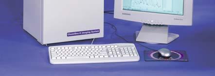
1 Chemiluminescence Detection Using UVP s ChemiDoc-It System

2 Protein Detection Agents Chemiluminescence Fluorescence Colorimetric Detection Radioisotopic Detection

3 Chemiluminescence Generation of light through sample s own light source by chemical reaction, without involvement of heat Chemiluminescence is widely used for protein blot detection Bioluminescence is chemiluminescence that takes place inside a living organism For purposes of limiting the discussion on chemiluminescence applications using the chemi doc-it, we will only cover chemiluminescent imaging of protein blots Its basic strength is its simplicity as a detection tool virtually no background or autofluorescence of samples to worry about; offers the same sensitivity as film, is less expensive than film and offers the speed which is a big drawback for film.


4 Fluorescence Absorption and emission of light using light energy When used in protein detection, it involves the use of fluorescent stains and transilluminated or epi illuminated light sources Just like chemiluminescence, fluorescence can be used for detecting signal inside a living organism (see ibox) Basic strength is the simplicity and speed of imaging and the richness of color; major weakness is the high background or autofluorescence of samples that can be addressed by proper filtering.

5 Colorimetric Detection Colorimetric or visible light imaging (from 480 nm to about 700 nm) using white light Generally makes use of stains such as coomassie blue and silver stain in imaging protein samples as Elisa/Blots. Uses white light to illuminate the samples
 End result is usually an X-Ray film which is")

6 Radioisotopic Imaging Use of radioisotopes (32p, 35s etc.) End result is usually an X-Ray film which is then read using white light illumination or a scanner Safety issues, cost and speed are typically its downside


 Southern Blots")
7 Chemiluminescence Applications For Proteins & DNA/RNA Western Blots (proteins) Dot/Slot Blots (proteins & DNA/RNA) Northern Blots (RNA) Southern Blots (DNA) Western Blot Southern Blot Dot Blot
8 Chemiluminescent Substrates Commonly Used Substrates: ECL Pierce Rockland FemtoMax(recommended substrate with femtogram level sensitivity) Compared with radioisotopes, chemi substrates will have the same sensitivity (down to femtogram level) & faster detection; these do not have any health and hazardous issues associated with the useand disposal of radioisotopes.
9 Chemiluminescent CCD Imaging Deeply cooled cameras with same sensitivity, higher dynamic range, higher linearity than film are currently available Super light-tight darkroom, necessary for low light imaging should also be a part of the system Capture and Analysis software to perform sequential images at same or varying exposure times as well as powerful analysis features should likewise be part of the imaging system
10 Chemiluminescence Detection Using UVP s ChemiDoc-It System Deeply Cooled, 2.0 megapixel monochrome Chemi 410 camera with highly sensitive optics & high quantum efficiency Light tight, super dark and compact darkroom Adjustable chemi tray for varying heights to obtain desired field of view VisionWorks Software For acquisition and analysis of chemi images
11 VisionWorks LS Software

12 Image Capture Straightforward Capabilities

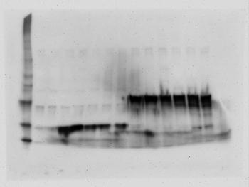
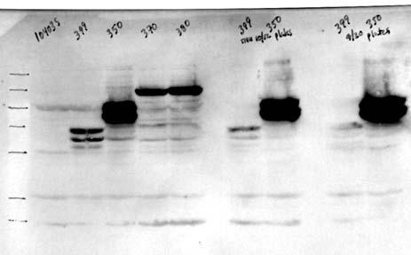
13 Image Capture Sequential Capture of Chemi Western Blot

14 Imaging Tools Using Invert & Histogram Tools to Optimize Chemi Western Blots

15 Imaging Tools Using Invert & Histogram Tools to Optimize Chemi Western Blots

16 Imaging Tools Recording & Saving Macros

17 Analysis Tools Intuitive, Easy to Use Analysis Tools Area Density Band Density and MW Calculations Scan Lanes for Purity

18 Area Density For Measuring Intensity


19 ID Analysis For Band Density & Molecular Weight Calculations

20 1D Analysis To Scan Lane for Purity

21 Analysis Results Exportable to Excel
Azure cseries. A new way to see the light. c600 c500 c400 c300 c200
 Azure cseries A new way to see the light. c6 c5 c4 c3 c2 Infrared Laser Excitation for Quantitative Western Blot Imaging in the NIR Improve Your Data Quality Imaging with infrared dyes offers signal stability
Azure cseries. A new way to see the light. c600 c500 c400 c300
 Azure cseries A new way to see the light. c600 c500 c400 c300 Infrared Laser Excitation for Quantitative Western Blot Imaging in the NIR Improve Your Data Quality Imaging with infrared dyes offers signal
Western Blotting. The complete range by biostep. It s all about Bio-Imaging.
 Western Blotting The complete range by biostep It s all about Bio-Imaging. www.biostep.de The way to Western Blots The perfect workflow with biostep instruments Lysis of samples in the homogenizer Bullet
Innovations To Meet Your Needs
 Innovations To Meet Your Needs Cooled CCD Camera 1340 x 1037 pixel resolution for greatest image quality 12-bit precision provides 3 orders of linear dynamic range Windows and Power Macintosh Software
MF-ChemiBIS. Today s most comprehensive solution for your bio-imaging needs and applications. Documenting Nature
 MF-ChemiBIS Today s most comprehensive solution for your bio-imaging needs and applications Documenting Nature MF-ChemiBIS Excellence in bio-imaging The DNR Advantage As pioneers in bio-imaging technologies
End of Year. Special Offers
 End of Year Special Offers 2018 Cell Biology TC20 Automated Cell Counter Automated and precise cell counts and viability assessment within 30 seconds Compatible with a broad range of cell sizes and types
GeneTools the Essential Software For Accurate DNA/RNA Gel and Blot Analysis
 Technical Note 27 GeneTools the Essential Software For Accurate DNA/RNA Gel and Blot Analysis Introduction Capturing an image of a DNA/RNA gel or blot with an image analysis system is only the start of
Azure Biosystems Western Blotting Workflow
 Azure Biosystems Western Blotting Workflow PROBE PLAN SEPARATE ANALYZE VISUALIZE PLAN Plan your experiment and choose your detection method Chemiluminescent Western Blotting The most common method for
Sapphire. Biomolecular Imager THE NEXT GENERATION OF LASER-BASED IMAGING
 Sapphire Biomolecular Imager THE NEXT GENERATION OF LASER-BASED IMAGING Breakthrough image capture and analysis The Sapphire Biomolecular Imager is a next generation laser scanning system that provides
OCTOPLUS QPLEX FLUORESCENCE IMAGER. for fast & powerful fast 2D Gel image acquisition
 OCTOPLUS QPLEX FLUORESCENCE IMAGER for fast & powerful fast 2D Gel image acquisition Octoplus QPLEX Fluorescence Imager The new Octoplus QPLEX fluorescence imager sets a novel standard fluorescence 2D
Instant-Bands Protein Sample Loading Buffer for SDS-PAGE. User s Manual. View Protein Bands in an SDS Gel. Instantly. EZBiolab.
 Instant-Bands Protein Sample Loading Buffer for SDS-PAGE User s Manual View Protein Bands in an SDS Gel Instantly www.ezbiolab.com 2 Instant-Bands User s Manual Table of Contents Introduction 3 Storage
Dolphin-Chemi Plus. Aim: To visualise and evaluate the performance of chemiluminescent immunoblots using Wealtec s Dolphin-Chemi plus image system
 Application Note 03 Dolphin-Chemi plus 8/22/2007 Dolphin-Chemi Plus Aim: To visualise and evaluate the performance of chemiluminescent immunoblots using Wealtec s Dolphin-Chemi plus image system INTRODUCTION
ChemiDoc MP Imaging System
 Imaging ChemiDoc MP Imaging System Confidence in every step of your imaging workflow. ChemiDoc MP Imaging System Because results always matter. Bring a new level of capability and efficiency to your experiments
Sale. Bio-Rad. See inside for an array of offers on: The
 Life Science Group The Bio-Rad 2013 Sale See inside for an array of offers on: Electrophoresis and Blotting Imaging Systems Thermal Cyclers Cell Counting PCR Reagents and Plastics Electroporation Immunoassay
NEWTON 7.0 BIOLUMINESCENCE & FLUORESCENCE IMAGING IN VIVO - IN VITRO IMAGING
 NEWTON 7.0 BIOLUMINESCENCE & FLUORESCENCE IMAGING IN VIVO - IN VITRO IMAGING The NEWTON s protocol driven image acquisition is as quick as it is intuitive: adjust your exposure, save, print or quantify.
NEWTON 7.0 BIOLUMINESCENCE & FLUORESCENCE IMAGING IN VIVO - IN VITRO IMAGING
 NEWTON 7.0 BIOLUMINESCENCE & FLUORESCENCE IMAGING IN VIVO - IN VITRO IMAGING SMART IMAGING SYSTEM The NEWTON 7.0 system combines high sensitivity with advanced animal-handling features and userfriendly
Sapphire. Biomolecular Imager THE NEXT GENERATION OF LASER-BASED IMAGING
 Sapphire Biomolecular Imager THE NEXT GENERATION OF LASER-BASED IMAGING Breakthrough image capture and analysis The Sapphire Biomolecular Imager is a next generation laser scanning system that provides
Wireless Gel Doc. Pacific Image Electronics Co. Booth #: Hall S1, Kevin Tseng (GM)
") Wireless Gel Doc. Pacific Image Electronics Co. Booth #: Hall S1, 1029 Kevin Tseng (GM) Pacific Image Electronics 20 years young company Image solutions Public company in Taiwan No.1 film scanner worldwide
Wireless Gel Doc. Pacific Image Electronics Co. Booth #: Hall S1, 1029 Kevin Tseng (GM) Pacific Image Electronics 20 years young company Image solutions Public company in Taiwan No.1 film scanner worldwide
Introduction. Important Product Information. Number Description. Table of Contents
 Super Signal Western ECL Substrate Number Description Super Signal Western ECL Substrate, Sufficient for 1000cm2 of membrane Kit Contents: Super Signal Western Solution A, 50mL Super Signal Western Solution
Reach greater highs. (and lows ) with new protein ladder choices. Fermentas now sold as. Thermo Scientific
 with new protein ladder choices. Fermentas now sold as. Thermo Scientific") Introducing Thermo Scientific Pierce Prestained and Unstained s for easy protein molecular weight determination. Fermentas now sold as Thermo Scientific Reach greater highs (and lows ) with new protein
Introducing Thermo Scientific Pierce Prestained and Unstained s for easy protein molecular weight determination. Fermentas now sold as Thermo Scientific Reach greater highs (and lows ) with new protein
Clarity and Clarity Max Western ECL Substrates
 Imaging and Max Western Substrates The Clear Choice in Chemiluminescent Western Blot Detection and Max Western Substrates Get. The Clear Choice in Chemiluminescent Western Blot Detection bio-rad.com/web/substrate
ctrophoresi April June % 30% 30% 40% Gel Documentation & IQ/OQ Western Blotting Fluorescence Dyes Protein Electrophoresis
 Acrylamide Solutions Constant quality for constant results 10687.02 Acrylamide/Bis Solution :1 (30 % w/v), 3.3 % C 10688.02 Acrylamide/Bis Solution 37.5:1 (30 % w/v), 2.6 % C 10680.02 Acrylamide/Bis Solution
NEWTON 7.0 BIOLUMINESCENCE & FLUORESCENCE IMAGING IN VIVO - IN VITRO IMAGING
 NEWTON 7.0 BIOLUMINESCENCE & FLUORESCENCE IMAGING IN VIVO - IN VITRO IMAGING The NEWTON s protocol driven image acquisition is as quick as it is intuitive: adjust your exposure, save, print or quantify.
SMART PROTEIN LAYERS. Quantitative and standardized protein gel and Western blot analysis
 SMART PROTEIN LAYERS Quantitative and standardized protein gel and Western blot analysis The challenge There is an increasing demand for quantitative, sensitive and reliable 1D gel and Western blot (WB)
Molecular Imager ChemiDoc XRS+ Systems
 Imaging Molecular Imager ChemiDoc XRS+ Systems Easy to Use, Reproducible, and Sensitive Solution for Western Blot and Gel Image Analysis The Trusted Leader in Easy to Use Systems for Reproducible Results
Focal Points. Application Note FP-153. Next Generation Gel Imaging with GelRed and GelGreen Dyes and GelDoc-It Imaging System
 Focal Points Application Note FP-153 MIDSCI 800.227.9997 636.227.9997 Gel Doc Equipment Next Generation Gel Imaging with GelRed and GelGreen Dyes and GelDoc-It Imaging System Introduction By using state-of-the-art
ChemiDoc Touch Imaging System
 Western Blotting ChemiDoc Touch Imaging System Sensitivity in detection, power in quantitation ChemiDoc Touch Imaging System Best-in-class performance Superior to film in signal-to-noise ratio Equal to
Electrophoresis and transfer
 Electrophoresis and transfer Electrophoresis Cation = positively charged ion, it moves toward the cathode (-) Anion = negatively charged ion, it moves toward the anode (+) Amphoteric substance = can have
Imaging. ChemiDoc XRS+ Imager. Effortless and Accurate Imaging and Analysis for Gels and Blots
 Imaging ChemiDoc XRS+ Imager Effortless and Accurate Imaging and Analysis for Gels and Blots Solve Complex Biological Questions with Simple and Reliable Imaging For more than two decades, Molecular Imager
About us. Cyanagen s.r.l. has a certified Quality System ISO QUALITY CERTIFIED. Green Stain DNA Loading Dye Rev00
 About us Cyanagen is a biotech company located in Bologna, dedicated to research, development and production of reagents for molecular diagnostic since 2003 and one of the leading companies in the field
GREEN STAIN DNA LOADING DYE DNA LOADING DYE CONTAINING FLUORESCENT STAIN FOR NUCLEIC ACID DETECTION IN GELS
 GREEN STAIN DNA LOADING DYE DNA LOADING DYE CONTAINING FLUORESCENT STAIN FOR NUCLEIC ACID DETECTION IN GELS About us Cyanagen is a biotech company located in Bologna, dedicated to research, development
ibox Explorer TM Imaging Microscope
 ibox Explorer TM Imaging Microscope Visible to NIR In Vivo Imaging for Macro to Micro Detection of Fluorescent Markers in Small Animals Capture images with the high sensitivity, cooled CCD camera and optics,
Notes to accompany the slidecast on theory of SDS PAGE and Western blotting
 S317 Biological science: from genes to species Notes to accompany the slidecast on theory of SDS PAGE and Western blotting SDS PAGE SDS PAGE is a standard technique for determining the molecular size of
BCH 462. Western Blot
 BCH 462 Western Blot Blotting Immunoassay: A test that uses antibody and antigen complexes [immuno-complexes] as a means of generating measurable results. Antigens [Ag]: A substance that when introduced
Amersham Western blotting
 Amersham Western blotting Selection guide gelifesciences.com Innovative solutions for your Western blotting Since 1990 when we first launched Amersham ECL, we have continued our commitment to providing
The world s first luminescent color development marker pen
 Reagent LuminolPenTM HRP system LuminolPenTM HRP system can provide the best way using luminescent to indicate target(s) while performing western blotting followed by luminescent color development..characteristics
PDIP46 (DNA polymerase δ interacting protein 46) is an activating factor for human DNA polymerase δ
 is an activating factor for human DNA polymerase δ") PDIP46 (DNA polymerase δ interacting protein 46) is an activating factor for human DNA polymerase δ Supplementary Material Figure S1. PDIP46 is associated with Pol isolated by immunoaffinity chromatography.
PDIP46 (DNA polymerase δ interacting protein 46) is an activating factor for human DNA polymerase δ Supplementary Material Figure S1. PDIP46 is associated with Pol isolated by immunoaffinity chromatography.
Optiblot ECL Ultra Detect Kit (1.2pg 2ng)
") ab133409 Optiblot ECL Ultra Detect Kit (1.2pg 2ng) Instructions for Use For enhanced chemiluminescent Western blotting. This product is for research use only and is not intended for diagnostic use. 1 Table
ab133409 Optiblot ECL Ultra Detect Kit (1.2pg 2ng) Instructions for Use For enhanced chemiluminescent Western blotting. This product is for research use only and is not intended for diagnostic use. 1 Table
d. Bands hidden by overexposure with chemiluminescence become clear when imaged with Odyssey.
 Core Equipment ID: 15922 Description: LI COR Biosciences, Odyssey Infrared Imaging System Room: B446 (Molecular & Biochemical Core) Champion: 1.0 Purpose Standardize the process for control, maintenance,
Amersham Western blotting
 Amersham Western blotting Selection guide www.gelifesciences.com Innovative solutions designed to meet your specific protein analysis needs Since 1990 when we first launched Amersham ECL we have continued
RASAQ NURUDEEN OLAJIDE
 RASAQ NURUDEEN OLAJIDE LECTURE CONTENT INTRODUCTION SOUTHERN BLOTTING WESTERN BLOTTING DNA PROFILING (DNA FINGERPRINTING) PARENTAL TESTING PROCEDURES INTRODUCTION Then the Lord said to Cain, Where is your
LED Illuminator VISIRAYS
 LED Illuminator VISIRAYS BLUE/ GREEN SHORT/ GREEN LONG AE-6935B VISIRAYS-B Cord No. Model Name Quantity 2008001 AE-6935B AE-6935B VISIRAYS-B 2008011 AE-6935GS AE-6935GS VISIRAYS-GS 2008012 AE-6935GL AE-6935GL
Solutions for Western Blotting
 dies Azure B Biosystems lot Develo C minescent Sub Solutions for Western Blotting ec ary Fluorescent An bodies Me branes Buffers Blot Develop rotein stains Chemiluminescent Sub Antibodies Secondary Fluorescent
Lumi-Phos Plus Chemiluminescent Reagent. Product Application Instructions
 1 Lumi-Phos Plus Chemiluminescent Reagent Product Application Instructions Catalog Number P-701 Contents Description 100 ml single ready-to-use formulation Lumi-Phos Plus reagent is recommended for either
Proteomics 6/4/2009 WESTERN BLOT ANALYSIS
 SDS-PAGE (PolyAcrylamide Gel Electrophoresis) Proteomics WESTERN BLOT ANALYSIS Presented by: Nuvee Prapasarakul Veterinary Microbiology Chulalongkorn University Proteomics has been said to be the next
Molecular Imager PharosFX Systems. Your Vision Ahead
 Molecular Imager PharosFX Systems 1 9 1 1 1 10 6 1 1 1 9 10 1 6 1 1 9 16 Your Vision Ahead Molecular Imager Systems Bio-Rad s line of Molecular Imager systems offers a unique selection of specially designed
Thermo Scientific GENESYS 10S Bio spectrophotometer
 PRODUCT SPECIFICATIONS GENESYS 10S Bio UV-Visible Spectrophotometer Thermo Scientific GENESYS 10S Bio spectrophotometer Accurate and convenient life science UV-Visible measurements Designed for performance
Chemiluminescent Substrates
 s Cost comparisons between various s Table 6. Blotting cost comparison between SuperSignal, GE Healthcare's ECL and Western Lightning Products. SuperSignal GE Healthcare (APB) Western West Pico ECL Lightning
Assays for gene expression and protein production
 Assays for gene expression and protein production Module 3, Lecture 5! 20.109 Spring 2011! Topics for Lecture 5 Measuring protein levels! Measuring transcript levels! Imaging assays! 2 Module overview:
Multiplex Fluorescent Western Blot Starter Kit for the Bio- Rad ChemiDoc MP
 Page 1 of 7 INSTRUCTIONS: Z-310 Multiplex Fluorescent Western Blot Starter Kit for the Bio- Rad ChemiDoc MP Rockland Immunochemicals and Bio-Rad Laboratories have jointly developed an easy to use multiplex
Western BLoT Ultra Sensitive HRP Substrate
 Cat. # T7104A For Research Use Western BLoT Ultra Sensitive Product Manual Table of Contents I. Description... 3 II. Components... 3 III. Storage... 3 IV. Materials Required but Not Provided... 3 V. Precautions...
Fluorescence Microscopy. Terms and concepts to know: 10/11/2011. Visible spectrum (of light) and energy
 and energy") Fluorescence Microscopy Louisiana Tech University Ruston, Louisiana Microscopy Workshop Dr. Mark DeCoster Associate Professor Biomedical Engineering 1 Terms and concepts to know: Signal to Noise Excitation
Fluorescence Microscopy Louisiana Tech University Ruston, Louisiana Microscopy Workshop Dr. Mark DeCoster Associate Professor Biomedical Engineering 1 Terms and concepts to know: Signal to Noise Excitation
Western Blotting Detection Reagents
 Electrophoresis Western Blotting Detection Reagents Maximize Western Blot Detection Solutions for Any Blotting Application Choose the Best Approach for Your Needs When it comes to western blot detection,
Ultra-Rapid and Ultra-Sensitive Detection of Proteins in Chemiluminescent Western
 Ultra-Rapid and Ultra-Sensitive Detection of Proteins in Chemiluminescent Western Page 1 of 5 David P. Chimento 1, Angela M. Zaorski 1, Lee Hedges 2, Carl A. Ascoli 1 1 Rockland Immunochemicals, Inc.,
Supplementary Figure 1. Antibody-induced cargo release studied by native PAGE. A clear band corresponding to the cargo strand (lane 1) is visible.
 is visible.") Supplementary Figure 1. Antibody-induced cargo release studied by native PAGE. A clear band corresponding to the cargo strand (lane 1) is visible. Because SYBR Gold is less sensitive to single stranded
Supplementary Figure 1. Antibody-induced cargo release studied by native PAGE. A clear band corresponding to the cargo strand (lane 1) is visible. Because SYBR Gold is less sensitive to single stranded
Western Blot Protocol
 Western Blot Protocol Western blotting (WB) is the most widely performed immunoassay and is the best initial validation technique used to identify proteins of interest within a tissue homogenate or cell
Quantum Dot applications in Fluorescence Imaging for Calibration and Molecular Imaging
 Quantum Dot applications in Fluorescence Imaging for Calibration and Molecular Imaging Introduction In this application note, we will discuss the application of quantum dots in fluorescence imaging, both
Imaging. Gel Doc XR+ Imager. Effortless and Accurate Gel Imaging and Analysis
 Imaging Gel Doc XR+ Imager Effortless and Accurate Gel Imaging and Analysis Fast, Automated, Reproducible Imaging for Perfect Images Every Time For more than two decades, the Molecular Imager systems from
IR-Blot Secondary antibodies Rev01
 0 About us Cyanagen is a biotech company located in Bologna, dedicated to research, development and production of reagents for molecular diagnostic since 2003 and one of the leading companies in the field
IR-Blot Secondary antibodies Rev00
 0 About us Cyanagen is a biotech company located in Bologna, dedicated to research, development and production of reagents for molecular diagnostic since 2003 and one of the leading companies in the field
VisiGlo and VisiGlo Plus
 VisiGlo and VisiGlo Plus Code Description Size N218-KIT N218-S-KIT N218-SAMPLE-KIT N219-KIT VisiGlo HRP Chemiluminscent Substrate Kit VisiGlo HRP Chemiluminscent Substrate Kit VisiGlo HRP Chemiluminscent
Fluorescent Nanoparticles for Western Blotting
 imaging tech note 3179 Fluorescent Nanoparticles for Western Blotting Kevin McDonald, Ahmed Elbaggari, and Marina Pekelis, Bio-Rad Laboratories, Inc., Hercules, CA 94547 USA Introduction Western blotting
Positively Charged Membrane
 BIOBOND NYLON MEMBRANES ProductInformation Technical Bulletin No. MB-570 June 1999 Size Quantity Positively Charged Membrane Neutral Membrane 30 cm x 3.5 m 1 roll N4781 N1031 30 cm x 12 m 1 roll N4906
Western BLoT Immuno Booster
 Cat. # T7111A For Research Use Western BLoT Product Manual Table of Contents I. Description... 3 II. Components... 3 III. Storage... 3 IV. Materials Required but Not Provided... 3 V. Precautions... 3 VI.
Company introduction CYANAGEN Reagents Bio-Technology w.cyanage co n. m
 Company introduction CYANAGEN is a company based in Bologna (Italy), dedicated to the Research, Synthesis, Development, Production of Reagents for the research in the Bio-Technology field Company History
Product Guide SPL Red Kit for 40 Western Blots
 Additional materials required: Smart Protein Layers Product Guide SPL Red Kit for 40 Western Blots Product No.: PR911-M, PR911-R, PR911-G; PR912-M, PR912-R, PR912-G Recommended combination product for
Illumatool ΤΜ Tunable Light System: A Non-Destructive Light Source For Molecular And Cellular Biology Applications. John Fox, Lightools Research.
 Illumatool ΤΜ Tunable Light System: A Non-Destructive Light Source For Molecular And Cellular Biology Applications. John Fox, Lightools Research. Fluorescent dyes and proteins are basic analytical tools
Odyssey Fc Imaging System Infrared fluorescent and chemiluminescent imaging in one system!
 Odyssey Infrared Imaging System A fundamental change in western blot analysis Odyssey Fc Imaging System Infrared fluorescent and chemiluminescent imaging in one system! Accurate Quantification Wide linear
cseries Imaging Systems
 cseries Imaging Systems SUPERIOR PERFORMANCE THROUGH INNOVATIVE DESIGN c600 c500 c400 c300 Big performance, small footprint, incomparable value Great science starts with high-quality data, and when it
Protein molecular weight marker and anti-marker antibody for chemiluminescent detection on Western blots.
 Roti -Mark WESTERN Set (Art. No. 0947.1) and MINI-Set (Art. No. 0947.2) Roti -Mark WESTERN Marker (Art. No. 0948.1) Roti -Mark WESTERN HRP-Conjugate (Art. No. 0949.1) Protein molecular weight marker and
Working Reagent Preparation 1:1,000 dilution in standard Alcoholic Ortho- Phosphoric acid staining solution
 www.smobio.com Product Information FluoroStain Protein Fluorescent Staining Dye (Red, 1,000X) PS1000 1 ml x 1 PS1001 1 ml x 5 Storage Protected from light -20 C for 24 months Working Reagent Preparation
Family of Imaging Systems
 Family of Imaging Systems QUANTITATIVE WESTERN BLOTS HIGH SENSITIVITY WIDE, LINEAR DYNAMIC RANGE NO FILM OR DARKROOM WIDE RANGE OF APPLICATIONS Family of Imaging Systems The Standard for Western Blot Technology
Biotin 3' End DNA Labeling Kit
 INSTRUCTIONS Biotin 3' End DNA Labeling Kit 3747 N. Meridian Road P.O. Box 117 Rockford, IL 61105 89818 1290.4 Number Description 89818 Biotin 3' End DNA Labeling Kit, sufficient reagents to perform 20
Western BLoT Quant HRP Substrate
 For Research Use Western BLoT Quant Product Manual Table of Contents I. Description... 3 II. Components... 3 III. Storage... 3 IV. Materials Required but Not Provided... 4 V. Precautions... 5 VI. Protocol...
Staining and Detection
 Third Quarter 2008 Volume 24, Number 3 A Quarterly Newsletter Featuring Product Information Published By AMRESCO Inc. Additonal Products Staining and Detection Non-mutagenic alternatives to Ethidium Bromide
Widefield Microscopy Bleed-Through
 In widefield microscopy the excitation wavelengths which illuminate the sample, and the emission wavelengths which reach the CCD camera are selected throughout a filter cube. A filter cube consists of
Measuring gene expression and protein production
 Measuring gene expression and protein production Module 3, Lecture 5! 20.109 Spring 2013! Lessons from Lecture 4 Always look for the recovery file! The myth of multitasking! task-switching taps processing
GE Healthcare Life Sciences. Laser. epi. trans CCD. epi. Imaging. Principles and Methods
 GE Healthcare Life Sciences 473 532 635 650 685 785 Laser UV IR epi trans CCD 312 UV W IR 365 460 520 630 710 epi Imaging Principles and Methods Handbooks from GE Healthcare GST Gene Fusion System Handbook
PRODUCT LINE 2018 EPO ANALYSIS. for standardized high sensitivity analysis of erythropoiesis stimulating agents MADE IN GERMANY
 E P O PRODUCT LINE 2018 EPO ANALYSIS for standardized high sensitivity analysis of erythropoiesis stimulating agents MADE IN GERMANY Product Line EPO Analysis Elevate your ESA analysis onto a new level
Sales Training. SmartView Transilluminator
 Sales Training SmartView Transilluminator Training Contents Introduction Pricing Key Features (Selling Points) Marketing Material Specification Certification Application After Sales Support Market Position
WESTERN BLOT. BCH462- Practical
 WESTERN BLOT BCH462- Practical What is Antigen [Ag]? What is Antibody [Ab]? Immunoassay: is a test that uses the highly specific and selective antigen-antibody reactions forming antibody and antigen complexes
Optiblot ECL Detect Kit (23 pg 187 ng)
") ab133406 Optiblot ECL Detect Kit (23 pg 187 ng) Instructions for Use For enhanced chemiluminescent Western blotting. This product is for research use only and is not intended for diagnostic use. 1 Table
ab133406 Optiblot ECL Detect Kit (23 pg 187 ng) Instructions for Use For enhanced chemiluminescent Western blotting. This product is for research use only and is not intended for diagnostic use. 1 Table
Western BLoT Quant HRP Substrate
 For Research Use Western BLoT Quant Product Manual Table of Contents I. Description... 3 II. Components... 3 III. Storage... 3 IV. Materials Required but Not Provided... 4 V. Precautions... 5 VI. Protocol...
Blot: a spot or stain, especially of ink on paper.
 Blotting technique Blot: a spot or stain, especially of ink on paper. 2/27 In molecular biology and genetics, a blot is a method of transferring proteins, DNA or RNA, onto a carrier (for example, a nitrocellulose,pvdf
Strep-tag Technology for Molecular Weight (MW) Determinations on Blots Using Precision Plus Protein Standards
 Determinations on Blots Using Precision Plus Protein Standards") blotting tech note 2847 Strep-tag Technology for Molecular Weight (MW) Determinations on Blots Using Precision Plus Protein Standards Introduction Bio-Rad has consistently provided innovative standards
blotting tech note 2847 Strep-tag Technology for Molecular Weight (MW) Determinations on Blots Using Precision Plus Protein Standards Introduction Bio-Rad has consistently provided innovative standards
Agarose Gel Electrophoresis of DNA. By: Sahar alsubaie
 Agarose Gel Electrophoresis of DNA By: Sahar alsubaie principle : Agarose Gel Electrophoresis uses electrical field to separate macromolecules (DNA and Protein) that differ in size, charge and configuration.
Western BLoT Hyper HRP Substrate
 For Research Use Western BLoT Hyper Product Manual Table of Contents I. Description... 3 II. Components... 3 III. Storage... 3 IV. Materials Required but Not Provided... 4 V. Precautions... 5 VI. Protocol...
EZ-Vision DNA Dye as Loading Buffer
 EZ-Vision DNA Dye as Loading Buffer Code Description Size N472-SAMPLE EZ-VIsion One, DNA Dye as Loading Buffer, 6X 0.3 ml N650-SAMPLE EZ-Vision Two, DNA Dye as Loading Buffer, 6X 0.3 ml N313-SAMPLE EZ-Vision
instructions product code i RPN6305PL Rev E 2004 Improved Protocol New
 instructions product code RPN6305 RPN6306 Warning For research use only. Not recommended or intended for diagnosis of disease in humans or animals. Do not use internally or externally in humans or animals.
WesternMAX Alkaline Phosphatase Chemiluminescent Detection Kits
 WesternMAX Alkaline Phosphatase Chemiluminescent Detection Kits Code N221-KIT N220-KIT Description WesternMAX Chemiluminescent AP Kit, Anti-Mouse Includes: Alkaline Phosphatase (AP) Conjugated Anti-Mouse
QImaging Camera Application Notes Multicolor Immunofluorescence Imaging
 QImaging Camera Application Notes Multicolor Immunofluorescence Imaging In order to image localization of intracellular proteins with high specificity, it is frequently necessary to multiplex antibody
Blotting and Immunodetection. Multiplex Fluorescent Blot Detection: A Troubleshooting Guide
 Blotting and Immunodetection? Multiplex Fluorescent Blot Detection: A Troubleshooting Guide Bio-Rad s Handy Introduction to Fluorescent Blotting This guide is intended as a brief introduction to fluorescent
Supplementary Online Material. Structural mimicry in transcription regulation of human RNA polymerase II by the. DNA helicase RECQL5
 Supplementary Online Material Structural mimicry in transcription regulation of human RNA polymerase II by the DNA helicase RECQL5 Susanne A. Kassube, Martin Jinek, Jie Fang, Susan Tsutakawa and Eva Nogales
Integrated Imaging Solutions for Reliable Results
 Integrated Imaging Solutions for Reliable Results Great Data is the Foundation of Discovery In the face of fast-evolving research best practices and reporting guidelines, an essential component of any
Lecture 5 Part 2 Visualisation. Oct 2011 SDMBT 1
 Lecture 5 Part 2 Visualisation 1 Staining vs Western blotting Western blot Use of antibodies to recognise protein(s) Use of enzymes (AP and HRP) linked to antibodies to visualise Staining Direct, specific
HYPERSPECTRAL MICROSCOPE PLATFORM FOR HIGHLY MULTIPLEX BIOLOGICAL IMAGING. Marc Verhaegen
 HYPERSPECTRAL MICROSCOPE PLATFORM FOR HIGHLY MULTIPLEX BIOLOGICAL IMAGING Marc Verhaegen CMCS, MONTREAL, MAY 11 th, 2017 OVERVIEW Hyperspectral Imaging Multiplex Biological Imaging Multiplex Single Particle
SpectraMax Paradigm Microplate Reader
 SpectraMax Paradigm Microplate Reader User-upgradeable Multi-Mode Detection Platform BENEFITS Flexible design allows for single or dual excitation and emission High speed detection and excellent sensitivity
WesternBright Quantum
 User Manual WesternBright Quantum Chemiluminescent HRP Substrate For Catalog Numbers K-12042-C20 20 ml, sufficient for 200 cm 2 K-12042-D10 100 ml, sufficient for 1000 cm 2 K-12042-D20 200 ml, sufficient
Western BLoT Chemiluminescence HRP Substrate
 Cat. # T7101A For Research Use Western BLoT Chemiluminescence Product Manual Table of Contents I. Description... 3 II. Components... 3 III. Storage... 3 IV. Materials Required but Not Provided... 4 V.
More on fluorescence
 More on fluorescence Last class Fluorescence Absorption emission Jablonski diagrams This class More on fluorescence Common fluorophores Jablonski diagrams to spectra Properties of fluorophores Excitation