NANO 243/CENG 207 Course Use Only
|
|
|
- Buddy Jackson
- 5 years ago
- Views:
Transcription
1 L5: Nanomedicine in Diagnostics and Bioimaging April 17, 2018
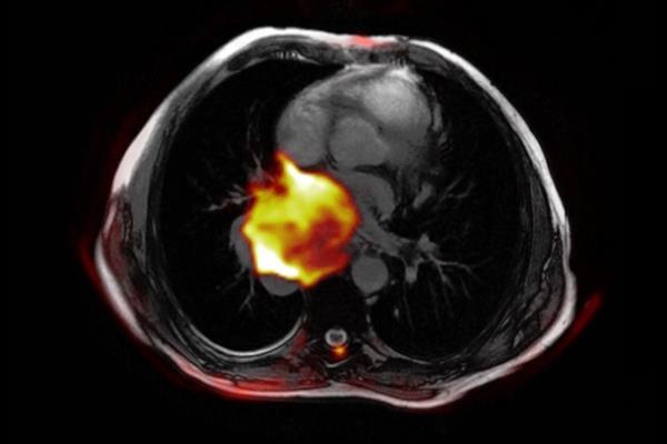
2 Cancer Diagnostics Diagnostics plays an important role throughout cancer treatment Before treatment, accurately locate tumors, stage the disease, and determine an appropriate combination of cancer treatments. Tumor molecular profiling helps identify the right chemotherapy or targeted therapy drugs before treatment, which reduces unnecessary toxicity and identifies an appropriate treatment approach. During treatment, track the size of the tumor, progression of the disease, and response to treatment, and modify treatment accordingly. Minimally invasive tools like navigational bronchoscopy and endoscopic ultrasound allow to find and reach very small tumors without the risks of surgery. After treatment, evaluate any possible symptoms, and schedule regular checkups to monitor for any signs of metastasis or recurrence. Technologies like PET/CT allow to accurately detect small lesions in areas of the body subject to movement, like the breasts, lungs, colon and prostate.


3 Overall Concept Jokerst UCSD


4 Content Diagnostics & bioimaging techniques Nanoparticles as contrast/imaging agents Examples and case studies

5 Technique 1: Fluorescence Imaging Fluorescence imaging is the visualization of fluorescent dyes or proteins as labels for molecular processes or structures. It enables a wide range of experimental observations including the location and dynamics of gene expression, protein expression and molecular interactions in cells and tissues.

6 Technique 2: Optical Imaging Optical imaging is a technique for noninvasively looking inside the body. It uses visible light and the special properties of photons to obtain detailed images of organs and tissues as well as smaller structures including cells and even molecules. Optical imaging includes a variety of techniques. The simplest and most widely recognized type of optical imaging is endoscopy. An endoscope consists of a flexible tube with a system to deliver light to illuminate an organ or tissue. For example, a physician can insert an endoscope through a patient s mouth to see the digestive cavity to find the cause of symptoms such as abdominal pain, difficulty swallowing, or gastrointestinal bleeding.


7 Technique 3: Ultrasound Imaging Ultrasound imaging (sonography) uses high-frequency sound waves to view inside the body. Because ultrasound images are captured in real-time, they can also show movement of the body's internal organs as well as blood flowing in the vessels. In an ultrasound exam, a transducer (probe) is placed directly on the skin or inside a body opening. A thin layer of gel is applied to the skin so that the ultrasound waves are transmitted from the transducer through the gel into the body. The ultrasound image is produced based on the reflection of the waves off of the body structures. The strength (amplitude) of the sound signal and the time it takes for the wave to travel through the body provide the information to produce an image.

8 Technique 4: Photoacoustic Imaging Photoacoustic imaging is a non-invasive imaging modality. It relies on the photoacoustic effect which describes conversion between light and acoustic waves due to absorption of electromagnetic waves and localized thermal excitation. Specifically, short pulses of electromagnetic radiation, mostly short laser pulses, are used to illuminate a sample. The local absorption of the light is followed by rapid heating, which subsequently leads to thermal expansion. Finally, broadband acoustic waves are generated. By recording the outgoing ultrasonic waves with adequate ultrasonic transducers outside of the sample the initial absorbed energy distribution can be recovered. Thus, photoacoustic imaging is a hybrid technique making use of optical absorption and ultrasonic wave propagation.
 scanner and an x-ray computed tomography (CT) scanner, so that images acquired from both devices can be taken sequentially, in the same")
, which depicts the spatial distribution of metabolic or biochemical activity in the body can be more precisely")
9 Technique 5: PET/CT Positron emission tomography computed tomography (better known as PET-CT or PET/CT) is a medical imaging technique using a device which combines both a positron emission tomography (PET) scanner and an x-ray computed tomography (CT) scanner, so that images acquired from both devices can be taken sequentially, in the same session, and combined into a single superposed image. Thus, functional imaging obtained by PET (using radiotracers), which depicts the spatial distribution of metabolic or biochemical activity in the body can be more precisely aligned or correlated with anatomic imaging obtained by CT scanning.
, which become aligned in a magnetic field. An MRI scanner applies a very strong magnetic field, which aligns the proton \"spins.")

10 Technique 6: Magnetic Resonance Imaging (MRI) Magnetic Resonance Imaging (MRI) is a simple, painless diagnostic procedure that produces images of anatomy without the use of radiation and there are no known side effects. Water molecules contain hydrogen nuclei (protons), which become aligned in a magnetic field. An MRI scanner applies a very strong magnetic field, which aligns the proton "spins. When the field is turned off, the protons gradually return to their normal spin (i.e., relax). The return process produces a radio signal that can be measured by receivers in the scanner and made into an image. Protons in different body tissues return to their normal spins at different rates, so the scanner can distinguish among tissues. The scanner settings can be adjusted to produce contrasts between different body tissues.
11 Fluorescent Nanoparticles vs. Molecular Fluorophores The availability of nanomaterials for purposes of imaging has generated a variety of methods for imaging, with features including improved brightness (defined as absorbance times quantum yield), inertness to their microenvironment and a more even distribution (unless targeted imaging of certain domains is desired, of course). Nanoparticles (NPs), in contrast to molecular probes, often are not cytotoxic and do not suffer from nonspecific binding by cellular biomacromolecules or unwanted sequestration. Chem. Soc. Rev., 2015, 44,


12 Polymeric nanoparticles Lipid nanoparticles Imaging Nanoparticles Fluorescently doped silica NPs and sol gels

 Carbon dots")

13 Imaging Nanoparticles Semiconducting (organic) polymer dots (P-dots) Carbon dots (C-dots) Metal chalcogenide quantum dots (classical Q-dots)


14 Imaging Nanoparticles Upconversion nanoparticles (UCNPs) Noble metal nanoparticles (e.g., gold, silver) Other nanomaterials (e.g., metal oxides, sulfides, tellurides)
15 Example 1

16 Remove Noise from Images During real-time photoacoustic (PA) data acquisition, a pulsed magnetic field is applied. Voxels within the imaging region experience a force induced by the local field and magnetization. When the field is on, MNP-gold NPs move as a result of their strong magnetization, creating a moving source within a PA image. When the fi eld is turned off, MNP-gold NPs return to their original positions. Non-magnetic PA sources do not move coherently with the applied field during this entire interval. Consequently, coherent motion processing of a PA image sequence can identify sources related to MNP-gold NPs and reject all background signals, whether from diffuse or localized sources. Such processing can greatly enhance the contrast specificity of the NP.

17 Synthesis of MNP-gold-coupled NP Schematic of MNP-gold core-shell NPs and mechanism of background suppression using mmpa. Key steps involved in hybrid NP synthesis. Monodisperse hydrophobic MNPs coated with oleic acids are first solubilized using amphiphilic PL-PEG-COOH. Poly-L-histidine (PLH), which is capable of chelating metal ions, is then adsorbed onto PL-PEG-COOH by electrostatic interaction. On addition of gold ions and a reducing reagent, thin gold shells form on the polypeptide template rather than directly on core nanoparticles.
. ( c ) T2-weighted MR images of bare and gold-coated MNPs at various dilutions.")
18 Multimodality Imaging Using MNP-gold hybrid NPs ( a, b ) Darkfield imaging of single MNPs spread on glass coverslips before and after gold nanoshell coating. The coated MNPs are readily detectable under current experimental conditions (insets show corresponding TEM images). ( c ) T2-weighted MR images of bare and gold-coated MNPs at various dilutions. Signal strength is indicated by the darkness of the images. At the same concentrations, the MRIs are indistinguishable between the two series, indicating unchanged magnetic properties before and after gold nanoshell coating. ( d, e ) Crosssectional PA images of a tube filled with ( d ) 5 nm MNPs and ( e ) 5 nm MNP-gold NPs on a db scale. A level of 0 db corresponds to the maximum signal level among both images. Note that a different dynamic range was used for better visualization. Signal-to-noise ratio can be improved by 1 order of magnitude (20 db) when MNP-gold NPs are used.

19 Modality, Magnetomotive Photoacustic (mmpa) Imaging A PVA phantom holds three 2-mmdiameter inclusions, in which the one on the left contains gold nanorods with absorption comparable to the 3 nm MNP-gold hybrid NPs placed in the centre inclusion, and the third inclusion on the right contains 3 nm MNPs. The gold nanorod inclusion on the left serves as a magnetic reference and mimics strong background tissue signals. The MNP inclusion on the right serves as an optical reference to MNPgold hybrid NPs. A conventional PA image of this phantom is presented in ( a ) on a logarithmic scale over a 40 db display range. Using a sequence of PA images acquired while a magnetic field was turned on and then off over a 10-s interval, the maximum displacement resulting from the action of the magnetic field is presented in ( b ) on a pixel pixel basis over a display range of (0 (dark), 30 (light)) μ m. ( c ) Three representative displacement traces and their fitted curves over the entire time interval of the experiment for pixels in different inclusions. Using these curves, velocity was computed over the full 10-s interval and the maximum positive and negative velocities, presented in ( d ) and ( e ) over a ( 20, 20) μm s 1 display range, were used to create a weighting image, presented in ( f ) over a (0, 1) display range, based on the magnitude of difference between peak positive velocity in the first half and peak negative velocity in the second half. ( g ) mmpa image produced from the product of ( a ) with ( f ) over a 40 db display range demonstrates that the gold nanorod inclusion mimicking a strong background signal is almost completely suppressed.

20 Example 2 Here we capitalized on the acidic, angiogenic tumour microenvironment to achieve the detection of tumour tissues in a wide variety of mouse cancer models. This was accomplished using ultra ph-sensitive fluorescent nanoprobes that have tunable, exponential fluorescence activation on encountering subtle, physiologically relevant ph transitions. These nanoprobes were silent in the circulation, and then strongly activated (>300- fold) in response to the neovasculature or to the low extracellular ph in tumours.
 in the tumour milieu, or endocytic organelles (ph i, 5.0 6.")
21 Ultra ph-sensitive (UPS) Nanoprobes A schematic of imaging the tumour microenvironment using UPS nanoprobes. The UPS nanoprobes stay OFF at ph 7.4 during blood circulation. After reaching tumours, the UPS nanoprobes are turned ON by acidic extracellular ph e ( ) in the tumour milieu, or endocytic organelles (ph i, ) in the tumour endothelial cells after receptor-mediated endocytosis.
. The UPS i can be activated inside acidic endocytic organelles (for example ph i =5:0 6.0). Cy5.")
22 Synthesis and Characterization of UPS Nanoprobes a, Structural composition of two types of nanoprobe, UPS e and UPS i, with ph transitions at 6.9 and 6.2, respectively. The UPS e is specifically designed to activate in acidic tumour extracellular fluid (ph e =6:5 6.8). The UPS i can be activated inside acidic endocytic organelles (for example ph i =5:0 6.0). Cy5.5 is used as the nearinfrared fluorophore in most of the animal studies. b, Normalized fluorescence intensity as a function of ph for UPS e and UPS i nanoprobes. At high ph (for example, 7.4), both probes stay silent. At ph below their transitions (that is 6.9 and 6.2), the nanoprobes can be activated as a result of micelle dissociation. The blue dashed line simulates the ph response of a small molecular ph sensor with a pka of 6.9 For UPS, the ph response is extremely sharp (<0.25 ph unit between ON/OFF states) with >100-fold signal amplification. In contrast, small molecular ph sensors require 3 ph units for a comparable signal change.
.")
23 Characterization of UPS Nanoprobes c, Fluorescent images of UPSe Cy5.5 nanoprobe solution in different ph buffers (Exitation/Emission= 675/710 nm). d, Transmission electron micrographs of UPSe nanoprobes at ph 7.4 and 6.7 (polymer concentration, 1 mg/ml; scale bars, 100 nm). e, UPSe nanoprobes remain stable in fresh mouse serum over 24 h at 37 ºC.

24 Imaging Angiogenic Tumor Vasculature a, Design of the crgd-upsi nanoprobe. b, Schematic of internalization and activation of crgd-upsi nanoprobes after α v β 3 -mediated endocytosis in tumour endothelial cells. The nanoprobes are accumulated in the endosomes or lysosomes, where the acidic ph activates the nanoprobes. c, Superimposed fluorescent images of A549-tumour-bearing mice at 6 h post-injection of UPSi nanoprobe, crgd-upsi or crgd+crgd- UPS i. Cy5.5 (red) and autofluorescence (green) are separately shown in the composite images. f, Representative images of ex vivo tumours, muscles and blood at 6 h post-injection of nanoprobes.
 and")
25 Broad Tumor Targeting Specificity b,c, Integrated crgd-ups e -Cy5.5 nanoprobes show broad tumour imaging specificity and efficacy in ten different tumour models of different cancer types (breast, prostate, head and neck, lung, brain and pancreatic cancers) and organ sites. In the 3LL lung cancer model (c), explanted lung was shown to illustrate the effective detection of small metastatic nodules (<1 mm). Scale bar, 2 mm. In each model, high T/N ratios were observed, demonstrating the success of targeting tumour microenvironment signals as a universal strategy to achieve broad tumour specificity
BME101 Introduction to Biomedical Engineering Medical Imaging Özlem BİRGÜL Ankara University Department of Biomedical Engineering
 BME101 Introduction to Biomedical Engineering Medical Imaging Özlem BİRGÜL Ankara University Department of Biomedical Engineering Outline What is Medical Imaging? History of Medical Imaging X-Ray Imaging
TARGETED IMAGING. Maureen Chan and Ruwani Mahathantila
 TARGETED IMAGING Maureen Chan and Ruwani Mahathantila Overview 2 Introduction to fluorescent imaging Fluorescent agents Quantum Dots Physical properties How QDs work In Vivo QD imaging Future Video What
Translational Multimodality Optical Imaging
 Translational Multimodality Optical Imaging Fred S. Azar Xavier Intes Editors 0 ARTECH H O U S E BOSTON LONDON artechhouse.com Contents Foreword Preface xv xvii CHAPTER1 Introduction to Clinical Optical
Nanomaterials for Imaging Technology. Nadeem A. Kizilbash, Ph.D. Assistant Professor Department of Chemistry Quaid-i-Azam University Islamabad
 Nanomaterials for Imaging Technology Nadeem A. Kizilbash, Ph.D. Assistant Professor Department of Chemistry Quaid-i-Azam University Islamabad Introduction Nanotechnology, most basically put, is the molecular
Molecular Imaging. by Magnetic Resonance. Silvio Aime. University of Torino (Italy) Department of Chemistry IFM & Molecular Imaging Center
 Department of Chemistry IFM & Molecular Imaging Center") Molecular Imaging by Magnetic Resonance Silvio Aime University of Torino (Italy) Department of Chemistry IFM & Molecular Imaging Center Molecular Imaging In vivo characterization and quantitative measurement
Molecular Imaging by Magnetic Resonance Silvio Aime University of Torino (Italy) Department of Chemistry IFM & Molecular Imaging Center Molecular Imaging In vivo characterization and quantitative measurement
Computer Assisted Surgery Basics of medical imaging
 Computer Assisted Surgery Basics of medical imaging Prof. Leo Joskowicz School of Engineering and Computer Science The Hebrew University of Jerusalem, ISRAEL Medical Image Processing Basics of medical
Medical instrumentationi 11/19/2010
 Medical instrumentationi BIOEN 302 11/19/2010 Medical instrumentation Definition: instrument for sensing, diagnostics, therapeutics or surgery of human being. 2 Medical instrumentation Definition: instrument
Biophotonics?? Biophotonics. technology in biomedical engineering. Advantages of the lightwave
 Biophotonics - Imaging: X-ray, OCT, polarimetry, DOT, TIRF, photon migration, endoscopy, confocal microscopy, multiphoton microscopy, multispectral imaging - Biosensing: IR spectroscopy, fluorescence,
Molecular Imaging: Definition, Overview and Goals
 This tutorial will define what is currently considered molecular imaging. It will provide history and an overview, discuss the goals and the advantages of molecular imaging. It will clarify what is and
Photoacoustic Imaging in Biomedicine Critical Review by Saurabh Vyas Group 9: Interventional Photoacoustic Ultrasound CIS II: 600.
 Photoacoustic Imaging in Biomedicine Critical Review by Saurabh Vyas Group 9: Interventional Photoacoustic Ultrasound CIS II: 600.446, Spring 2011 Introduction Photoacoustic imaging (PA Imaging) is the
Miniature fibre optic probe for minimally invasive photoacoustic sensing
 Miniature fibre optic probe for minimally invasive photoacoustic sensing Sunish J. Mathews*, Edward Z. Zhang, Adrien E. Desjardins and Paul C. Beard Department of Medical Physics and Biomedical Engineering,
SURFACE ENHANCED RAMAN SCATTERING NANOPARTICLES AS AN ALTERNATIVE TO FLUORESCENT PROBES AN EVALUATION
 APPLICATION NOTE SURFACE ENHANCED RAMAN SCATTERING NANOPARTICLES AS AN ALTERNATIVE TO FLUORESCENT PROBES AN EVALUATION Summary: Interest in using nanoparticles specifically, Surface Enhanced Raman Scattering
Absorption of an electromagnetic wave
 In vivo optical imaging?? Absorption of an electromagnetic wave Tissue absorption spectrum Extinction = Absorption + Scattering Absorption of an electromagnetic wave Scattering of an electromagnetic wave
Principles of translational medicine: imaging, biomarker imaging, theranostics
 Principles of translational medicine: imaging, biomarker imaging, theranostics Compiled by: Endre Mikus PhD, CEO Budapest, 21/9/2015 Imaging and imaging biomarkers An imaging biomarker is an anatomic,
Basic principles of quantification using optical techniques
 Contents Basic principles of quantification using optical techniques Adrian Taruttis Helmholtz Zentrum München Chair for Biological Imaging Technische Universität München Light/ tissue interactions Planar
1st Faculty of Medicine, Charles University in Prague Center for Advanced Preclinical Imaging (CAPI)
") ADVANTAGES Optical Imaging OI Optical Imaging is based on the detection of weak light by a highly sensitive and high resolution CCD camera DISADVANTAGES High sensitivity Limited penetration depth Easy
ADVANTAGES Optical Imaging OI Optical Imaging is based on the detection of weak light by a highly sensitive and high resolution CCD camera DISADVANTAGES High sensitivity Limited penetration depth Easy
Nanomaterials for Molecular Imaging: Nanotheranostics. 7 September 2018 Bodø, Norge. Ola Åberg, PhD, researcher
 The Nanoscale Nanomaterials for Molecular Imaging: Nanotheranostics 7 September 2018 Bodø, Norge Ola Åberg, PhD, researcher ola.aberg@pet.medchem.uu.se Uppsala University Department of Medicinal Chemistry
Photoacoustic imaging of vascular networks in transgenic mice
 Photoacoustic imaging of vascular networks in transgenic mice J.G. Laufer 1, J.O. Cleary 1,2, E.Z. Zhang 1, M.F. Lythgoe 2, P.C. Beard 1 1. Department of Medical Physics and Bioengineering, University
UNIVERSITY OF ROME LA SAPIENZA NANOTECHNOLOGIES ENGINEERING NANOPARTICLES IN BIOMEDICINE
 UNIVERSITY OF ROME LA SAPIENZA NANOTECHNOLOGIES ENGINEERING NANOPARTICLES IN BIOMEDICINE Challenges The challenges are: in-situ analysis and in vivo at micro level recognize biomolecules functionality
PREPARED FOR: U.S. Army Medical Research and Materiel Command Fort Detrick, Maryland
 AD Award Number: W81XWH-07-1-0231 TITLE: Spectroscopic Photoacoustic Tomography of Prostate Cancer PRINCIPAL INVESTIGATOR: Xueding Wang CONTRACTING ORGANIZATION: University Of Michigan Ann Arbor, MI 48109-1274
PREPARED FOR: U.S. Army Medical Research and Materiel Command Fort Detrick, Maryland
 AD Award Number: W81XWH-07-1-0231 TITLE: Spectroscopic Photoacoustic Tomography of Prostate Cancer PRINCIPAL INVESTIGATOR: Xueding Wang CONTRACTING ORGANIZATION: University Of Michigan Ann Arbor, MI 48109-1274
Diagnostic Medical Image Processing
 Diagnostic Medical Image Processing Introduction WS 2010/11 Joachim Hornegger, Dietrich Paulus, Markus Kowarschik Lehrstuhl für Mustererkennung (Informatik 5) Friedrich-Alexander-Universität Erlangen-Nürnberg
Optical Observation - Hyperspectral Characterization of Nano-scale Materials In-situ
 Optical Observation - Hyperspectral Characterization of Nano-scale Materials In-situ Research at the nanoscale is more effective, when research teams can quickly and easily observe and characterize a wide
KILLING THROMBUS WITH
 KILLING THROMBUS WITH D. Dash Department of Biochemistry Institute of Medical Sciences Banaras Hindu University Thrombus has two components: (1) Protein Component composed of Insoluble Fibrin Clot (2)
2.3 Quantum Dots (QDs)
") 2.3 Quantum Dots (QDs) QDs are inorganic nanocrystals, approximately 1 10 nm in size, with unique optical properties of broad excitation, narrow size-tunable emission spectra, high photochemical stability,
2.3 Quantum Dots (QDs) QDs are inorganic nanocrystals, approximately 1 10 nm in size, with unique optical properties of broad excitation, narrow size-tunable emission spectra, high photochemical stability,
Spectroscopy and Imaging IV
 PROGRESS IN BIOMEDICAL OPTICS AND IMAGING Vol. 16 No. 55 Clinical and Biomedical Spectroscopy and Imaging IV J. Quincy Brown Volker Decked Edifors 22-24 June 2015 Munich, Germany Sponsored by SPIE (United
1 Ultrasound targeting
 This lecture deals with the concepts involved in ultrasound targeting and the challenges involved therein. A snapshot of the challenges involved in targeting the brain acrosss the blood-brain barrier (BBB)
MEDICAL PHYSICS (MED PHYS)
") Medical Physics (MED PHYS) 1 MEDICAL PHYSICS (MED PHYS) MED PHYS/PHYSICS 265 INTRODUCTION TO MEDICAL PHYSICS Primarily for premeds and other students in the medical and biological sciences. Applications
Medical Physics (MED PHYS) 1 MEDICAL PHYSICS (MED PHYS) MED PHYS/PHYSICS 265 INTRODUCTION TO MEDICAL PHYSICS Primarily for premeds and other students in the medical and biological sciences. Applications
Contents Preface xiii Introduction Fabrication and manufacturing technology for optical MEMS
 Contents Preface xiii 1 Introduction 1 1.1 Optical MEMS and optofluidics 1 1.2 History 1 1.2.1 Processes and materials 1 1.2.2 Early devices and systems 2 1.3 Progress in optical MEMS and optofluidics
OUR WISH LIST RESEARCH EQUIPMENT
 OUR WISH LIST RESEARCH EQUIPMENT YOU CAN MAKE A TANGIBLE DIFFERENCE! The South Australian Health and Medical Research Institute (SAHMRI) is one of the most exciting developments in the field of health
OUR WISH LIST RESEARCH EQUIPMENT
 OUR WISH LIST RESEARCH EQUIPMENT WITH YOUR PHILANTHROPIC SUPPORT, WE CAN WORK TOGETHER TO COMPLETE OUR FULLY-FUNCTIONAL FACILITY BY PURCHASING THE CUTTING-EDGE EQUIPMENT AND RESOURCES TO SUPPORT SAHMRI
Spectra Chacracterizations of Optical Nanoparticles
 THAI NGUYEN UNIVERSITY OF EDUCATION Spectra Chacracterizations of Optical Nanoparticles Chu Viet Ha Department of Physics 18/2018 1 THAI NGUYEN UNIVERSITY OF EDUCATION Address 20 Luong Ngoc Quyen Street,
Simple, intuitive and accessible MRI solution for preclinical research. M-Series Compact MRI Systems
 Simple, intuitive and accessible MRI solution for preclinical research M-Series Compact MRI Systems Application Oriented Imaging Anatomy and Morphology In vivo soft tissue imaging for morphological characterization.
Pre-Clinical Optical Molecular Imager
 Pre-Clinical Optical Molecular Imager ART Advanced Research Technologies Inc. 2300 Alfred-Nobel Blvd. Saint-Laurent, QC Canada, H4S 2A4 T 514.832.0777 / 1.888.278.7888 F 514.832.0778 E info@art.ca Table
Simple, intuitive and accessible MRI solution for preclinical research. M-Series Compact MRI Systems
 Simple, intuitive and accessible MRI solution for preclinical research M-Series Compact MRI Systems Application Oriented Imaging Molecular Imaging Using Contrast Agents Detection and quantification of
Applications of Nanotechnology in Medical Device Design James Marti, Ph.D. Minnesota Nano Center
 Applications of Nanotechnology in Medical Device Design James Marti, Ph.D. Minnesota Nano Center November 4, 2015 The University of Minnesota Nano Center An open-use nanotechnology lab with tools for fabricating
RADIATION ONCOLOGY RESIDENCY PROGRAM Competency Evaluation of Resident
 Resident s Name: RADIATION ONCOLOGY RESIDENCY PROGRAM Competency Evaluation of Resident Rotation: PHYS 705: Clinical Rotation 3 Inclusive dates of rotation: Aug. 25, 2015 Feb. 25, 2016 Director or Associate
TRANSLATIONAL NANOMEDICINE
 TRANSLATIONAL NANOMEDICINE St Louis, MO. CORTEX Building Center of Research, Technology, and Entrepreneurial Exchange C-TRAIN @ Washington University Consortium For Translational Research in Advanced Imaging
Patrick Vilela, a Afaf El-Sagheer, c,d Timothy M. Millar, e Tom Brown, c Otto L. Muskens, a,b and Antonios G. Kanaras a,b,*
 Patrick Vilela, a Afaf El-Sagheer, c,d Timothy M. Millar, e Tom Brown, c Otto L. Muskens, a,b and Antonios G. Kanaras a,b,* a Physics and Astronomy, Faculty of Physical Sciences and Engineering, b Institute
262 Chapter 16. Figure Fiber-optic-enabled arrays using fluorescence for high-speed screening. 1
 262 Chapter 16 Figure 16.13 Fiber-optic-enabled arrays using fluorescence for high-speed screening. 1 Figure 16.14 Fluorescent array microsphere sensors. 10 to have low background fluorescence to maximize
Quantum Dot applications in Fluorescence Imaging for Calibration and Molecular Imaging
 Quantum Dot applications in Fluorescence Imaging for Calibration and Molecular Imaging Introduction In this application note, we will discuss the application of quantum dots in fluorescence imaging, both
Fluorescence Microscopy. Terms and concepts to know: 10/11/2011. Visible spectrum (of light) and energy
 and energy") Fluorescence Microscopy Louisiana Tech University Ruston, Louisiana Microscopy Workshop Dr. Mark DeCoster Associate Professor Biomedical Engineering 1 Terms and concepts to know: Signal to Noise Excitation
Fluorescence Microscopy Louisiana Tech University Ruston, Louisiana Microscopy Workshop Dr. Mark DeCoster Associate Professor Biomedical Engineering 1 Terms and concepts to know: Signal to Noise Excitation
Cellular imaging using Nano- Materials. A Case-Study based approach Arun Murali, Srivats V
 Cellular imaging using Nano- Materials A Case-Study based approach Arun Murali, Srivats V Agenda Discuss a few papers Explain a couple of new imaging techniques and their benefits over conventional imaging
Kazuki N. Sugahara, Tambet Teesalu, Priya Prakash Karmali, Venkata Ramana Kotamraju, Lilach
 Cancer Cell, Volume 16 Supplemental Data Tissue-Penetrating Delivery of Compounds and Nanoparticles into Tumors Kazuki N. Sugahara, Tambet Teesalu, Priya Prakash Karmali, Venkata Ramana Kotamraju, Lilach
Supplementary Information
 Supplementary Information Biomimetic nanoflowers by self-assembly of nanozymes to induce intracellular oxidative damage against hypoxic tumors Wang et al. Supplementary Figure 1. The effect of Pt/Co ratio
PRINCIPLES OF CT AND MR IMAGING Marc-André d Anjou, DMV, DACVR Faculty of Veterinary Medicine, University of Montreal Saint-Hyacinthe, Quebec, Canada
 PRINCIPLES OF CT AND MR IMAGING Marc-André d Anjou, DMV, DACVR Faculty of Veterinary Medicine, University of Montreal Saint-Hyacinthe, Quebec, Canada CT and MR imaging offer superior diagnostic possibilities
Supplementary Figure 1. Thin layer chromatography of R18 salts with different counterions. The mobility of the R18 salts with TPB counterions is much
 Supplementary Figure 1. Thin layer chromatography of R18 salts with different counterions. The mobility of the R18 salts with TPB counterions is much higher with perchlorate, showing their much higher
2018 REVIEW CATEGORIES
 2018 REVIEW CATEGORIES 100 Neuro 101 Neuro: Acquisition 102 Neuro: Processing 103 Neuro: Neonatal & Pediatric - Normal Development 104 Neuro: Neonatal & Pediatric - Clinical Studies 105 Neuro: Normal Aging
Department of Chemistry, University of California, Davis, California 95616, USA 2
 Enhance Solar Water Splitting Performance by Utilizing Near Infrared Radiation with Composite Films of Hematite and Rare Earth Doped Upconversion Materials Ming Zhang, 1 Yongjing Lin, 2 Thomas J. Mullen,
CREOL, The College of Optics & Photonics, University of Central Florida
 Metal Substrate Induced Control of Ag Nanoparticle Plasmon Resonances for Tunable SERS Substrates Pieter G. Kik 1, Amitabh Ghoshal 1, Manuel Marquez 2 and Min Hu 1 1 CREOL, The College of Optics and Photonics,
Self-assembly of oligonucleotides
 Self-assembly of oligonucleotides Dr. K. Uma Maheswari Professor, School of Chemical & Biotechnology SASTRA University Joint Initiative of IITs and IISc Funded by MHRD Page 1 of 9 Table of Contents 1 APPLICATIONS
Future Areas of Technology Convergence
 Future Areas of Technology Convergence Dr J Malcolm Wilkinson Managing Director Technology For Industry Ltd Cambridgeshire, UK Medilink Yorkshire & Humberside, 8 December 2005 1 Technology For Industry
Date: May 26, 2015 Page 1
 Part I. Answer these questions by marking the best answer among the choices given: [2 points each] 1. Ethics and morals differ in that a. Only one of them should be followed b. Ethics are for professionals
Supplementary Information. Trojan Horse Nanotheranostics with Dual Transformability and Multifunctionality. for Highly Effective Cancer Treatment
 Supplementary Information Trojan Horse Nanotheranostics with Dual Transformability and Multifunctionality for Highly Effective Cancer Treatment Xue et. al. Supplementary Figure 1. Synthetic route of PhD
Biophotonics. Light Matter Interactions & Lasers. NPTEL Biophotonics 1
 Biophotonics Light Matter Interactions & Lasers NPTEL Biophotonics 1 Overview In this lecture you will learn, Light matter interactions: absorption, emission, stimulated emission Lasers and some laser
DNA Biosensors. Anand Jagota 16 November 2015
 DNA Biosensors Anand Jagota 16 November 2015 1 Market, Unmet Needs Worldwide In-vitro diagnostics ~$ 50 Billion and growing Nucleic Acid diagnostics ~$9 Billion Health, Security, Pathogen Detection, etc.
Quantifying biological forces involved in the process of cell-mediated cytolysis
 Quantifying biological forces involved in the process of cell-mediated cytolysis Experimentation based on responses of living cells (Cell-based assays) represents an important stratum in biological and
Small Nodule Localization. Bernard Park, M.D. Attending Surgeon, Thoracic Service Memorial Sloan Kettering Cancer Center
 Small Nodule Localization Bernard Park, M.D. Attending Surgeon, Thoracic Service Memorial Sloan Kettering Cancer Center Disclosures Intuitive Surgical: honoraria Medtronic: consulting Image guidance in
Class 7: Methods in Research By: Ray
 Class 7: Methods in Research By: Ray Method in Brain Research 1. Non-Invasive (Human) o Imaging Computerized (Axial) Tomography (X-rays). Static pictures and high spatial resolution. Horizontal plane only.
K-Space analysis for plane-wave ultrasound imaging. Georg Schmitz
 K-Space analysis for plane-wave ultrasound imaging Georg Schmitz Abstract: Ultrasound imaging can be considered as an inverse scattering problem in which the unknown distribution of the material parameters
@01-258_via99-011_hepatoma_onscreen
 Lei Xing, Ph.D. & Jacob Haimson Professor Department of Radiation Oncology & MIPS Stanford University School of Medicine Case study TrueBeam 6 MV FFF RapidArc Tx of a Lung SBRT 3D modeling Treatment planning
Revised Examination Guidelines for Industrially Applicable Inventions (Provisional Translation)
") Revised Examination Guidelines for Industrially Applicable Inventions (Provisional Translation) Examination Guidelines for Patent and Utility Model Part II: REQUIREMENTS FOR PATENTABILITY Chapter 1 Industrially
Revised Examination Guidelines for Industrially Applicable Inventions (Provisional Translation) Examination Guidelines for Patent and Utility Model Part II: REQUIREMENTS FOR PATENTABILITY Chapter 1 Industrially
Abstract: Keywords: Gold nanoparticle, Folic acid, Folate, Hela cell, Mcf7 cell, Targeting, 4-aminothiophenol, 6- mercapto-1-hexanol
 Abstract: Cancer Nanotechnology Treatment through Folate Conjugated Gold Nanoparticles ( * ) Ali Shakeri-Zadeh and G.Ali Mansoori ( ** ) University of Illinois at Chicago (*) Invited paper. (**) Corresponding
3.1.4 DNA Microarray Technology
 3.1.4 DNA Microarray Technology Scientists have discovered that one of the differences between healthy and cancer is which genes are turned on in each. Scientists can compare the gene expression patterns
Bioengineering (BIOE)
") Bioengineering (BIOE) 1 Bioengineering (BIOE) Courses BIOE 5301. Biosignals. 3 Credit Hours. This course offers a deep overview of the signals in the Biomedical fields. Signals are studied in several modalities,
Bioengineering (BIOE) 1 Bioengineering (BIOE) Courses BIOE 5301. Biosignals. 3 Credit Hours. This course offers a deep overview of the signals in the Biomedical fields. Signals are studied in several modalities,
Biosensors. DNA Microarrays (for chemical analysis) Protein Sensors (for identifying viruses)
 Protein Sensors (for identifying viruses)") Biosensors DNA Microarrays (for chemical analysis) Protein Sensors (for identifying viruses) DNA Microarrays 40 000 detectors in parallel, each detecting a specific DNA sequence. Combinatorial Chemistry
Biosensors DNA Microarrays (for chemical analysis) Protein Sensors (for identifying viruses) DNA Microarrays 40 000 detectors in parallel, each detecting a specific DNA sequence. Combinatorial Chemistry
Solar Cells and Photosensors.
 Designing Photonic Crystals in Strongly Absorbing Material for Applications in Solar Cells and Photosensors. Minda Wagenmaker 1, Ebuka S. Arinze 2, Botong Qiu 2, Susanna M. Thon 2 1 Mechanical Engineering
Master of Molecular Imaging Course Outline
 Master of Molecular Imaging Course Outline Graduate Outcomes On completion of the course, graduates will have achieved the following skills, knowledge and attributes: chemistry/pharmacy physics/engineering
Biomedical Applications of Molecular Spectroscopy
 Biomedical Applications of Molecular Spectroscopy Mike Kayat B&W Tek, Inc 19 Shea Way Newark, DE 19713 United States of America +1 302 368 7824 mikek@bwtek.com 1 Overview Molecular spectroscopy is a large
Noninvasive and Targeted Brain Drug Delivery using Transcranial Focused Ultrasound
 Noninvasive and Targeted Brain Drug Delivery using Transcranial Focused Ultrasound Muna Aryal, PhD Mentors: Drs. Raag Airan & Jeremy Dahl SCIT Seminar August 29 th 2018 1/21 Outline Introduction Focused
Prof. Steven S. Saliterman
 Department of Biomedical Engineering, University of Minnesota http://saliterman.umn.edu/ Prof. Angela Panoskaltsis-Mortari s BMEn 5361, 3D Bioprinting Tissue engineering Bioprinting Design considerations
Measuring gene expression and protein production
 Measuring gene expression and protein production Module 3, Lecture 5! 20.109 Spring 2013! Lessons from Lecture 4 Always look for the recovery file! The myth of multitasking! task-switching taps processing
Nanotechnology in Medicine
 25 Prajnan O Sadhona Nanotechnology in Medicine Prajnamoy Pal * Nanomedicine involves utilization of nanotechnology for the benefit of human health and well-being. The use of nanotechnology in various
Un Sistema di Navigazione basato su Mixed Reality per Trattamenti HIFU
 La Medicina Incontra la Realtà Virtuale 2011- MIMOS Un Sistema di Navigazione basato su Mixed Reality per Trattamenti HIFU C. Freschi, V. Ferrari*, F. Porcelli, A. Peri, L. Pugliese, L. Morelli, M. Ferrari,
Motivation: Biofunctional Composite Nanoparticles
 1 Motivation: Biofunctional Composite Nanoparticles Iron Oxide Core Magnetic core Alternating magnetic field Brownian and Neél relaxation Bi-Material Composites MRI contrast enhancement and RF thermal
Nanodiamond-Based Therapeutic Delivery Films For the Treatment of Cancer and Inflammation
 Nanodiamond-Based Therapeutic Delivery Films For the Treatment of Cancer and Inflammation Dean Ho, Ph.D. Assistant Professor Departments of Biomedical and Mechanical Engineering Lurie Comprehensive Cancer
ENDOTOFPET-US: Project Status Update
 ENDOTOFPET-US: Project Status Update Alessandro Silenzi Hamburg 14.11.2011 Outline > ENDOTOFPET US: project introduction > Technology Challenges > Project Design > Outlook for collaboration > Outlook for
Novel Kidney Imaging. Objectives. Imaging surveillance in VHL. Disclosures. From nephrology perspective 10/11/2018
 Disclosures Novel Kidney Imaging Contract with Lantheus Medical Imaging (Definity) Contrast-enhanced ultrasound Emily Chang, MD Nephrology October 4, 2018 13 th International VHL Symposium 10/11/2018 2
Nanoscale Materials Inspires Innovation and Drives Economic Development
 Nanoscale Materials Inspires Innovation and Drives Economic Development 5nm Greg Salamo & Alex Biris InAs Quantum Dots What is Nanoscience? The effort to understand and design structures at the nano size
Photon Upconversion Sensitized Nanoprobes for
 Electronic Supplementary Material (ESI) for Nanoscale. This journal is The Royal Society of Chemistry 2014 Supporting Information Photon Upconversion Sensitized Nanoprobes for Sensing and Imaging of ph
Real-Time Monitoring of Arsenic Trioxide Release and Delivery
 Real-Time Monitoring of Arsenic Trioxide Release and Delivery by Activatable T 1 Imaging Zhenghuan Zhao, Xiaomin Wang, Zongjun Zhang, Hui Zhang, Hanyu Liu, Xianglong Zhu, Hui Li, Xiaoqin Chi, Zhenyu Yin,
Plasmonics using Metal Nanoparticles. Tammy K. Lee and Parama Pal ECE 580 Nano-Electro-Opto-Bio
 Plasmonics using Metal Nanoparticles Tammy K. Lee and Parama Pal ECE 580 Nano-Electro-Opto-Bio April 1, 2007 Motivation Why study plasmonics? Miniaturization of optics and photonics to subwavelength scales
Engineering Quantum Dots for Live-Cell Single-Molecule Imaging
 Engineering Quantum Dots for Live-Cell Single-Molecule Imaging Andrew M. Smith and Shuming Nie Georgia Tech and Emory University Department of Biomedical Engineering 2011 NSF Nanoscale Science and Engineering
Supporting Information for Particle-size Dependent Förster Resonance Energy Transfer from Upconversion Nanoparticles to Organic Dyes
 Supporting Information for Particle-size Dependent Förster Resonance Energy Transfer from Upconversion Nanoparticles to Organic Dyes Verena Muhr a, Christian Würth b, Marco Kraft b, Markus Buchner a, Antje
Supplementary Figure 1. CryoTEM images of the barcoded nanoparticles (a) and
 and") a b Supplementary Figure 1. CryoTEM images of the barcoded nanoparticles (a) and size measurements of the particles (b). Liposomes were loaded with DNA barcodes and were imaged using cryo-tem and measured
a b Supplementary Figure 1. CryoTEM images of the barcoded nanoparticles (a) and size measurements of the particles (b). Liposomes were loaded with DNA barcodes and were imaged using cryo-tem and measured
European Technology Platform for Nanomedicine. Inês Mendes Pinto, PharmD, PhD Portugal
 European Technology Platform for Nanomedicine Inês Mendes Pinto, PharmD, PhD Portugal INL International Iberian Nanotechnology Laboratory General presentation of the Institute 1) Location: Braga- Portugal
Magnetic Resonance Imaging of concrete. Coring. Location of flaws. Assessment of Concrete Structures. Systems to monitor concrete modulus
 Magnetic Resonance Imaging of concrete Dr Chris Burgoyne Department of Engineering University of Cambridge Assessment of Concrete Structures How can we tell what is going on inside concrete? We would like
Small Animal Imaging Facility Newsletter
 Small Animal Imaging Facility Newsletter Biomedical Research Imaging Center Volume 2. Issue. 2 SAI Facility News and Updates 1. The BRIC website has a new look (http://bric.unc.edu). There were some minor
In-vivo Targeting of Liver Lesions with a Navigation System based on Fiducial Needles
 In-vivo Targeting of Liver Lesions with a Navigation System based on Fiducial Needles L. Maier-Hein 1, A. Tekbas 2, A. Seitel 1, F. Pianka 2, S. A. Müller 2, S. Schawo 3, B. Radeleff 3, R. Tetzlaff 1,4,
APPLICATION NOTE Rev. 7/2017, v4.0 Fluorescent Nanodiamonds: Bio-applications. Physical and Fluorescence Properties
 APPLICATION NOTE Rev. 7/2017, v4.0 Fluorescent Nanodiamonds: Bio-applications Fluorescent nanodiamonds (FNDs) offer a unique alternative to currently existing fluorescent biomarkers. With exceptional photo
MST Starting Guide Monolith NT.115
 MST Starting Guide Monolith NT.115 Contents 1. How to design an experiment 2. Before you start 3. Assay Setup Pretests 4. Assay Setup 5. MST-experiment using temperature control 6. Data Interpretation
Liposomes: New Therapeutics in Medicine
 Liposomes: New Therapeutics in Medicine BY: MASOUMEH ZAHMATKESHAN PHD OF NANOMEDICINE 1396.6.13 Outline 2 Liposomes Types Preparation methods Advantages Applications Liposomes on market or in clinical
Folic Acid-Conjugated Chitosan Functionalized Gold Nanoparticles for Targeted Delivery of 5-Fluorouracil in Breast Cancer
 Proceedings of the 3 rd World Congress on Recent Advances in Nanotechnology (RAN'18) Budapest, Hungary April 10-12, 2018 Paper No. NDDTE 103 DOI: 10.11159/nddte18.103 Folic Acid-Conjugated Chitosan Functionalized
Biomarker Discovery using Surface Plasmon Resonance Imaging
 F e a t u r e A r t i c l e Feature Article Biomarker Discovery using Surface Plasmon Resonance Imaging Elodie LY-MORIN, Sophie BELLON, Géraldine MÉLIZZI, Chiraz FRYDMAN Surface Plasmon Resonance (SPR)
Femtosecond micromachining in polymers
 Femtosecond micromachining in polymers Prof. Dr Cleber R. Mendonca Daniel S. Corrêa Prakriti Tayalia Dr. Tobias Voss Dr. Tommaso Baldacchini Prof. Dr. Eric Mazur fs-micromachining focus laser beam inside
Cell analysis and bioimaging technology illustrated
 Cell analysis and bioimaging technology illustrated The Cell Analysis Center Scientific Bulletin Part 1 Sysmex has been studying and exploring principles of automated haematology analysers, making full
IsoFluxTM. System. The next generation of CTC Analysis is here
 IsoFluxTM System The next generation of CTC Analysis is here Product Overview IsoFlux System The next generation of circulating tumor cell analysis is here The IsoFlux System enriches intact rare cells
Exploring metallic nanoparticles in Biophotonics
 Exploring metallic nanoparticles in Biophotonics Renato E. de Araujo Laboratório de Óptica Biomédica e Imagem, Universidade Federal de Pernambuco Recife, PE, Brazil. renato.earaujo@ufpe.br LABORATÓRIO
Enhanced Light Trapping in Periodic Aluminum Nanorod Arrays as Cavity Resonator
 Enhanced Light Trapping in Periodic Aluminum Nanorod Arrays as Cavity Resonator Rosure B. Abdulrahman, Arif S. Alagoz, Tansel Karabacak Department of Applied Science, University of Arkansas at Little Rock,
REVOLUTIONIZING EARLY CANCER DETECTION
 cover story Written by candace o connor Photos by geoff story IMAGING WITH LIGHT AND SOUND: REVOLUTIONIZING EARLY CANCER DETECTION For years, the field of optical imaging in biological tissue had languished,