X-ray imaging of synchrotrons & lab sources. Janos Kirz
|
|
|
- Abel Carson
- 5 years ago
- Views:
Transcription
1 X-ray imaging of synchrotrons & lab sources Janos Kirz EuXFEL 11/2017

2 W. C. Röntgen 1895 Origins Nov 8 first observation Nov 9 Dec 27 experimentation, write-up Dec 28 manuscript submitted Sitzungsberichte der Physikalischen-medizinischen Gesellschaft zu Würzburg See bones in hand shadowgraphs Instant sensation around the world Radiology absorption contrast Works well for bone fracture air vs fluid in lung tooth decay

3 3D Imaging: CAT scans 3D imaging based on many projections 1979 Nobel prize Godfrey Hounsfield & Allan McLeod Cormack Resolution: several mm Limitations: breathing, beating heart, digestion,

 Phase contrast tomography A. Momose et al.")
4 Better radiology? Hundreds of images: high radiation dose! Absorption contrast in soft tissue poor To reduce dose improve contrast! Extract phase shift from interference pattern Bonse-Hart interferometer Appl. Phys. Lett 6, 155 (1965) Phase contrast tomography A. Momose et al. Nature Med. 2, 473 (1996)
 Spring-8 source for coherence Object distorts Moire pattern Talbot-Lau interferometer Third grating for use with ordinary X-ray tube F. Pfeiffer et al. Nature Phys.")
5 Interferometry without crystals Grating-based phase measurement Using Talbot (1836) effect (self-image) A. Momose et al. Jpn. J Appl. Phy. 42, L866 (2003) Spring-8 source for coherence Object distorts Moire pattern Talbot-Lau interferometer Third grating for use with ordinary X-ray tube F. Pfeiffer et al. Nature Phys. 2, 258 (2006) Intensive development worldwide

6 Absorption & Phase contrast F. Pfeiffer et al. Nature Phys. 2, 258 (2006)
")
7 Application to mammography Phase contrast Absorption contrast (From Konica Minolta)

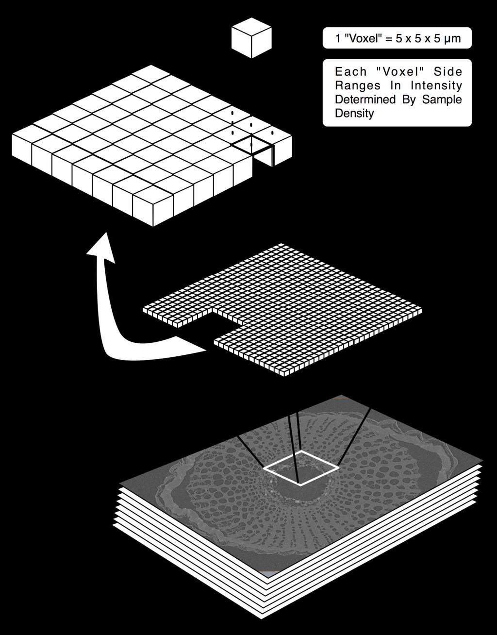


8 X-ray micro-tomography-als Lots of Math
9 Microtomography Simple projection onto detector Resolution: ~ 1-5 µm Commercial microscopes Lab sources or synchrotrons Zeiss (formerly XRADIA) Brucker (formerly Skyscan) 3D Studies of bone structure, seeds, small animals


10 Examples of Grapevine Xylem


11 Get insights in insect flight control Investigate the biomechanics underlying flight manoeuvres and gaze shifts à CT following the dynamics of 100+ Hz wing beat! Need: single-shot propagation-based phase contrast high-speed X-ray tomographic microscopy SLS beam Requires: Coherence and Flux BRIGHTNESS So, this is a perfect task for a synchrotron! S. M. Walker et al. P{LOS Biology 12, e (2014) 11
12 Toward higher resolution Microscopes with zone plate optics Resolution ~ 20 nm (50 nm in 3D) Radiation damage becomes limitation! Cryo preserves morphology Instruments at BESSY II ALS ALBA


13 Cryo Soft X-ray Tomography of Cells at the MISTRAL beamline E. Pereiro et al. J. Synchr. Rad. 16, (2009) reconstructed slice 1 µm A. SorrenBno et al. J. Synchr. Rad. 22, (2015)


14 The ultimate challenge Radiation damage in biological samples Frozen hydrated state of protein Howells et al. JESRP 170, 4 (2009) Resolution limit? Inverse fourth power law of dose vs resolution: Dose ~ 1/resolution-size 4


15 Scanning X-ray diffracbon microscopy Ptychography with a focused X-ray probe J. Rodenburg Pilatus 2M Reconstruct bith amplitude and phase P. Thibault, M. Dierolf, A. Menzel, O. Bunk, C. David, F. Pfeiffer, Science, 321, (2008). 15
 Sarah Shamoradian Quickly biopsy-punched,")

16 Frozen hydrated unstained brain tissue Locate, extract and further study intact hallmarks of Parkinson disease Large volumes High resolubon As close to nabve state as possible (no staining) Sarah Shamoradian Quickly biopsy-punched, infiltrated with cryo protectant, mounted on pin and gradually frozen Trimmed with cryo ultramicrotome Cryo transferred to OMNY ~ 80 micron at the base, ~ 100 nm 3D resolubon 446 projecbons 17 hour measurement Shamoradian et al., Sci. Rep (2017) Page 16
17 3D Elemental mapping by XRF 3D scan as for tomography Record fluorescence spectrum for each point Perform tomographic recontstruction for each element

18 3D elemental microtomography of Cyclotella meneghiana M. de Jonge, et al., PNAS 107, 15676, (2010)
19 Changing Landscape of X-ray facilities ~ dedicated storage rings 30+ years: a revolution in X-ray analysis ~ 2010 Toward higher brightness, coherence MBA lattice, MAX IV, Sirius, storage ring upgrades FELs LCLS, FLASH, FERMI, SACLA, EuXFEL, Older sources: DORIS, NSLS, Daresbury, shut down Fewer stations available for routine measurements


20 New initiatives in Lab-scale sources to fill the gap EXCILLUM liquid metal jet Lyncean back-scattered Compton Sigray microstructured anode in diamond substrate
21 Conclusions I X-rays are great! Penetrate opaque objects Rich spectra allow elemental & chemical info Radiation damage is a concern One way to mitigate radiation damage: cryo Other ways: many copies, as in crystals or diffract & destroy... but ptychography not compatible
22 Conclusions II A bit of humility: There is competition! MRI for imaging humans Cryo EM for high resolution on thin samples Super resolution visible light microscopy
23 Acknowledgments Thanks to colleages who provided material for this talk: Manuel Guizar Sicarios Eva Pereiro Marco Stampanoni Andrew McElrone
24 Thank you
Recent Developments In Scanning Microscopy at Stony Brook
 Recent Developments In Scanning Microscopy at Stony Brook C. Jacobsen, S. Abend, T. Beetz, M. Carlucci-Dayton, M. Feser, K. Kaznacheyev, J. Kirz, J. Maser 1, U. Neuhäusler 2, A. Osanna, A. Stein, C. Vaa,
Three dimensional visualisation of barley corn seed and fish tissue using Talbot Lau grating interferometer.
 Three dimensional visualisation of barley corn seed and fish tissue using Talbot Lau grating interferometer. Guruprasad Rao, Christian Gusenbauer, Sascha Senck, Johann Kastner University of Applied Sciences
SLS Symposium on Coherent X-Ray Imaging
 SLS Symposium on Coherent X-Ray Imaging Tuesday, October 5, 2010 10:00 to 12:15, WBGB/019 10:00 X-Ray Diffraction Microscopy: turning the rather esotheric into a usable technique Andreas Menzel, M. Dierolf,
X-ray Grating-based Phase Contrast CT for Non-Destructive Testing and Evaluation
 X-ray Grating-based Phase Contrast CT for Non-Destructive Testing and Evaluation Christian Kottler 1, Vincent Revol 1, Rolf Kaufmann 1, Philippe Niedermann 1, Francis Cardot 1, Alex Dommann 1 1 Centre
On dose related issues in XFELs vs. ERLs
 On dose related issues in XFELs vs. ERLs Outline: Motivation Overview of processes involved Conventional protein damage in crystallography Cryoprotected X-ray microscopy I.V. Bazarov, On dose related issues
Ideas for microfluidics experiments at MID
 Ideas for microfluidics experiments at MID Sarah Köster, Tim Salditt Institut für Röntgenphysik, Georg-August-Universität Göttingen Early Science Workshop @ MID 27.01.2015 Cellular imaging light microscopy
Life science imaging applications at future XFEL sources
 7 October 2008 Life science imaging applications at future XFEL sources Franz Pfeiffer 1,2, Oliver Bunk 1, Cameron Kewish 1 1 Swiss Light Source, Paul Scherrer Institut 2 École Polytechnique Fédérale de
Challenges for grating interferometer X-ray computed tomography for practical applications in industry
 Challenges for grating interferometer X-ray computed tomography for practical applications in industry More info about this article: http://www.ndt.net/?id=22948 Johann Kastner 1, Christian Gusenbauer
Small-angle X-ray scattering (SAXS) with synchrotron radiation
 with synchrotron radiation") Small-angle X-ray scattering (SAXS) with synchrotron radiation Martin Müller Institut für Experimentelle und Angewandte Physik der Christian-Albrechts-Universität zu Kiel Introduction to small-angle scattering
Small-angle X-ray scattering (SAXS) with synchrotron radiation Martin Müller Institut für Experimentelle und Angewandte Physik der Christian-Albrechts-Universität zu Kiel Introduction to small-angle scattering
Synchrotron Imaging Techniques
 Synchrotron Imaging Techniques Applications in materials science W. Ludwig 1,2, P. Cloetens 1, L. Helfen 1, P. Bleuet 1, M. Di Michiel 1 1 ESRF, Grenoble, France 2 Mateis, INSA de Lyon, France Outline:
X-ray production and applications. by: Dr. Ahmed M. Maghraby
 X-ray production and applications by: Dr. Ahmed M. Maghraby I - Discovery During the early 1890 s many physicists had been studying electrical conduction in gases at low pressures. Wilhelm Conrad Roentgen
Multiscale investigations on tracheids and ray. parenchyma cells of spruce
 Multiscale investigations on tracheids and ray Kari Pirkkalainen www.helsinki.fi/yliopisto 5.10.2010 1 The people behind the work Kari Pirkkalainen Pekka Saranpää Kirsi Leppänen Prof. Ritva Serimaa Marko
Microstructural Characterization of Materials
 Microstructural Characterization of Materials 2nd Edition DAVID BRANDON AND WAYNE D. KAPLAN Technion, Israel Institute of Technology, Israel John Wiley & Sons, Ltd Contents Preface to the Second Edition
MEDICAL PHYSICS (MED PHYS)
") Medical Physics (MED PHYS) 1 MEDICAL PHYSICS (MED PHYS) MED PHYS/PHYSICS 265 INTRODUCTION TO MEDICAL PHYSICS Primarily for premeds and other students in the medical and biological sciences. Applications
Medical Physics (MED PHYS) 1 MEDICAL PHYSICS (MED PHYS) MED PHYS/PHYSICS 265 INTRODUCTION TO MEDICAL PHYSICS Primarily for premeds and other students in the medical and biological sciences. Applications
Looking Forward to NSLS-II and Future Capabilities
 Looking Forward to NSLS-II and Future Capabilities Qun Shen Director, Experimental Facilities Division NSLS-II Workshop on Applications in Glass Research April 6, 2009 1 BROOKHAVEN SCIENCE ASSOCIATES NSLS-II:
Optical Observation - Hyperspectral Characterization of Nano-scale Materials In-situ
 Optical Observation - Hyperspectral Characterization of Nano-scale Materials In-situ Research at the nanoscale is more effective, when research teams can quickly and easily observe and characterize a wide
BME101 Introduction to Biomedical Engineering Medical Imaging Özlem BİRGÜL Ankara University Department of Biomedical Engineering
 BME101 Introduction to Biomedical Engineering Medical Imaging Özlem BİRGÜL Ankara University Department of Biomedical Engineering Outline What is Medical Imaging? History of Medical Imaging X-Ray Imaging
On Filtration for High-Energy Phase-Contrast X-ray Imaging
 On Filtration for High-Energy Phase-Contrast X-ray Imaging Christian Riess a, Ashraf Mohamed b, Waldo Hinshaw a and Rebecca Fahrig a a Radiological Sciences Laboratory, Stanford University, 121 Welch Road,
Study Guide Imaging Physics and Biophysics for the Master-Study Programmes
 Study Guide Imaging Physics and Biophysics for the Master-Study Programmes Imaging Physics is one of the main areas of research of the Faculty for Physics and Astronomy at the Julius-Maximilians-University
Introduction to Histology
 Introduction to Histology The name "Histology" is derived from the Greek word for a tissue "Histos", and "-logos" = the study of It is tightly bounded to molecular biology, genetics, immunology and other
MICROSCOPIC X-RAY FLUORESCENCE ANALYSIS
 MICROSCOPIC X-RAY FLUORESCENCE ANALYSIS Edited by Koen H.A. Janssens, Freddy C.V. Adams Department of Chemistry, University of Antwerp, Belgium Anders Rindby Department of Physics, Chalmers University
Transmission Electron Microscopy (TEM) Prof.Dr.Figen KAYA
 Prof.Dr.Figen KAYA") Transmission Electron Microscopy (TEM) Prof.Dr.Figen KAYA Transmission Electron Microscope A transmission electron microscope, similar to a transmission light microscope, has the following components along
Transmission Electron Microscopy (TEM) Prof.Dr.Figen KAYA Transmission Electron Microscope A transmission electron microscope, similar to a transmission light microscope, has the following components along
World-class beamlines for Australian science
 World-class beamlines for Australian science 2 First tranche beamlines 3 In just 11 years of operation, the Australian Synchrotron has emerged as one of Australia s most important pieces of landmark scientific
1st Faculty of Medicine, Charles University in Prague Center for Advanced Preclinical Imaging (CAPI)
") ADVANTAGES Optical Imaging OI Optical Imaging is based on the detection of weak light by a highly sensitive and high resolution CCD camera DISADVANTAGES High sensitivity Limited penetration depth Easy
ADVANTAGES Optical Imaging OI Optical Imaging is based on the detection of weak light by a highly sensitive and high resolution CCD camera DISADVANTAGES High sensitivity Limited penetration depth Easy
Synchrotron-radiation based microtomography of new materials for lightweight construction
 Synchrotron-radiation based microtomography of new materials for lightweight construction Felix Beckmann, Tilman Donath, Thomas Lippmann, Andreas Schreyer, Helmut Clemens: GKSS-Forschungszentrum, Geesthacht,
Diffraction Contrast Tomography. Unlocking Crystallographic Information from Laboratory X-ray Microscopy. Technical Note
 Diffraction Contrast Tomography Unlocking Crystallographic Information from Laboratory X-ray Microscopy Technical Note Diffraction Contrast Tomography Unlocking Crystallographic Information from Laboratory
More Thin Film X-ray Scattering and X-ray Reflectivity
 Stanford Synchrotron Radiation Laboratory More Thin Film X-ray Scattering and X-ray Reflectivity Mike Toney, SSRL 1. Introduction (real space reciprocal space) 2. Polycrystalline film (no texture) RuPt
Pyrite Form of Group-14 Element Pernitrides Synthesized at High Pressure and High Temperature
 Electronic Supplementary Material (ESI) for Dalton Transactions. This journal is The Royal Society of Chemistry 2017 Supporting information figures Pyrite Form of Group-14 Element Pernitrides Synthesized
MICRO-CT ANALYSIS. Related instrument: Leica EM CPD300 MATERIAL RESEARCH LIFE SCIENCE RESEARCH INDUSTRIAL MEDICAL MANU RESEARCH FACTURING
 MATERIAL RESEARCH LIFE SCIENCE RESEARCH Application Booklet MICRO-CT ANALYSIS Related instrument: Leica EM CPD300 INDUSTRIAL MEDICAL MANU RESEARCH FACTURING NATURAL RESOURCES 2. 3 X-RAY MICRO-COMPUTED
ORGANIZING THE MEDICAL IMAGING DEPARTMENT THE MEDICAL IMAGING DEPARTMENT 1
 ORGANIZING THE MEDICAL IMAGING DEPARTMENT THE MEDICAL IMAGING DEPARTMENT 1 Modern Medical Imaging methods Modern Medical Imaging includes a lot of methods: Conventional and Digital Radiology. Nuclear Medicine
Super Resolution Microscopy - Breaking the Diffraction Limit Radiological Research Accelerator Facility
 Super Resolution Microscopy - Breaking the Diffraction Limit Radiological Research Accelerator Facility Sabrina Campelo, Dr. Andrew Harken Outline Motivation Fluorescence Microscopy -Multiphoton Imaging
Lesson 1 Good Diffraction Data
 Lesson 1 Good Diffraction Data Nicola Döbelin RMS Foundation, Bettlach, Switzerland Digital Diffractometers Transmission Geometry Debye-Scherrer Geometry Reflective Geometry Bragg-Brentano Geometry Glass
Cryopreservation of Structural Integrity under High Pressure
 Cryopreservation of Structural Integrity under High Pressure Chae Un Kim Cornell High Energy Synchrotron Source Science at the Hard X-ray Diffraction Limit June 6, 2011 1 High Pressure Cryocooling & X-ray
Evolution of the microstructure of frozen foods during storage: Application on plant tissue
 Evolution of the microstructure of frozen foods during storage: Application on plant tissue Victor Vicent Graciela Alvarez Fatou Toutie Ndoye Prof. Bart Nicolaï Pieter Verboven Outline Introduction Objectives
GEOLOGY 333 LAB 14. Lab Final Exam See information sheet for details
 GEOLOGY 333 LAB 14 X-RAY DIFFRACTION OF EVERYDAY MATERIALS Lab Final Exam See information sheet for details! Next week during Lab (10 am - noon, May 2, 69 CAB).! 25% of Lab grade, out of 65 points plus
Positron Emission Tomography Present status and future prospects
 Positron Emission Tomography Present status and future prospects S. Tavernier VRIJE UNIVERSITEIT BRUSSEL July 2011 NDIP Lyon 1 What is PET Positron Emission Tomography is a non invasive method for imaging
Two-photon microscopy in plant research
 Femto2D MULTIPHOTON LASER SCANNING MICROSCOPE Two-photon microscopy in plant research Femtonics Kft. Tűzoltó u. 59. H-1094 Budapest Hungary www.femtonics.eu info@femtonics.eu +36-1-2103349 Advantages of
Elettra Sincrotrone Trieste
 Elettra Sincrotrone Trieste Elettra Synchrotron Radiation Facility 400 employees 34 beamlines 5000 hrs/yr each 12 support labs >1000 scientists/yr from more than 50 countries FERMI seeded Free Electron
Tomography with synchrotron light and the structure of multiscale porous materials
 Tomography with synchrotron light and the structure of multiscale porous materials Timm Weitkamp Synchrotron Soleil, Gif-sur-Yvette, France weitkamp@synchrotron-soleil.fr 5 th Marseille Winter School on
Lorentz Workshop on Advanced X-ray Tomography Abstracts of Lectures
 Lorentz Workshop on Advanced X-ray Tomography Abstracts of Lectures Monday, February 10 Monday, February 10, 10.15-11.15 Challenges in data intensive science at synchrotron based 3D X-ray imaging facilities
> 3d imaging of ceramic CMC for numerical modelling - 91st annual meeting of the DKG - 8. March 2016
 www.dlr.de Chart 1 > 3d imaging of ceramic CMC for numerical modelling - 91st annual meeting of the DKG - 8. March 2016 3-dimensional microstructure characterization of porous ceramic matrix composites
Electronic Supplementary Information
 Electronic Supplementary Information The Effect of Pore Connectivity on Li Dendrite Propagation Within LLZO Electrolytes Observed with Synchrotron X-Ray Tomography Fengyu Shen 1,2*, Marm Dixit 2*, Xianghui
Supporting Information
 Supporting Information Jackson et al. 10.1073/pnas.1417456111 Fig. S1. Geologic sketch map of the Roman region, showing the Pozzolane Rosse quarries at Castel di Leva, Corcolle and Fossignano and Tufo
A Comparative Study of the Ultrastructure of Living Cells of the Green Alga Chlamydomonas
 A Comparative Study of the Ultrastructure of Living Cells of the Green Alga Chlamydomonas Using Both Soft X-Ray Contact and Direct Imaging Systems and an Evaluation of Possible Radiation Damage T.W. Ford
Carbon nanotube field emission based imaging and irradiation technology development for cancer research and treatment
 Carbon nanotube field emission based imaging and irradiation technology development for cancer research and treatment Sha Chang 1 and Otto Zhou 2 Dept. of Radiation Oncology 1 and Physics & Astronomy 2
JSM-7800F Field Emission Scanning Electron Microscope
 JSM-7800F catalogue JSM-7800F Field Emission Scanning Electron Microscope We provide high performance The Ultimate Research Tool for Multi-Disciplinary Research Institutions Extreme resolution The super
Basic principles of quantification using optical techniques
 Contents Basic principles of quantification using optical techniques Adrian Taruttis Helmholtz Zentrum München Chair for Biological Imaging Technische Universität München Light/ tissue interactions Planar
RADIATION ONCOLOGY RESIDENCY PROGRAM Competency Evaluation of Resident
 Resident s Name: RADIATION ONCOLOGY RESIDENCY PROGRAM Competency Evaluation of Resident Rotation: PHYS 705: Clinical Rotation 3 Inclusive dates of rotation: Aug. 25, 2015 Feb. 25, 2016 Director or Associate
X-RAY DIFFRACTION IN SEMICONDUCTOR INDUSTRY AND RESEARCH
 X-RAY DIFFRACTION IN SEMICONDUCTOR INDUSTRY AND RESEARCH M. Leszczyński High Pressure Research Center UNIPRESS, Sokolowska 29/37, 01 142 Warsaw, Poland, e-mail: mike@unipress.waw.pl ABSTRACT The paper
X-ray diffraction. Talián Csaba Gábor University of Pécs, Medical School Department of Biophysics
 X-ray diffraction Talián Csaba Gábor University of Pécs, Medical School Department of Biophysics 2012.10.11. Outline of the lecture X-ray radiation Interference, diffraction Crystal structure X-ray diffraction
TOWARDS 3-D NEAR FIELD MICROSCOPY
 TOWARDS 3-D NEAR FIELD MICROSCOPY Gael Moneron, Alexandra Fragola, Florian Formanek, Laurent Williame, Arnaud Dubois, Lionel Aigouy, Yannick de Wilde, Samuel Grésillon and Claude Boccara Laboratoire d
In situ microscopic structural investigations with a three-dimensional X-ray microscope: nano3dx
 Technical articles In situ microscopic structural investigations with a three-dimensional X-ray microscope: nano3dx Kazuhiko Omote*, Yoshihiro Takeda*, Raita Hirose* and Joseph D. Ferrara** 1. Introduction
Sample Injection Technology
 Sample Injection Technology Status and Perspective A reasons for sample injection 2 Highest repetition rate in the world Requires fast sample change Another reasons for sample injection 3 Lensless imaging
POROSITY AND PORE SIZE DISTRIBUTION DETERMINATION OF TUMBLAGOODA FORMATION SANDSTONE BY X-RAY MICROTOMOGRAPHY
 2007 International Nuclear Atlantic Conference - INAC 2007 Santos, SP, Brazil, September 30 to October 5, 2007 ASSOCIAÇÃO BRASILEIRA DE ENERGIA NUCLEAR - ABEN ISBN: 978-85-99141-02-1 POROSITY AND PORE
Synchrotron Research for Bio4Energy Researchers Nils Skoglund, UMU Mikael Thyrel, SLU
 Synchrotron Research for Bio4Energy Researchers Nils Skoglund, UMU Mikael Thyrel, SLU David Castor, https://commons.wikimedia.org/w/index.php?curid=37567445 Why use synchrotron light? Enables studies of
Computer Assisted Surgery Basics of medical imaging
 Computer Assisted Surgery Basics of medical imaging Prof. Leo Joskowicz School of Engineering and Computer Science The Hebrew University of Jerusalem, ISRAEL Medical Image Processing Basics of medical
Monday: Y42 G53 Tuesday: Y42 G53 Wednesday: Y42 J11
 Locations: Irchel building 42, Level H and F Locations: Irchel building 42, Level H and F Self-study sessions: Monday: Y42 G53 Tuesday: Y42 G53 Wednesday: Y42 J11 1 Center for Microscopy and Image Analysis
In situ Insight: Getting the best from in situ methods at Diamond
 In situ Insight: Getting the best from in situ methods at Diamond Danny Axford, DLS CCP4 Workshop 10/12/14 Overview: What do we want from in situ? Aims Characterise early crystal hits, salt? protein? my
Coherent Diffraction Methods in Corrosion Science
 Coherent Diffraction Methods in Corrosion Science I. K. Robinson London Centre for Nanotechnology & Diamond Light Source Corrosion Chemistry in Pits, Crevices and Cracks Mansfield College, Oxford, 20th
Stress Mitigation of X-ray Beamline Monochromators using a Topography Test Unit
 128 Stress Mitigation of X-ray Beamline Monochromators using a Topography Test Unit J. Maj 1, G. Waldschmidt 1 and A. Macrander 1, I. Koshelev 2, R. Huang 2, L. Maj 3, A. Maj 4 1 Argonne National Laboratory,
ECE280: Nano-Plasmonics and Its Applications. Week5. Extraordinary Optical Transmission (EOT)
") ECE280: Nano-Plasmonics and Its Applications Week5 Extraordinary Optical Transmission (EOT) Introduction Sub-wavelength apertures in metal films provide light confinement beyond the fundamental diffraction
ECE280: Nano-Plasmonics and Its Applications Week5 Extraordinary Optical Transmission (EOT) Introduction Sub-wavelength apertures in metal films provide light confinement beyond the fundamental diffraction
Phase Contrast Microradiography of Mouse Lung Using Synchrotron X-ray: Correlation with Optical Microscopy
 Original Article DOI 10.3349/ymj.2009.50.3.422 pissn: 0513-5796, eissn: 1976-2437 Yonsei Med J 50(3): 422-426, 2009 Phase Contrast Microradiography of Mouse Lung Using Synchrotron X-ray: Correlation with
TEM and Electron Diffraction Keith Leonard, PhD (1999) U. Cincinnati
 U. Cincinnati") TEM and Electron Diffraction Keith Leonard, PhD (1999) U. Cincinnati Electron Microscopes: Electron microscopes, such as the scanning electron microscope (SEM) and transmission electron microscope (TEM)
TEM and Electron Diffraction Keith Leonard, PhD (1999) U. Cincinnati Electron Microscopes: Electron microscopes, such as the scanning electron microscope (SEM) and transmission electron microscope (TEM)
Available Imaging Lei Xing, Ph.D., Jacob Haimson Profssor
 sensitivity Lei Xing, Ph.D., Jacob Haimson Profssor Department of Radiation Oncology Molecular Imaging Program at Stanford (MIPS) Department of Electrical Engineering Stanford University School of Medicine
Thin Film Scattering: Epitaxial Layers
 Thin Film Scattering: Epitaxial Layers 6th Annual SSRL Workshop on Synchrotron X-ray Scattering Techniques in Materials and Environmental Sciences: Theory and Application May 29-31, 2012 Thin films. Epitaxial
Searching for new API Polymorphs Crystal Structure Determination of Pharmaceutical crystals using Electron Diffraction Tomography
 Searching for new API Polymorphs Crystal Structure Determination of Pharmaceutical crystals using Electron Diffraction Tomography Dr. Partha Pratim Das Application Specialist NanoMEGAS SPRL, Belgium This
Lecture C4b Microscopic to Macroscopic, Part 4: X-Ray Diffraction and Crystal Packing
 Lecture C4b Microscopic to Macroscopic, Part 4: X-Ray Diffraction and Crystal Packing X-ray Diffraction Max von Laue won the 1914 Nobel Prize for his discovery of the diffraction of x-rays by crystals.
[CRYSTALLIZATION AND DIFFRACTION] Fall Weeks Course. Crystallization and Diffraction GOALS
![[CRYSTALLIZATION AND DIFFRACTION] Fall Weeks Course. Crystallization and Diffraction GOALS](/thumbs/83/87620905.jpg "[CRYSTALLIZATION AND DIFFRACTION] Fall Weeks Course. Crystallization and Diffraction GOALS") Crystallization and Diffraction Practical course in Protein Crystallization and X-Ray Diffraction Session: Lecture: 60 min; Lab: 90 min; Q&A: 30 min; 3 weeks 2 sessions/week GOALS The practical course
Crystallization and Diffraction Practical course in Protein Crystallization and X-Ray Diffraction Session: Lecture: 60 min; Lab: 90 min; Q&A: 30 min; 3 weeks 2 sessions/week GOALS The practical course
A Brief History of Light Microscopy And How It Transformed Biomedical Research
 A Brief History of Light Microscopy And How It Transformed Biomedical Research Suewei Lin Office: Interdisciplinary Research Building 8A08 Email: sueweilin@gate.sinica.edu.tw TEL: 2789-9315 Microscope
Dr. Altaf H. Carim. Scientific User Facilities Division Office of Basic Energy Sciences Office of Science U.S. Department of Energy
 OFFICE OF SCIENCE Status and Further Development of Synchrotrons / Light Sources Dr. Altaf H. Carim Scientific User Facilities Division Office of Basic Energy Sciences Office of Science U.S. Department
Pink-beam and monochromatic micro-x-ray fluorescence analysis at the beamline L
 Pink-beam and monochromatic micro-x-ray fluorescence analysis at the beamline L Introduction G. Falkenberg and K. Rickers Micro-X-ray fluorescence analysis has been introduced at HASYLAB beamline L almost
Preparation of tissues for study
 Preparation of tissues for study HISTOLOGY : It is the branch of science which deals with the microscopic study of normal tissue HISTOPATHOLOGY : It is the branch of science which deals with the microscopic
Preparation and characterization of Co BaTiO 3 nano-composite films by the pulsed laser deposition
 Journal of Crystal Growth 289 (26) 48 413 www.elsevier.com/locate/jcrysgro Preparation and characterization of Co BaTiO 3 nano-composite films by the pulsed laser deposition Wu Weidong a,b,, He Yingjie
Figure 6. Rare-gas atom-beam diffraction patterns. These results were obtained by Wieland Schöllkopf and Peter Toennies at the Max-Planck Institute
 Figure 6. Rare-gas atom-beam diffraction patterns. These results were obtained by Wieland Schöllkopf and Peter Toennies at the Max-Planck Institute in Göttingen, Germany, using a freestanding, 100nm-period
Comparison between Traditional Non-Destructive Techniques and Phase Contrast X-Ray Imaging applied to Aeronautical Carbon Fibre Reinforced Polymer
 19 th World Conference on Non-Destructive Testing 2016 Comparison between Traditional Non-Destructive Techniques and Phase Contrast X-Ray Imaging applied to Aeronautical Carbon Fibre Reinforced Polymer
Lecture C4a Microscopic to Macroscopic, Part 4: X-Ray Diffraction and Crystal Packing
 Lecture C4a Microscopic to Macroscopic, Part 4: X-Ray Diffraction and Crystal Packing X-ray Diffraction Max von Laue won the 1914 Nobel Prize for his discovery of the diffraction of x-rays by crystals.
Synchrotron radiation X-ray microfluorescence techniques and biological applications
 PRAMANA c Indian Academy of Sciences Vol. 76, No. 2 journal of February 2011 physics pp. 271 279 Synchrotron radiation X-ray microfluorescence techniques and biological applications RTLOPES 1,,I LIMA 1,2,
Carbon Nanofiber Arrays: Cell Penetrant Probes for Manipulation and Monitoring of Molecular Scale Events
 Carbon Nanofiber Arrays: Cell Penetrant Probes for Manipulation and Monitoring of Molecular Scale Events T. McKnight, A. Melechko, M. Simpson, J. Morrell-Falvey, U.C. Kalluri Oak Ridge National Laboratory
Photoacoustic Imaging in Biomedicine Critical Review by Saurabh Vyas Group 9: Interventional Photoacoustic Ultrasound CIS II: 600.
 Photoacoustic Imaging in Biomedicine Critical Review by Saurabh Vyas Group 9: Interventional Photoacoustic Ultrasound CIS II: 600.446, Spring 2011 Introduction Photoacoustic imaging (PA Imaging) is the
Full report available at genomicscience.energy.gov/userfacilities/structuralbiologyworkshop/
 Full report available at genomicscience.energy.gov/userfacilities/structuralbiologyworkshop/ a Applications of New DOE National User Facilities in Biology Workshop May 2011 Convened by U.S. Department
In Situ X-ray Fluorescence Measurements During Atomic Layer Deposition: Nucleation and
 In Situ X-ray Fluorescence Measurements During Atomic Layer Deposition: Nucleation and Growth of TiO 2 on Planar Substrates and in Nanoporous Films 1 and In-Situ Synchrotron X-Ray Scattering Study of Thin
BIOLOGICAL SAMPLE PREPARATION FOR TEM OBSERVATION. TEM Seminar Nov 16, 2017 Astari Dwiranti, Ph.D
 BIOLOGICAL SAMPLE PREPARATION FOR TEM OBSERVATION TEM Seminar Nov 16, 2017 Astari Dwiranti, Ph.D Why do we need EM for biological samples? (O'Connor and Adams, 2010) Why do we need EM for biological samples?
Imaging facilities at WUR
 Imaging facilities at WUR Advanced light microscopy facilities at Wageningen UR Programme Thursday 13 June 2013 Lunch meeting organized by Cat-Agro Food 12.00 Welcome and sandwich lunch 12.10 Introduction
Fundamentals of X-ray diffraction and scattering
 Fundamentals of X-ray diffraction and scattering Don Savage dsavage@wisc.edu 1231 Engineering Research Building (608) 263-0831 X-ray diffraction and X-ray scattering Involves the elastic scattering of
Introduction to Electron Microscopy Andres Kaech
 Center for Microscopy and Image Analysis Introduction to Electron Microscopy Andres Kaech The types of electron microscopes Transmission electron microscope (TEM) Scanning electron microscope (SEM) 1 The
MIRacle Single Reflection ATR: Design and Performance Features
 MIRacle Single Reflection ATR: Design and Performance Features The single reflection attenuated total reflectance (ATR) accessory has become one of the most popular means of sampling for Fourier transform
Crystallographic Textures Measurement
 Crystallographic Textures Measurement D. V. Subramanya Sarma Department of Metallurgical and Materials Engineering Indian Institute of Technology Madras E-mail: vsarma@iitm.ac.in Macrotexture through pole
Sapphire. Biomolecular Imager THE NEXT GENERATION OF LASER-BASED IMAGING
 Sapphire Biomolecular Imager THE NEXT GENERATION OF LASER-BASED IMAGING Breakthrough image capture and analysis The Sapphire Biomolecular Imager is a next generation laser scanning system that provides
Structure Analysis of -phase in Sb-Te Alloys by HRTEM* 1
 Materials Transactions, Vol. 45, No. 8 (2004) pp. 2673 to 2677 #2004 The Japan Institute of Metals Structure Analysis of -phase in Sb-Te Alloys by HRTEM* 1 Yoshiyuki Nakata 1, Takehito Suenaga 1; * 2,
Plasmonics using Metal Nanoparticles. Tammy K. Lee and Parama Pal ECE 580 Nano-Electro-Opto-Bio
 Plasmonics using Metal Nanoparticles Tammy K. Lee and Parama Pal ECE 580 Nano-Electro-Opto-Bio April 1, 2007 Motivation Why study plasmonics? Miniaturization of optics and photonics to subwavelength scales
Supporting Information. Solution-Processed 2D PbS Nanoplates with Residual Cu 2 S. Exhibiting Low Resistivity and High Infrared Responsivity
 Supporting Information Solution-Processed 2D PbS Nanoplates with Residual Cu 2 S Exhibiting Low Resistivity and High Infrared Responsivity Wen-Ya Wu, Sabyasachi Chakrabortty, Asim Guchhait, Gloria Yan
A national user laboratory hosted by Lund University
 A national user laboratory hosted by Lund University MAX IV Laboratory Data Management & Scientific Computing Swedish e-science Academy - Arlanda Tomas Lundqvist 14 October 2015 Content MAX IV what we
Atomic resolution data collection with the PILATUS3 R 1M detector on a metaljet X-ray source. Application Note 29-May-18
 Atomic resolution data collection with the PILATUS3 R 1M detector on a metaljet X-ray source Claudio Klein, GmbH, Norderstedt 1 1. Introduction Home X-ray sources have limited capabilities compared to
SUPPLEMENTARY INFORMATION
 Measuring subwavelength spatial coherence with plasmonic interferometry Drew Morrill, Dongfang Li, and Domenico Pacifici School of Engineering, Brown University, Providence, RI 02912, United States List
The Partners 05/03/2015. Florent Bernaudat scientific coordinator. International Institutions: Institut Laue Langevin
 Florent Bernaudat scientific coordinator The Partners International Institutions: Institut Laue Langevin European Synchrotron Radiation Facility European Molecular Biology Laboratory National Institution:
The liquid-metal-jet anode provides unprecedented brightness. Oscar Hemberg
 The liquid-metal-jet anode provides unprecedented brightness Oscar Hemberg 2010-09-22 Agenda Introduction History and development of X-ray source Excillum s liquid-metal-jet-anode x-ray source Performance
Scanning thermal microscopy probe capable of simultaneous electrical imaging and the addition of a diamond tip
 Scanning thermal microscopy probe capable of simultaneous electrical imaging and the addition of a diamond tip E Brown, L Hao, D C Cox and J C Gallop National Physical Laboratory, Hampton Road, Teddington,
QUANTIFICATION OF PORE STRUCTURE CHARACTERISTICS FOR DETERIORATED MORTAR DUE TO CALCIUM LEACHING BY SYNCHROTRON MICROTOMOGRAPHY
 QUANTIFICATION OF PORE STRUCTURE CHARACTERISTICS FOR DETERIORATED MORTAR DUE TO CALCIUM LEACHING BY SYNCHROTRON MICROTOMOGRAPHY Takafumi SUGIYAMA (Hokkaido Univ.) Michael A. B. PROMENTILLA (Hokkaido Univ.)