A Brief History of Light Microscopy And How It Transformed Biomedical Research
|
|
|
- Maude Joseph
- 5 years ago
- Views:
Transcription
1 A Brief History of Light Microscopy And How It Transformed Biomedical Research Suewei Lin Office: Interdisciplinary Research Building 8A08 TEL:
2 Microscope = To View Small

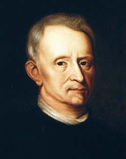
3 Antony van Leeuwenhoek ( )

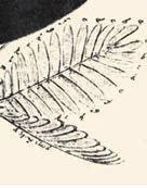
4 Robert Hook ( )
5 How simple lens microscope works
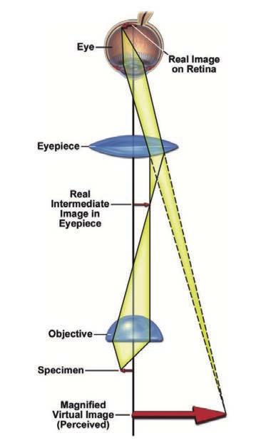
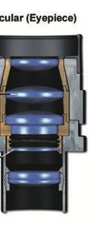
6 Compound Microscope


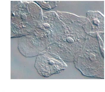
7 Light as a probe of matter!="# $=%"=%!/#

8 Light interacts with matter
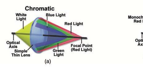
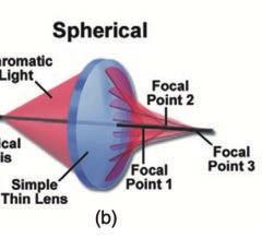
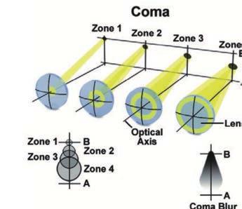
9 Aberrations of a simple lens


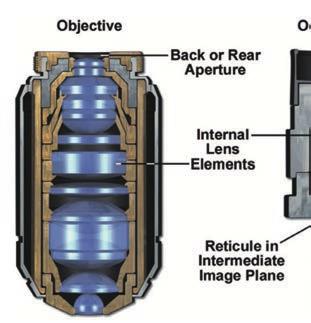
10 Objective lens designs Cheap Red-blue corrected Less expensive color-corrected Bright good resolution Very expensive Highly color-corrected Bright High resolution
11
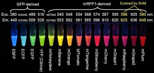
")

 Air:")

12 Numerical aperture (NA) resolving power = #/2NA Refractive index (RI) Air: 1.0 Water: 1.3 Glycerol: 1.47 Glass: 1.5 Oil: 1.52


13 Bright field Phase contrast Differential interference Stained

14 An unstained brain

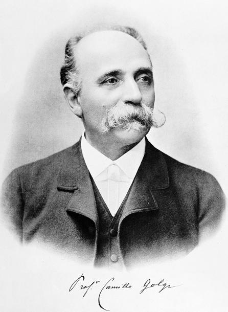
15 Camillo Golgi ( )

16 Santiago Ramón y Cajal ( )

17 Neuron theory

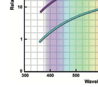
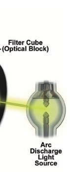
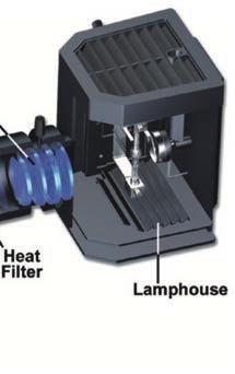
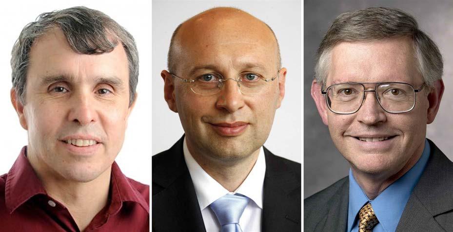
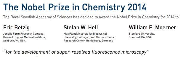
18 Nobel laureates in chemistry 2008


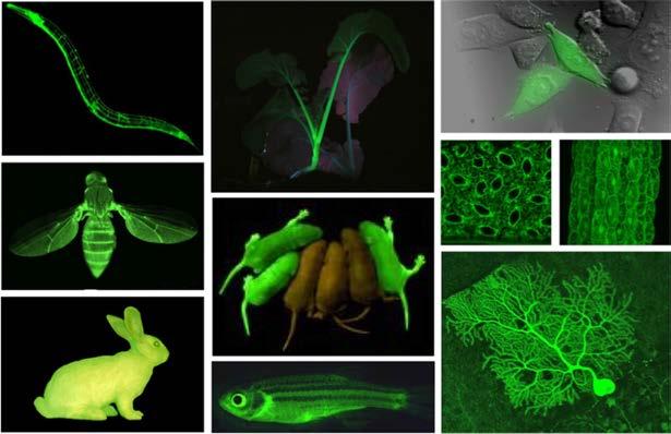
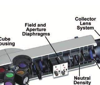
19 Aequorea victoria Osamu Purified Cloned & seq Douglas Prasher Robert Mutated Martin Expressed Improved

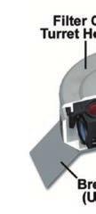
20 The power of differential labelling
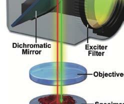
21

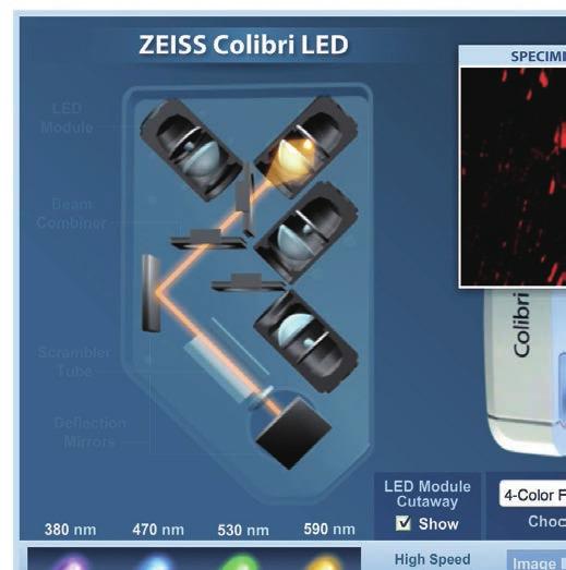
22 Seeing signaling pathway & protein-protein interaction Seeing cell-cell interaction Fluorescence Resonance Energy Transfer Seeing neural activity Seeing protein modification

23 Observing protein-protein interaction with FRET
24 Observing functioning synapses


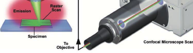
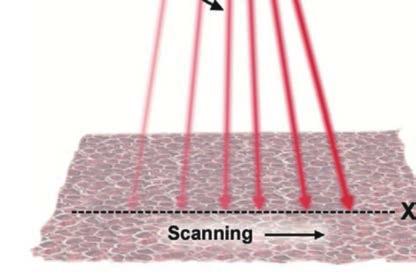
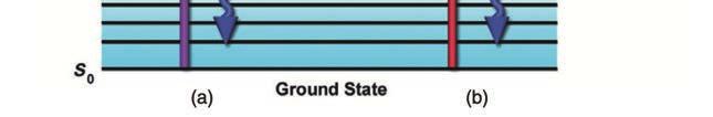
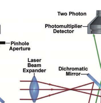
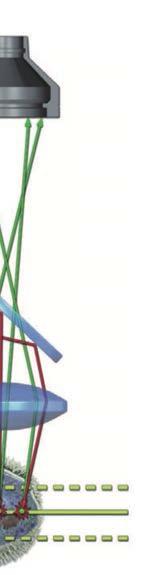
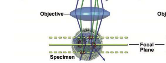
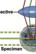
25 Physical basis of fluorescence upward arrow: absorption downward arrow: fluorescence emission wavy lines: heat
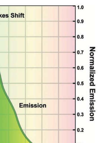
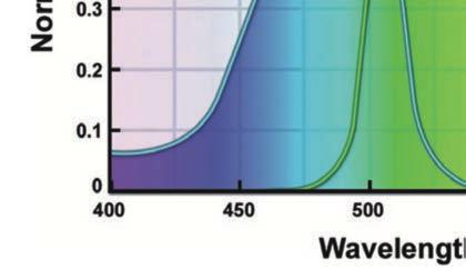
26 Absorption and emission spectra of fluorescein

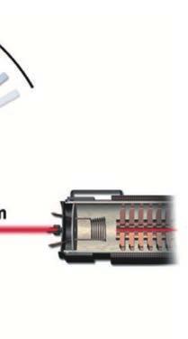
27 Light source


28 Filters can be used to isolated specific wavelength

29 Filter modules


30 The operation of filter cubes
31 Light-emitting diode (LED)
32 A four-color LED setup

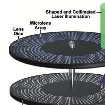
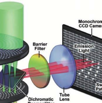
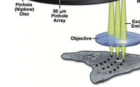
33 The thickness problem


34 Confocal laser scanning microscopy


35 Pinhole is the main mechanism for optical sectioning in confocal microscopy

36 Confocal laser scanning microscopy

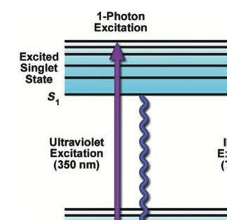
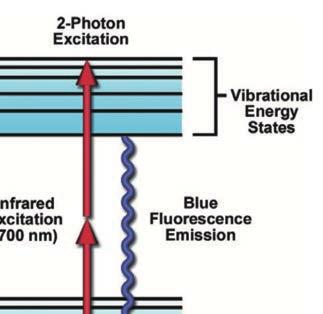
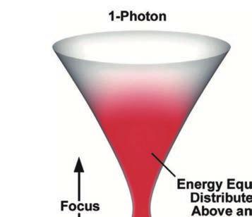
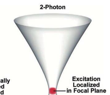
37 Scanning control mechanism

38 Confocal vs. Wildefield Microscopy


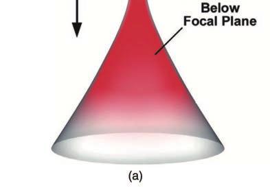
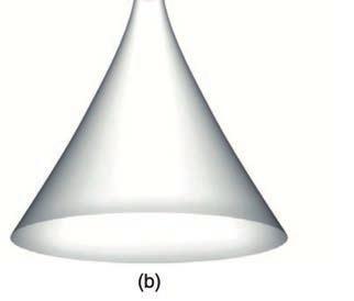
39 Effect of confocal parameters on image quality

40 3D reconstruction of a fly brain
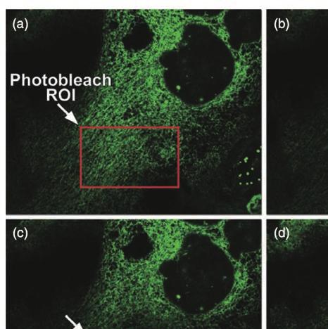
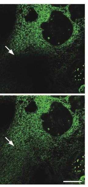
41 Fluorescence recovery after photobleaching (FRAP)
42 Increasing speed by spinning disks

43 Increasing speed by spinning disks


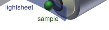
44 Eric Betzig Light sheet Microscopy


45 Whole brain activity imaging
46 Two-photon excitation Maria Göppert-Mayer ( )

47 Two photon vs confocal
48 Localised excitation
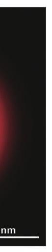
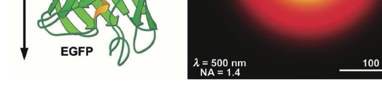
49 Localized excitation
50 Advantages and disadvantages of two-photon microscopy Near-infrared radiation penetrates tissues better: good for imaging thick specimens. The single-spot excitation causes less photodamage overall. Good for inducing photochemical reactions only on the focal plane: e.g. photoactivation of fluorescence proteins. Lower resolution compared to confocal microscopy

51 A specific subset of dopaminergic neurons responds to water


52 Less photon toxicity is the key Holtmaat & Svoboda 2009

53 Marching from high-resolution to super-resolution
54 Ernst Abbe ( ) Theoretical resolution limit for light microscopy & = #/2NA


55 Light microscopes only allow us to see a small portion of the world


56 Airy disc formation

57 Two airy discs


58 Image of a single GFP protein
59


60 Stefan Hell The RESOLFT concept REversible Saturable Optical Fluorescence Transitions

61 STED microscopy STimulated Emission Depletion microscopy

62 STED depletion lasers

63 STED Microscopy


64 PALM/STORM microscopy Eric Betzig PALM: photo activated localization microscopy STORM: stochastic optical reconstruction microscopy

65 PALM/STORM microscopy

66 Current microscopy limit Electron microscopy Light microscopy Superresolution microscopy Unaided eye Human height Length of some nerve and muscle cells Chicken egg Frog egg Human egg Nucleus Most plant and animal cells Most bacteria Mitochondrion Smallest bacteria Viruses Ribosomes Proteins Lipids Small molecules Atoms 10 m 1 m 0.1 m 1 cm 1 mm 100!m 10!m 1!m 100 nm 10 nm 1 nm
Cell Structure and Function
 Cell Structure and Function Dead White Men Who Discovered (and were made of) Cells: Anton Van Leeuwenhoek Robert Hooke Where the Magic Happened Schleiden Cell Theory All plants are made of cells Schwann
Introduction to Computational Fluorescence Microscopy!
 Introduction to Computational Fluorescence Microscopy! EE367/CS448I: Computational Imaging and Display! stanford.edu/class/ee367! Lecture 13! Gordon Wetzstein! Stanford University! Midterm! Tuesday, Feb
Special Techniques 1. Mark Scott FILM Facility
 Special Techniques 1 Mark Scott FILM Facility SPECIAL TECHNIQUES Multi-photon microscopy Second Harmonic Generation FRAP FRET FLIM In-vivo imaging TWO-PHOTON MICROSCOPY Alternative to confocal and deconvolution
Final Exam, 176 points PMB 185: Techniques in Light Microscopy
 Final Exam, 176 points Name PMB 185: Techniques in Light Microscopy Point value is in parentheses at the end of each question. 1) Order the steps in setting up Köhler illumination. It is not necessary
PALM/STORM, BALM, STED
 PALM/STORM, BALM, STED Last class 2-photon Intro to PALM/STORM Cyanine dyes/dronpa This class Finish localization super-res BALM STED Localization microscopy Intensity Bins = pixels xx 2 = ss2 + aa 2 /12
Bi177 - Lecture 13 Microscopy Outside the Box. Fluorescence Nanoscopy TIRF 4-pi STED STORM/PALM
 Bi177 - Lecture 13 Microscopy Outside the Box Fluorescence Nanoscopy TIRF 4-pi STED STORM/PALM The diffraction limit: Abbe s law The Problem Diffraction limit 100x larger than molecular scale! Green Fluorescent
Confocal Microscopy & Imaging Technology. Yan Wu
 Confocal Microscopy & Imaging Technology Yan Wu Dec. 05, 2014 Cells under the microscope What we use to see the details of the cell? Light and Electron Microscopy - Bright light / fluorescence microscopy
Microscopy. CS/CME/BioE/Biophys/BMI 279 Nov. 2, 2017 Ron Dror
 Microscopy CS/CME/BioE/Biophys/BMI 279 Nov. 2, 2017 Ron Dror 1 Outline Microscopy: the basics Fluorescence microscopy Resolution limits The diffraction limit Beating the diffraction limit 2 Microscopy:
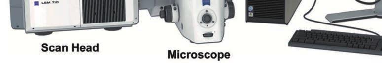
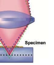
Confocal Microscopes. Evolution of Imaging
 Confocal Microscopes and Evolution of Imaging Judi Reilly Hans Richter Massachusetts Institute of Technology Environment, Health & Safety Office Radiation Protection What is Confocal? Pinhole diaphragm
STORM/PALM. Super Resolution Microscopy 10/31/2011. Looking into microscopic world of life
 Super Resolution Microscopy STORM/PALM Bo Huang Department of Pharmaceutical Chemistry, UCSF CSHL Quantitative Microscopy, 1/31/211 Looking into microscopic world of life 1 µm 1 µm 1 nm 1 nm 1 nm 1 Å Naked
Two-Photon Microscopy for Deep Tissue Imaging of Living Specimens
 for Deep Tissue Imaging of Living Specimens Tilman Franke* and Sebastian Rhode TILL Photonics GmbH, an FEI company, Lochhamer Schlag 21, D-82166 Gräfelfing, Germany *tilman.franke@fei.com Introduction
EuBI application for access
 * Title * First name * Last name * Email address * Institution/Company URL of the Institution/Company * Phone number Country code Phone * Street address * Zip Code * City * Country * Position * Not a Principal
Visualizing Cells Molecular Biology of the Cell - Chapter 9
 Visualizing Cells Molecular Biology of the Cell - Chapter 9 Resolution, Detection Magnification Interaction of Light with matter: Absorbtion, Refraction, Reflection, Fluorescence Light Microscopy Absorbtion
Super-resolution Microscopy
 Semr oc kwhi t epaperser i es : 1. Introduction Super-resolution Microscopy Fluorescence microscopy has revolutionized the study of biological samples. Ever since the invention of fluorescence microscopy
Super Resolution Microscopy - Breaking the Diffraction Limit Radiological Research Accelerator Facility
 Super Resolution Microscopy - Breaking the Diffraction Limit Radiological Research Accelerator Facility Sabrina Campelo, Dr. Andrew Harken Outline Motivation Fluorescence Microscopy -Multiphoton Imaging
Fluorescence Light Microscopy for Cell Biology
 Fluorescence Light Microscopy for Cell Biology Why use light microscopy? Traditional questions that light microscopy has addressed: Structure within a cell Locations of specific molecules within a cell
Resolution of Microscopes Visible light is nm Dry lens(0.5na), green(530nm light)=0.65µm=650nm for oil lens (1.4NA) UV light (300nm) = 0.13µm f
, green(530nm light)=0.65µm=650nm for oil lens (1.4NA) UV light (300nm) = 0.13µm f") Microscopes and Microscopy MCB 380 Good information sources: Alberts-Molecular Biology of the Cell http://micro.magnet.fsu.edu/primer/ http://www.microscopyu.com/ Approaches to Problems in Cell Biology
Microscopes and Microscopy MCB 380 Good information sources: Alberts-Molecular Biology of the Cell http://micro.magnet.fsu.edu/primer/ http://www.microscopyu.com/ Approaches to Problems in Cell Biology
Sample region with fluorescent labeled molecules
 FLUORESCENCE IMAGING I. Fluorescence-imaging with diffraction limited spots The resolution in optical microscopy has been hampered by the smallest spot possible (~ λ/2) that can be achieved by conventional
5/11/2015 MICROSCOPIC TECHNIQUES 2. Fluorescence microscopy SPECIAL TECHNIQUES BASED ON FLUORESCENCE MICROSCOPY
 UNIVERSITY OF PÉCS MEDICAL SCHOOL www.medchool.pte.hu MICROSCOPIC TECHNIQUES 2 SPECIAL TECHNIQUES BASED ON FLUORESCENCE MICROSCOPY BIOPHYSICS 2. 2015 25th March Dr. Beáta Bugyi Department of Biophyic Fluorecence
Fluorescence microscopy
 Fluorescence microscopy 1 Fluorescence microscopies basic fluorescence, fluorophores Deconvolution Confocal Two-photon/multi-photon 4Pi Light sheet Total internal reflection STED FRAP/FLIP/FCS FRET PALM/STORM/iPALM
Fluorescence Nanoscopy
 Fluorescence Nanoscopy Keith A. Lidke University of New Mexico panda3.phys.unm.edu/~klidke/index.html Optical Microscopy http://en.wikipedia.org/wiki/k%c3%b6hler_illumination 30 µm Fluorescent Probes Michalet
Dino-Lite knowledge & education. Fluorescence Microscopes
 Dino-Lite knowledge & education Fluorescence Microscopes Dino-Lite Fluorescence models Smallest fluorescence microscope in the world Revolution to biomedical and educational applications Flexible Easy
Cellular imaging using Nano- Materials. A Case-Study based approach Arun Murali, Srivats V
 Cellular imaging using Nano- Materials A Case-Study based approach Arun Murali, Srivats V Agenda Discuss a few papers Explain a couple of new imaging techniques and their benefits over conventional imaging
BIO 315 Lab Exam I. Section #: Name:
 Section #: Name: Also provide this information on the computer grid sheet given to you. (Section # in special code box) BIO 315 Lab Exam I 1. In labeling the parts of a standard compound light microscope
Winter College on Micro and Nano Photonics for Life Sciences February General Overview
 1932-15 Winter College on Micro and Nano Photonics for Life Sciences 11-22 February 2008 General Overview Martina Havenith Ruhr University Bochum Bochum, Germany Microscopy- An Overview M. Havenith Ruhr-University
Imaging facilities at WUR
 Imaging facilities at WUR Advanced light microscopy facilities at Wageningen UR Programme Thursday 13 June 2013 Lunch meeting organized by Cat-Agro Food 12.00 Welcome and sandwich lunch 12.10 Introduction
FLUORESCENCE. Matyas Molnar and Dirk Pacholsky
 FLUORESCENCE Matyas Molnar and Dirk Pacholsky 1 Information This lecture contains images and information from the following internet homepages http://micro.magnet.fsu.edu/primer/index.html http://www.microscopyu.com/
Confocal Microscopy of Electronic Devices. James Saczuk. Consumer Optical Electronics EE594 02/22/2000
 Confocal Microscopy of Electronic Devices James Saczuk Consumer Optical Electronics EE594 02/22/2000 Introduction! Review of confocal principles! Why is CM used to examine electronics?! Several methods
Multiphoton Microscopy: Seeing deeper and clearer
 Multiphoton Microscopy: Seeing deeper and clearer Since the invention of simple microscope by Leuwenhoek and Hooke in the 17th century, different types of light microscopy techniques (such as phase contrast,
Fluorescence Microscopy
 Fluorescence Microscopy Dr. Arne Seitz Swiss Institute of Technology (EPFL) Faculty of Life Sciences Head of BIOIMAGING AND OPTICS BIOP arne.seitz@epfl.ch Fluorescence Microscopy Why do we need fluorescence
The most extensively used technique for tissue analysis is light microscopy.
 Fluorescence Theory Quantum yield Wavelength shift Ligand interactions Membrane interactions Using quenchning effects Fluorescence in-vivo Localization Distance measurements FRET The most extensively used
Fluorescence Microscopy
 Fluorescence Microscopy Dr. Arne Seitz Swiss Institute of Technology (EPFL) Faculty of Life Sciences Head of BIOIMAGING AND OPTICS BIOP arne.seitz@epfl.ch Fluorescence Microscopy Why do we need fluorescence
Methods of Culturing Microorganisms. Chapter 3. Five Basic Techniques of Culturing Bacteria. Topics
 Chapter 3 Topics Methods of Culturing Microorganisms Microscope (History, Types, Definitions) Staining (Gram s) Methods of Culturing Microorganisms Five basic techniques of culturing Media Microbial growth
BIO 315 Lab Exam I. Section #: Name:
 Section #: Name: Also provide this information on the computer grid sheet given to you. (Section # in special code box) BIO 315 Lab Exam I 1. In labeling the parts of a standard compound light microscope
Fluorescence Microscopy: A Biological Perspective
 Fluorescence Microscopy: A Biological Perspective From nanometre to metre: the scale of life Instrumentation and accessible scale limits the questions that can be addressed in biology Why are there limits?
Lasers for Microscopy: Major Trends
 Lasers for Microscopy: Major Trends Marco Arrigoni, Nigel Gallaher, Darryl McCoy, Volker Pfeufer and Matthias Schulze, Coherent Inc. Laser development for the microscopy market continues to be driven by
In spite of its long history, optical
 Major Trends Laser development for the microscopy market continues to be driven by key trends in applications, which currently include superresolution techniques, multiphoton applications in optogenetics
Fluorescence Microscopy. Terms and concepts to know: 10/11/2011. Visible spectrum (of light) and energy
 and energy") Fluorescence Microscopy Louisiana Tech University Ruston, Louisiana Microscopy Workshop Dr. Mark DeCoster Associate Professor Biomedical Engineering 1 Terms and concepts to know: Signal to Noise Excitation
Fluorescence Microscopy Louisiana Tech University Ruston, Louisiana Microscopy Workshop Dr. Mark DeCoster Associate Professor Biomedical Engineering 1 Terms and concepts to know: Signal to Noise Excitation
Fluorescence Nanoscopy 高甫仁 ) Institute of Biophotonics, National Yang Ming University. Outline
 Institute of Biophotonics, National Yang Ming University. Outline") Fluorescence Nanoscopy 高甫仁 ) Fu-Jen Kao ( 高甫仁 Institute of Biophotonics, National Yang Ming University Outline The Abbe s (diffraction) limit and nanoscopy Fundamentals and opportunities of FLIM/FRET Visualizing
Fluorescence Nanoscopy 高甫仁 ) Fu-Jen Kao ( 高甫仁 Institute of Biophotonics, National Yang Ming University Outline The Abbe s (diffraction) limit and nanoscopy Fundamentals and opportunities of FLIM/FRET Visualizing
Simultaneous multi-color, multiphoton fluorophore excitation using dual-color fiber lasers
 Multiphoton Microscopy / Fiber Laser Simultaneous multi-color, multiphoton fluorophore excitation using dual-color fiber lasers Matthias Handloser, Tim Paasch-Colberg, Bernhard Wolfring TOPTICA Photonics
a) JOURNAL OF BIOLOGICAL CHEMISTRY b) PNAS c) NATURE
 JOURNAL OF BIOLOGICAL CHEMISTRY b) PNAS c) NATURE") a) JOURNAL OF BIOLOGICAL CHEMISTRY b) c) d) ........................ JOURNAL OF BIOLOGICAL CHEMISTRY MOLECULAR PHARMACOLOGY TRENDS IN PHARMACOLOGICAL S AMERICAN JOURNAL OF PHYSIOLOGY-HEART AND CIRCULATORY
a) JOURNAL OF BIOLOGICAL CHEMISTRY b) c) d) ........................ JOURNAL OF BIOLOGICAL CHEMISTRY MOLECULAR PHARMACOLOGY TRENDS IN PHARMACOLOGICAL S AMERICAN JOURNAL OF PHYSIOLOGY-HEART AND CIRCULATORY
New developments in STED Microscopy
 New developments in STED Microscopy Arnold Giske*, Jochen Sieber, Hilmar Gugel, Marcus Dyba, Volker Seyfried, Dietmar Gnass Leica Microsystems CMS, Am Friedensplatz 3, 68126 Mannheim, Germany ABSTRACT
 Page 1 of 9 Fundamentals and Applications in Multiphoton Excitation Microscopy Two-photon excitation microscopy (also referred to as non-linear, multiphoton, or two-photon laser scanning microscopy) is
Workshop advanced light microscopy
 Workshop advanced light microscopy Multi-mode confocal laser scanning microscope Jan Willem Borst Laboratory of Biochemistry Biomolecular Networks www.bic.wur.nl MicroSpectroscopy Centre Wageningen Microspectroscopy
How to use the SP5 confocal microscope
 How to use the SP5 confocal microscope Mailfert Sébastien (mailfert@ciml.univ-mrs.fr) Tel : 9126 Imaging Immunity (ImagImm) photonic microscopy facility Centre d Immunologie de Marseille-Luminy 2016 /
Wednesday, October 8. Today: Last Time: Changes in T & P Units Equilibrium calculations: some examples. Readings: Chang & Thoman:
 Wednesday, October 8 Last Time: Entropy of mixing Chemical potential and equilibrium The equilibrium constant Today: Changes in T & P Units Equilibrium calculations: some examples Readings: Chang & Thoman:
Live cell microscopy
 Live cell microscopy 1. Why do live cell microscopy? 2. Maintaining living cells on a microscope stage. 3. Considerations for imaging living cells. 4. Fluorescence labeling of living cells. 5. Imaging
Rice/TCU REU on Computational Neuroscience. Fundamentals of Molecular Imaging
 Rice/TCU REU on Computational Neuroscience Fundamentals of Molecular Imaging June 2, 2009 Neal Waxham 713-500-5621 m.n.waxham@uth.tmc.edu Objectives Introduction to resolution in light microscopy Brief
Total Internal Reflection Fluorescence Microscopy
 Total Internal Reflection Microscopy Nicole O Neil Indiana University October 24, 2005 Agenda Why use TIRFM? Theory behind TIR Snell s Law Instrumentation Evanescent Wave Excitation of Fluorophores Advantages/Disadvantages
Multiplexed 3D FRET imaging in deep tissue of live embryos Ming Zhao, Xiaoyang Wan, Yu Li, Weibin Zhou and Leilei Peng
 Scientific Reports Multiplexed 3D FRET imaging in deep tissue of live embryos Ming Zhao, Xiaoyang Wan, Yu Li, Weibin Zhou and Leilei Peng 1 Supplementary figures and notes Supplementary Figure S1 Volumetric
Janos Szabad Department of Biology University of Szeged 6720 Szeged, Somogyi str
 Janos Szabad Department of Biology University of Szeged 6720 Szeged, Somogyi str. 4. E-mail: szabad.janos@med.u-szeged.hu - Through the use of antibodies - against the protein - against a fusion partner
Absorption of an electromagnetic wave
 In vivo optical imaging?? Absorption of an electromagnetic wave Tissue absorption spectrum Extinction = Absorption + Scattering Absorption of an electromagnetic wave Scattering of an electromagnetic wave
ADVANCED LIGHT MICROSCOPY TECHNOLOGY SPECIFIC REVIEW CRITERIA FOR EURO-BIOIMAGING NODE
 ADVANCED LIGHT MICROSCOPY TECHNOLOGY SPECIFIC REVIEW CRITERIA FOR EURO-BIOIMAGING NODE October 15, 2012 TABLE OF CONTENTS Introduction... 3 Common Technology Review Criteria for Advanced Light Microscopy
SAPIENZA Università di Roma Laurea magistrale in Ingegneria delle Nanotecnologie A.A Biophotonics Laboratory Course
 SAPIENZA Università di Roma Laurea magistrale in Ingegneria delle Nanotecnologie A.A. 2016-2017 Biophotonics Laboratory Course Prof. Francesco Michelotti SAPIENZA Università di Roma Facoltà di Ingegneria
Super Resolution Imaging Solution Provider. Imaging Future
 Super Resolution Imaging Solution Provider Imaging Future Imaging Solution More Than Equipment NanoBioImaging(NBI) is the Industrial Partner of HKUST Super Resolution Imaging Center (SRIC). NBI aims to
Practical light microscopy: an introduction
 Practical light microscopy: an introduction Dr. Mark Leake, Oxford University www.physics.ox.ac.uk/users/leake Aim of today s talk: Explanation of the very (very) basics of how a light microscope works
Microscopy, Staining, and Classification
 CSLO CHECK CSLO1. Describe distinctive characteristics and diverse growth requirements of prokaryotic organisms compared to eukaryotic organisms. PowerPoint Lecture Presentations prepared by Mindy Miller-Kittrell,
MICROSCOPY. "micro" (small) "scopeo" (to watch)
 scopeo (to watch)") MICROSCOPY "micro" (small) "scopeo" (to watch) THE RELATIVE SIZES OF MOLECULES, CELLS AND ORGANISMS THE RELATIVE SIZES OF MOLECULES, CELLS AND ORGANISMS MICROSCOPY 1590 2012 MICROSCOPY THE LIGHT Light:
MICROSCOPY "micro" (small) "scopeo" (to watch) THE RELATIVE SIZES OF MOLECULES, CELLS AND ORGANISMS THE RELATIVE SIZES OF MOLECULES, CELLS AND ORGANISMS MICROSCOPY 1590 2012 MICROSCOPY THE LIGHT Light:
STED microscopy with single light source. TeodoraŞcheul
 STED microscopy with single light source TeodoraŞcheul Dr. Iréne Wang, Dr. Jean-Claude Vial LIPhy, Grenoble, France Summary I. Introduction to STED microscopy II. STED with one laser source 1. Two-photon
HYPERSPECTRAL MICROSCOPE PLATFORM FOR HIGHLY MULTIPLEX BIOLOGICAL IMAGING. Marc Verhaegen
 HYPERSPECTRAL MICROSCOPE PLATFORM FOR HIGHLY MULTIPLEX BIOLOGICAL IMAGING Marc Verhaegen CMCS, MONTREAL, MAY 11 th, 2017 OVERVIEW Hyperspectral Imaging Multiplex Biological Imaging Multiplex Single Particle
Biophotonics?? Biophotonics. technology in biomedical engineering. Advantages of the lightwave
 Biophotonics - Imaging: X-ray, OCT, polarimetry, DOT, TIRF, photon migration, endoscopy, confocal microscopy, multiphoton microscopy, multispectral imaging - Biosensing: IR spectroscopy, fluorescence,
More on fluorescence
 More on fluorescence Last class Fluorescence Absorption emission Jablonski diagrams This class More on fluorescence Common fluorophores Jablonski diagrams to spectra Properties of fluorophores Excitation
Nanoscale measurement examples in biology: Sub-diffractive imaging & the fly brain challenge
 Nanoscale measurement examples in biology: Sub-diffractive imaging & the fly brain challenge Harald Hess, HHMI Janelia Farm Introduction to HHMI Janelia Farms Sample: Nerves, Worm Brains, Fly Brains, Rodent
SUPER-RESOLUTION MICROSCOPY. Dr. Nathalie Garin
 SUPER-RESOLUTION MICROSCOPY Dr. Nathalie Garin Content Motivation for superresolution Superresolution, nanoscopy, : definition Structured Illumination Microscopy (SIM) Localization microscopy STimulated
Imaging of endocrine organs
 Imaging of endocrine organs Helen Christian Department of Physiology, Anatomy & Genetics St Anne s College, University of Oxford Diabetesforum, Stockholm 2017 Islets of Langerhan Pituitary gland Renin
Concept review: Fluorescence
 16 Concept review: Fluorescence Some definitions: Chromophore. The structural feature of a molecule responsible for the absorption of UV or visible light. Fluorophore. A chromophore that remits an absorbed
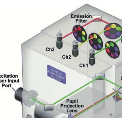
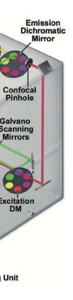
Imaging & analysis with the LSM780 NLO Discover the secrets beyond the twilight zone
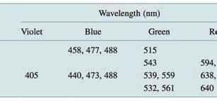
 Imaging & analysis with the LSM780 NLO Discover the secrets beyond the twilight zone Sven Terclavers LSM780 System overview The Scan Module - Core of the LSM 780 1 V/tunable PTC laser ports (405/440, cw/ps;
CENTER FOR BRAIN EXPERIMENT
 CENTER FOR BRAIN EXPERIMENT Section of Brain Structure Associate Professor: ARII, Tatsuo, PhD 1967 Graduated from Tohoku University, Faculty of Science. Completed the doctoral course in Engineering, Nagoya
BIOCHEMIST ALL IN ONE ARTICLE
 BIOCHEMIST ALL IN ONE ARTICLE Bringing ease-of-use to microscopy From the Philosopher s Stone to the Researcher s Dream Although naturally occurring luminescence has been observed for many centuries, the
High Throughput Whole Organ Imaging Based on Multifocal Multiphoton Microscope
 High Throughput Whole Organ Imaging Based on Multifocal Multiphoton Microscope LBRC researchers: Peter So, Jae Won Cha, Elijah Yew, Vijay Singh External technology collaborators: Prof. Hanry Yu (University
Introduction CHAPTER 1
 CHAPTER 1 Introduction 1.1 Light Microscopy The light microscope is one of the significant inventions in the history of humankind that, along with the telescope, played a central role in the Scientific
Genetically targeted all-optical electrophysiology with a transgenic Credependent
 Genetically targeted all-optical electrophysiology with a transgenic Credependent Optopatch mouse Short title: Transgenic Optopatch mouse Shan Lou 1, Yoav Adam 1, Eli N. Weinstein 1,4, Erika Williams 2,
FLIM Fluorescence Lifetime IMaging
 FLIM Fluorescence Lifetime IMaging Fluorescence lifetime t I(t) = F0 exp( ) τ 1 τ = k f + k nr k nr = k IC + k ISC + k bl Batiaens et al, Trends in Cell Biology, 1999 τ τ = fluorescence lifetime (~ns to
Design for Manufacturability (DFM) in the Life Sciences
 in the Life Sciences") T E C H N I C A L N O T E Design for Manufacturability (DFM) in the Life Sciences Fluorescence Spectroscopy Product Platform Realized with TracePro TM Suite of Opto-Mechanical Design Software Tools Authors:
T E C H N I C A L N O T E Design for Manufacturability (DFM) in the Life Sciences Fluorescence Spectroscopy Product Platform Realized with TracePro TM Suite of Opto-Mechanical Design Software Tools Authors:
Satoshi Kawata. Near-Field Optic s and Surface Plasmon Polaritons
 Satoshi Kawata Near-Field Optic s and Surface Plasmon Polaritons Near-Field Optics and the Surface Plasmon Polariton Dieter W. Pohl 1 1. Introduction 1 2. Back to the Roots 1 2.1. Rayleigh and Mie Scattering
SIM SSIM. nanoscopy RESOLFT. Super-resolution STORM GSDIM. dstorm PALMIRA FPALM PALM PAINT SPRAIPAINT SOFI BALM CALM. Bo Huang
 STEDGSD STORM SOFI nanoscopy GSDIM PALMIRA SMACM BBB PAINT SPRAIPAINT CALM RESOLFT BALM SIM SSIM Super-resolution Bo Huang 2013.08.01 dstorm FPALM PALM 50 years to extend the resolution Confocal microscopy
Foundations in Microbiology Seventh Edition
 Lecture PowerPoint to accompany Foundations in Microbiology Seventh Edition Talaro Chapter 3 Tools of the Laboratory: The Methods for Studying Microorganisms Copyright The McGraw-Hill Companies, Inc. Permission
Confocal Microscopy Analyzes Cells
 Choosing Filters for Fluorescence A Laurin Publication Photonic Solutions for Biotechnology and Medicine November 2002 Confocal Microscopy Analyzes Cells Reprinted from the November 2002 issue of Biophotonics
PEER REVIEW FILE. Reviewers' comments: Reviewer #1 (Remarks to the Author):
:") PEER REVIEW FILE Reviewers' comments: Reviewer #1 (Remarks to the Author): General In its beginnings in the mid 1990s, localization microscopy based on optical isolation of fluorescent point targets was
PEER REVIEW FILE Reviewers' comments: Reviewer #1 (Remarks to the Author): General In its beginnings in the mid 1990s, localization microscopy based on optical isolation of fluorescent point targets was
Microscopy from Carl Zeiss. DirectFRAP. News from the Cell. The New Class of Laser Manipulation for the Analysis of Cell Dynamics
 Microscopy from Carl Zeiss DirectFRAP News from the Cell The New Class of Laser Manipulation for the Analysis of Cell Dynamics DirectFRAP. New Insights into Cell Dynamics. Fluorescence breaks new ground:
CHARACTERIZATION OF MOLECULAR ORIENTATION IN SUPER-RESOLUTION FLUORESCENCE MICROSCOPY
 Master Erasmus Mundus in Photonics Engineering, Nanophotonics and Biophotonics Europhotonics MASTER THESIS WORK CHARACTERIZATION OF MOLECULAR ORIENTATION IN SUPER-RESOLUTION FLUORESCENCE MICROSCOPY Yibing
In vivo fast imaging and optogenetic manipulation using genetically-encoded fluorescent indicators and actuators. Serena Bovetti
 In vivo fast imaging and optogenetic manipulation using genetically-encoded fluorescent indicators and actuators Serena Bovetti Istituto Italiano di Tecnologia Genova, Italy Bogliasco, June 6-8 2016 Analyzing
Sub-micron scale patterning of fluorescent. silver nanoclusters using low-power laser
 Sub-micron scale patterning of fluorescent silver nanoclusters using low-power laser Puskal Kunwar 1,*, Jukka Hassinen 2, Godofredo Bautista 1, Robin H. A. Ras 2, and Juha Toivonen 1 1 Tampere University
A simple introduction to multiphoton microscopy
 Journal of Microscopy, Vol. 243, Pt 3 2011, pp. 221 226 Received 29 April 2011; accepted 28 June 2011 doi: 10.1111/j.1365-2818.2011.03532.x A simple introduction to multiphoton microscopy A. USTIONE &
Introduction. (b) (a)
 (a)") Introduction Whispering Gallery modes (WGMs) in dielectric micro-cavities are resonant electromagnetic modes that are of considerable current interest because of their extremely high Q values leading to
Introduction Whispering Gallery modes (WGMs) in dielectric micro-cavities are resonant electromagnetic modes that are of considerable current interest because of their extremely high Q values leading to
Microstructural Characterization of Materials
 Microstructural Characterization of Materials 2nd Edition DAVID BRANDON AND WAYNE D. KAPLAN Technion, Israel Institute of Technology, Israel John Wiley & Sons, Ltd Contents Preface to the Second Edition
8:00 pm David A. Agard, HHMI/University of California, San Francisco Welcome and introduction to the meeting
 Sunday, May 20 th 3:00 pm Check-in 6:00 pm Reception 7:00 pm Dinner 8:00 pm Session 1: Introduction 8:00 pm David A. Agard, HHMI/University of California, San Francisco Welcome and introduction to the
cell and tissue imaging by fluorescence microscopy
 cell and tissue imaging by fluorescence microscopy Steven NEDELLEC Plateforme Micropicell SFR Santé François Bonamy Nantes 1 A matter of size Limit of resolution 0.15mm aims: building the image of an object
High Power Diode Lasers and Multi Laser Engines, Expanding the Range of Biophotonics Applications. Konstantin Birngruber TOPTICA Photonics AG
 High Power Diode Lasers and Multi Laser Engines, Expanding the Range of Biophotonics Applications Konstantin Birngruber TOPTICA Photonics AG TOPTICA Photonics AG Company facts Founded 1998 180 employees
Confocal Microscopy. Alberto Diaspro Mario Faretta Paolo Sapuppo
 Confocal Microscopy Alberto Diaspro Mario Faretta Paolo Sapuppo Confocal Book Alberto Diaspro LAMBS-MicroScoBio, Department of Physics, University of Genoa, Genoa, Italy Istituto FIRC di Oncologia Molecolare
Methods of Characterizing Neural Networks
 Methods of Characterizing Neural Networks Ashley Nord University of Minnesota Minneapolis, MN 55414 Advisors: Katsushi Arisaka, Adrian Cheng University of California Los Angeles Los Angeles, CA 90024 September
Femtosecond micromachining in polymers
 Femtosecond micromachining in polymers Prof. Dr Cleber R. Mendonca Daniel S. Corrêa Prakriti Tayalia Dr. Tobias Voss Dr. Tommaso Baldacchini Prof. Dr. Eric Mazur fs-micromachining focus laser beam inside
Supplementary Table 1. Components of an FCS setup (1PE and 2PE)
") Supplementary Table 1. Components of an FCS setup (1PE and 2PE) Component and function Laser source Excitation of fluorophores Microscope with xy-translation stage mounted on vibration isolated optical
Supplementary Table 1. Components of an FCS setup (1PE and 2PE) Component and function Laser source Excitation of fluorophores Microscope with xy-translation stage mounted on vibration isolated optical
A cost-effective fluorescence detection system for pulsed laser analysis
 Susquehanna University Scholarly Commons Chemistry Faculty Publications 2-2015 A cost-effective fluorescence detection system for pulsed laser analysis J. W. Lafferty Susquehanna University N. A. Fox Susquehanna
NV High Brightness Series
 PRODUCT SHEET Rev. 8/17, v9 NV High Brightness Series The NV-High Brightness Series features fluorescent nanodiamonds ranging in size from 20 nm up to 150µm containing nitrogen vacancy (NV) centers with
Super-resolution imaging: early days w/ Video-enhanced DIC, TIRF, PALM, STORM, etc.
 15/05/2012 Super-resolution imaging: early days w/ Video-enhanced DIC, TIRF, PALM, STORM, etc. Prof. Dr. Rainer Duden duden@bio.uni-luebeck.de 1 Using conventional light microscopy resolution is limited
Research area in the Strategic Objective Development of optical control technologies and elucidation of biological mechanisms
 Research area in the Strategic Objective Development of optical control technologies and elucidation of biological mechanisms 6.1.5 Development and application of optical technology for spatiotemporal
The new LSM 700 from Carl Zeiss
 The new LSM 00 from Carl Zeiss Olaf Selchow, Bernhard Goetze To cite this version: Olaf Selchow, Bernhard Goetze. The new LSM 00 from Carl Zeiss. Biotechnology Journal, Wiley- VCH Verlag, 0, (), pp.. .
Introduction to N-STORM
 Introduction to N-STORM Dan Metcalf Advanced Imaging Manager Outline Introduction Principles of STORM Applications N-STORM overview Biological Scale Mitochondrion Microtubule Amino Acid 1Å Kinesin 1nm
Euro-BioImaging European Research Infrastructure for Imaging Technologies in Biological and Biomedical Sciences
 Euro-BioImaging 262023 D6.3 Definition of technical standards Euro-BioImaging European Research Infrastructure for Imaging Technologies in Biological and Biomedical Sciences WP6 Advanced Light Microscopy