unc-33/crmp and ankyrin organize microtubules and localize kinesin to polarize
|
|
|
- Margaret Harvey
- 6 years ago
- Views:
Transcription
1 Supplementary Information unc-33/crmp and ankyrin organize microtubules and localize kinesin to polarize axon-dendrite sorting Tapan A. Maniar 1, Miriam Kaplan 1, George J. Wang 2, Kang Shen 2, Li Wei 3, Jocelyn E. Shaw 3, Sandhya P. Koushika 4, Cornelia I. Bargmann 1* 1 Laboratory of Neural Circuits and Behavior, Howard Hughes Medical Institute, The Rockefeller University, 1230 York Avenue, New York, NY Department of Biology, Howard Hughes Medical Institute, Stanford University, 385 Serra Mall, California 94305, USA 3 Department of Genetics, Cell Biology and Development, University of Minnesota, Minneapolis, MN 55406, USA 4 NCBS-TIFR, Bellary Road, Bangalore , India *Author for correspondence: cori@rockefeller.edu This document includes seven Supplementary Figures and Legends, and details of plasmid construction. 1
2 2
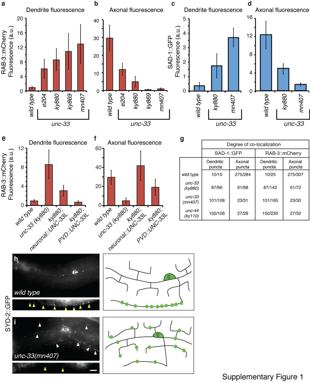
3 Supplementary Figure 1. unc-33 mutants mislocalize RAB-3::mCherry, SAD-1::GFP, and the active zone component SYD-2::GFP (liprin-α) to PVD dendrites (a-d) Alternative quantification of axonal localization defects (a, c) and dendritic mislocalization defects (b, d) of RAB-3::mCherry (a-b) and SAD-1::GFP (c-d) in PVDs of unc-33 mutants, scored as total average fluorescence intensity per cell (n=10-17 animals/genotype). (e,f) Rescue of axonal marker localization defects of unc-33(ky880) mutant through panneuronal and PVD-specific expression of UNC-33L. All error bars indicate s.e.m. (g) Quantification of co-localization of SAD-1::GFP and RAB-3::mCherry in PVD axons and dendrites of wild type, unc-33 and unc-44 animals. Fractions of SAD-1::GFP puncta co-localizing with RAB-3::mCherry are shown in the first two columns, and fractions of RAB-3::mCherry puncta co-localizing with SAD-1::GFP are shown in the third and fourth columns. (h,i) SYD-2::GFP localization in PVD neurons of wild type and unc-33(mn407) animals, with schematics. For each set of fluorescence micrographs, the top panel is the maximum intensity projection of dendritic focal planes and the bottom panel is the maximum intensity projection for axonal focal planes. Yellow and white arrowheads indicate axonal and dendritic puncta, respectively; cb denotes the PVD cell body and asterisks indicate gut autofluorescence. Anterior is at left and dorsal is up in all panels. Scale bar, 10 µm. 3
4 4
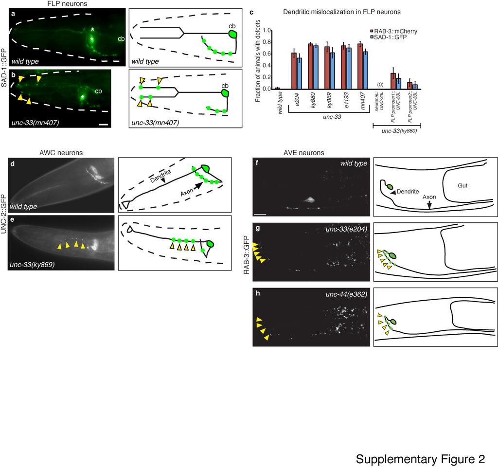
5 Supplementary Figure 2. unc-33 mutants mislocalize presynaptic proteins in multiple neuronal cell types (a,b) SAD-1::GFP distribution in FLP neurons of wild type and unc-33(mn407) animals, with schematics. cb indicates FLP cell bodies; yellow arrowheads point to SAD-1::GFP puncta mislocalized to FLP dendrites. Asterisks denote labeling of AWC neuron cell bodies with an odr-1::dsred marker. Anterior is at left and dorsal is up in all panels. (c) Dendritic mislocalization defects of RAB-3::mCherry and SAD-1::GFP in FLPs of unc-33 mutants, and rescue of unc-33 mutants through pan-neuronal and FLP-specific expression of UNC-33L. The fraction of animals with qualitative defects is shown (n>30 animals/genotype). Error bars indicate s.e.p. tag-168p, pan-neuronal promoter; mec-3p, promoter expressed in PVD, FLP, and six mechanosensory neurons; des-2p, promoter expressed in PVD and FLP. (d,e) Distribution of UNC-2::GFP 1, an α-subunit of a presynaptic voltage-gated calcium channel, in AWC and AWB chemosensory neurons of wild type (d) and unc-33(ky869) (e) animals, with schematics. Yellow arrowheads indicate mistargeting of UNC-2::GFP to dendrites. Scale bar, 10 µm. (f-h) Distribution of GFP::RAB-3 in AVE interneurons of wild type (f), unc-33(e204) (g) and unc-44(e362) (h) animals, with corresponding diagrams. Yellow arrowheads indicate mistargeting of GFP::RAB-3 to dendrites, observed in 65% (unc-33, n=222) or 23% (unc-44, n=92) of animals. Scale bar, 10 µm. 5

6 6
7 Supplementary Figure 3. unc-33 and unc-34/enabled mutants have short FLP and PVD axons, but unc-34 mutants do not mislocalize axonal markers (a) Premature termination of FLP axons in wild type, unc-33(ky880) and unc-34(e315). (b,c) Representative maximum projection fluorescence images of des-2::myristoyl::gfp showing FLP axon in a wild type animal (b), and a prematurely terminated FLP axon in an unc-34(e315) mutant (c). cb indicates the FLP cell body; white brackets indicate axons. Anterior is at left and dorsal is up in all panels. (d) Dendritic mislocalization of RAB-3::mCherry and SAD-1::GFP in FLPs of wild type and unc-34(e315) animals. Compare unc-33, Supplementary Fig. 2c. (e,f) Representative maximum projection fluorescence images showing SAD-1::GFP localization in a wild type FLP (e) and in a FLP with a prematurely terminated axon in an unc-34 (e315) animal (f). White arrowheads point to SAD-1::GFP axonal puncta. Asterisks denote AWC cell bodies labeled with an odr-1::dsred marker. Scale bars, 10 µm. (g) Quantification of animals with PVD axons shorter than the vulva spatial landmark. (h,i) Axonal localization defects (h) and dendritic mislocalization defects (i) of RAB- 3::mCherry and SAD-1::GFP in PVDs of wild type and unc-34(e315) animals (n>30 animals/genotype). Compare unc-33, Fig. 1k,l. All error bars indicate s.e.p. 7
 Representative images demonstrating the distribution of")
8 Supplementary Figure 4. Polarized localization of presynaptic proteins RAB- 3::mCherry and SAD-1::GFP can be detected in PVD neurons in late L3 larval stage (a,b) Representative images demonstrating the distribution of RAB-3::mCherry (a) and SAD-1::GFP (b) in PVD neurons of late L3 stage wild-type animals. For each set of fluorescence micrographs, the top panel is the maximum intensity projection of dendritic focal planes and the bottom panel is the maximum intensity projection for axonal focal planes. White arrowheads indicate axonal puncta, cb marks the PVD cell body, and asterisks mark gut autofluorescence. Anterior is at left and dorsal is up in all panels. Scale bar, 10 µm. 8
 Immunostaining against UNC-104 in the tail regions of wild type (a) and unc- 33(mn407) animals, with")
9 Supplementary Figure 5. Kinesin UNC-104 mislocalization to dendrites in multiple neurons in unc-33 mutants (a,b) Immunostaining against UNC-104 in the tail regions of wild type (a) and unc- 33(mn407) animals, with corresponding schematic diagrams. Yellow arrowheads point to UNC-104 immunoreactivity in tail dendrite regions. vnc, ventral nerve cord. (c,d) Localization of UNC-104::GFP in FLP neurons of wild type (c) and unc-33(mn407) (d) animals, with schematic diagrams. White arrowheads indicate UNC-104::GFP enrichment in FLP axons; yellow arrowheads indicate UNC-104::GFP mislocalized to FLP dendrite endings. (e) Quantification of animals with detectable UNC-104::GFP fluorescence in FLP dendrites (n>25 animals/genotype). Error bars indicate s.e.p. Scale bars, 10 µm. 9
 Representative maximum projection fluorescence images showing ODR-10::GFP localization in AWB neurons of odr-4 (a), unc-101 (c), odr-4; unc-33 (b) and unc-101; unc-33 (d) animals.")
10 Supplementary Figure 6. odr-4 and unc-101 regulate the distribution of ODR-10::GFP in wild type and unc-33 animals (a) Schematic diagram of AWB chemosensory neurons in the head. (b-e) Representative maximum projection fluorescence images showing ODR-10::GFP localization in AWB neurons of odr-4 (a), unc-101 (c), odr-4; unc-33 (b) and unc-101; unc-33 (d) animals. Anterior is at left and dorsal is up in all images. Scale bar, 10 µm. 10
11 Comparisons with Fig 5a-c show that the double mutants odr-4; unc-33 and unc-101; unc-33 resemble odr-4 and unc-101 single mutants, respectively. (f) Quantification of animals with axonal ODR-10::GFP fluorescence, demonstrating that both the normal cilia localization and the axonal mislocalization of ODR-10::GFP in unc- 33 mutants are odr-4-dependent (n>30 animals per genotype). Error bars indicate s.e.p. Since odr-4 is required for the exit of ODR-10::GFP from the ER, the observation that axonal ODR-10::GFP in unc-33 mutants also requires odr-4 function implies that the axonal mislocalization is due to the misregulation of a post-er step in trafficking. 11
12 12
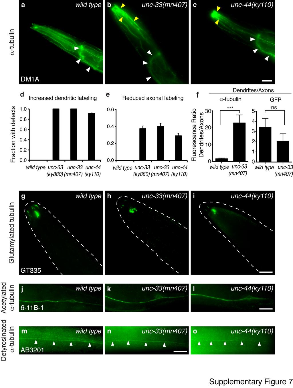
13 Supplementary Figure 7. Distribution of tubulin post-translational modifications in unc- 33 null and unc-44(ky110) mutants (a-c) Maximum intensity projections of α-tubulin immunoreactivity in head neurons of wild-type, unc-33(mn407) and unc-44(ky110) animals. White arrowheads indicate axonrich nerve ring and ventral nerve cord regions; yellow arrowheads indicate distal sensory dendrites. (d,e) Quantification of increased dendritic labeling (d) and reduced axonal labeling defects (e) upon immunostaining against α-tubulin (n>45 animals/genotype). Fraction of animals with qualitative defects is shown. Error bars indicate s.e.p. (f) Quantification of the ratio of dendritic fluorescence intensities versus axonal fluorescence for α-tubulin immunostaining, compared to that for soluble GFP immunofluorescence in head neurons of wild type animals (n=10) and unc-33(mn407) mutants (n=10). Error bars indicate s.e.m. GFP was expressed under the ceh-23 promoter, which labels a representative subset of sensory neurons and a few other neurons. *** p < according to the Mann Whitney test, ns, not significant. (g-i) Immunostaining of glutamylated tubulin detects neuronal cilia in wild type (g), unc- 33 (h) and unc-44 (i) animals. (j-o) Immunostaining of acetylated α-tubulin (j-l) and detyrosinated α-tubulin (m-o) in the PLM mechanosensory neurons of wild type (j,m), unc-33(mn407) (k,n) and unc- 44(ky110) (l,o) animals. Scale bars, 10 µm. 13
14 Plasmid construction Most expression clones were made using FseI-AscI restriction sites in psm or psm-gfp vectors (S. McCarroll and C. I. Bargmann). The heat shock promoter plasmid was ppd49.83 (a gift from A. Fire). cdnas were amplified from N2 cdna and cloned using restriction sites as indicated by their primer names. Plasmids were sequenced to verify the integrity of cloned PCR fragments. des-2 promoter The PVD and FLP neuron-specific des-2 promoter fragment was generated by combining a 0.43 kb fragment located approximately 2.7 kb upstream of the start codon with a 0.4 kb fragment immediately upstream of the start site. Primers used to amplify the 0.43 kb fragment: FseI-F, ATCCTAGGCCGGCCAGATCTCAAAGTACATACATTCTT Fusion-R, AGTTCTGACAGGTATGTTGTGCCTTGTTGCACACACTTTTCGAC Primers for amplifying the 0.4 kb fragment : Fusion-F, GTCGAAAAGTGTGTGCAACAAGGCACAACATACCTGTCAGAACT AscI-R, AGACATGGCGCGCCTGTGTTGTGTGGACCAAAAAGT tag-168 promoter primers FseI-F, GAATCGTAGGCCGGCCTCTCCTTGAAGCTCATCCAGACGTCGCTG AscI-R, GATTGGAAGGCGCGCCACACGGGCCAGAGCTGCAGCTGGATGGCA mec-3 promoter primers FseI-F, GAGAGAGGCCGGCCCCAAGCTTGCATGCCTGCAGG AscI-R, GTATATGGCGCGCCGGGGATCCGATTTCCGTAG 14
15 des-2::myristoyl::gfp A des-2 promoter fragment was cloned into psm-gfp bearing a KpnI-XhoI fragment from unc-86::myrisotyl::gfp 2. SAD-1 cdna primers (des-2::sad-1::gfp) NheI-F, CAGAATGCTAGCATGTTTGAAGCTCTCAAGGAAGTC KpnI-R, TTGTTCGGTACCACGACTTTATCAGCCTTGTTGAACAT des-2::mcherry::rab-3 The des-2 promoter was sub-cloned into a psm-mcherry::rab-3 construct 3. SYD-2 cdna primers (des-2::syd-2::gfp) NheI-F, CAGAATGCTAGCATGAGCTACAGCAATGGAAACATAA KpnI-R, TTGTTCGGTACCCAGGTATATAAATGAAACTCGTAGGATT UNC-104a cdna primers (des-2::unc-104::gfp) NheI-F, CAGAATGCTAGCATGTCATCGGTTAAAGTAGCTGTAC KpnI-R, TTGTTCGGTACCACTGAAGCAGCAATTGAAGATGATGATG psm-n-terminal-mcherry The mcherry sequence from psm-mcherry vector was cloned into the NheI site of psm. SpeI-F, CATTCTACTAGTATGGTGAGCAAGGGCGAGGAG NheI-R, ATGCTAGCTAGCCTTGTACAGCTCGTCCATGCC TBA-1 cdna primers (des-2::mcherry::tba-1) NheI-F, CAGAATGCTAGCATGCGTGAGGTCATCTCCATCC NheI-R, TTGTTCGCTAGCTTAATACTCTTCTCCTTCCTCCTC EBP-2 cdna primers (des-2::ebp-2::gfp) NheI-F, CAGAATGCTAGCATGGTCGTCAACGTGTTCATCTC 15
16 KpnI-R, TTGTTCGGTACCACGAAAGTCTCCGTATCGTCCAGA UNC-33L cdna primers (des-2::unc-33l and tag-168::unc-33l and mec- 3::UNC-33L) UNC-33L-NheI-F, CAGAATGCTAGCATGTTCCCGTTTTTAGCACCAATT SalI-R, TTGTTCGTCGACGCTACCAAAACCCTGTAGTCCG UNC-33M cdna primers (des-2::unc-33m and tag-168::unc-33m) UNC-33M-NheI-F, GAGCCAGCTAGCATGGATATGACGGATATTGAACTAC Reverse primer same as that used for UNC-33L cloning above (Sal I-R). UNC-33L cdna primers (hsp16-41::unc-33l) UNC-33L-NheI-F, CAGAATGCTAGCATGTTCCCGTTTTTAGCACCAATT NcoI-R, TAGTTGCCATGGCTACCAAAACCCTGTAGTCCGT UNC-33S, UNC-33L and GFP primers (tag-168::unc-33l::gfp, des-2::unc- 33L::GFP and des-2::unc-33s::gfp) GFP was inserted into UNC-33L immediately before the beginning of the sequence shared with UNC-33S. Primers were designed such that a GGGGS linker was added before GFP and a GGGGE linker was added after GFP. The first step involved cloning a fusion PCR product containing the GFP sequence followed by the UNC-33S sequence, using NheI and SalI restriction sites. This yielded the UNC-33S::GFP construct. Next, the N-terminus of UNC-33L was PCR amplified and subsequently cloned in the correct orientation using the NheI site upstream of the UNC-33S::GFP, resulting in the desired UNC-33L::GFP construct. GFP primers 16
17 GFP-NheI-F, GTAAATGCTAGCGGAGGAGGAGGATCTATGAGTAAAGGAGAAGAACTTTTC AC GFP-Linker-R, AATCGACATTTCACCGCCGCCTCCAGATTTGTATAGTTCATCCATGCCATGTG Unc-33S-Linker-F, CACATGGCATGGATGAACTATACAAATCTGGAGGCGGCGGTGAAATGTCGAT SalI-R, TTGTTCGTCGACGCTACCAAAACCCTGTAGTCCG UNC-33L N-terminus primers UNC-33L-NheI-F (mentioned above) N-terminus-NheI-R, ATAAATGCTAGCGTTGCCGTCGTCTCCACTATTCTT Supplementary References 1. Saheki, Y. & Bargmann, C.I. Presynaptic CaV2 calcium channel traffic requires CALF- 1 and the alpha(2)delta subunit UNC- 36. Nat Neurosci 12, (2009). 2. Adler, C.E., Fetter, R.D. & Bargmann, C.I. UNC- 6/Netrin induces neuronal asymmetry and defines the site of axon formation. Nat Neurosci 9, (2006). 3. Patel, M.R., et al. Hierarchical assembly of presynaptic components in defined C. elegans synapses. Nat Neurosci 9, (2006). 17
T H E J O U R N A L O F C E L L B I O L O G Y
 Supplemental material Wang et al., http://www.jcb.org/cgi/content/full/jcb.201405026/dc1 T H E J O U R N A L O F C E L L B I O L O G Y Figure S1. Generation and characterization of unc-40 alleles. (A and
(A) Schematic depicting morphology of cholinergic DA and DB motor neurons in an L1
 Schematic depicting morphology of cholinergic DA and DB motor neurons in an L1") SUPPLEMENTAL MATERIALS Supplemental Figure 1 Figure S1. Expression of the non-alpha nachr subunit ACR-2. (A) Schematic depicting morphology of cholinergic DA and DB motor neurons in an L1 animal. Wide-field
SUPPLEMENTAL MATERIALS Supplemental Figure 1 Figure S1. Expression of the non-alpha nachr subunit ACR-2. (A) Schematic depicting morphology of cholinergic DA and DB motor neurons in an L1 animal. Wide-field
SUPPLEMENTARY INFORMATION
 doi: 1.138/nature7866 1.8 Fraction Bordering Fraction Aggregating.6.4.2 1.8.6.4.2 Promoter Head neurons expressing npr-1 cdna Genotype WT - - URX AQR ALN AVM gcy-35 sax-7 gcy-32+ gpa-3 RMG ILs URX AQR
mod-1::mcherry unc-47::gfp RME ser-4::gfp vm2 vm2 vm2 vm2 VNC
 A mod-1::mcherry unc-47::gfp RME B ser-4::gfp vm2 vm2 VNC vm2 vm2 VN Figure S1 Further details of mod- 1 and ser- 4 reporter expression patterns. (A) Adult head region showing mod- 1::mCherry and unc-
Neuronal Activity and CaMKII Regulate Kinesin-Mediated Transport of Synaptic AMPARs
 Neuron Supplemental Information Neuronal Activity and CaMKII Regulate Kinesin-Mediated Transport of Synaptic AMPARs Frédéric J. Hoerndli, Rui Wang, Jerry E. Mellem, Angy Kallarackal, Penelope J. Brockie,
Figure S1. USP-46 is expressed in several tissues including the nervous system
 Supplemental Figure legends Figure S1. USP-46 is expressed in several tissues including the nervous system Transgenic animals expressing a transcriptional reporter (P::GFP) were imaged using epifluorescence
Supplementary Figure 1. Time Line of C. elegans Embryonic Development. (a) Proliferation occurs from min post fertilization (m.p.f.
 Proliferation occurs from min post fertilization (m.p.f.") a b Supplementary Figure 1. Time Line of C. elegans Embryonic Development. (a) Proliferation occurs from 0-360 min post fertilization (m.p.f.) and morphogenesis begins at the bean stage (360 m.p.f.), when
a b Supplementary Figure 1. Time Line of C. elegans Embryonic Development. (a) Proliferation occurs from 0-360 min post fertilization (m.p.f.) and morphogenesis begins at the bean stage (360 m.p.f.), when
Nature Biotechnology: doi: /nbt.4166
 Supplementary Figure 1 Validation of correct targeting at targeted locus. (a) by immunofluorescence staining of 2C-HR-CRISPR microinjected embryos cultured to the blastocyst stage. Embryos were stained
Two classes of silencing RNAs move between Caenorhabditis elegans tissues.
 Two classes of silencing RNAs move between Caenorhabditis elegans tissues. Antony M Jose, Giancarlo A Garcia, and Craig P Hunter. Supplementary Figures, Figure Legends, and Tables. Supplementary Figure
SUPPLEMENTARY INFORMATION
 DOI:.38/ncb327 a b Sequence coverage (%) 4 3 2 IP: -GFP isoform IP: GFP IP: -GFP IP: GFP Sequence coverage (%) 4 3 2 IP: -GFP IP: GFP 33 52 58 isoform 2 33 49 47 IP: Control IP: Peptide Sequence Start
Flybow genetic multicolor cell labeling for neural circuit analysis in Drosophila melanogaster
 Nature Methods Flybow genetic multicolor cell labeling for neural circuit analysis in Drosophila melanogaster Dafni Hadjieconomou, Shay Rotkopf, Cyrille Alexandre, Donald M Bell, Barry J Dickson & Iris
Liu et al., http :// /cgi /content /full /jcb /DC1
 Supplemental material JCB Liu et al., http ://www.jcb.org /cgi /content /full /jcb.201506081 /DC1 THE JOU RNAL OF CELL BIO LOGY Figure S1. Identification of sorf-1 and sorf-2. (A) RNAi of ZK563.5 increases
Nature Methods: doi: /nmeth Supplementary Figure 1. Neither driver nor effector alone displays expression of GFP
 Supplementary Figure 1 Neither driver nor effector alone displays expression of GFP Comparison of GFP fluorescence in a driver- only strain (a) or an effector-only strain (b) to their driver + effector
Supporting information
 Supporting information Construction of strains and plasmids To create ptc67, a PCR product obtained with primers cc2570-162f (gcatgggcaagcttgaggacggcgtcatgt) and cc2570+512f (gaggccgtggtaccatagaggcgggcg),
SUPPLEMENTARY INFORMATION
 DOI: 10.1038/ncb3374 Supplementary Figure 1 staccato mutations affect a late step of the tube fusion process. (a) Immunostaining of stage 15 embryos labeled with anti- Dys (green in merge) and a probe
Supporting Online Material for
 www.sciencemag.org/cgi/content/full/323/5920/1500/dc1 Supporting Online Material for RSY-1 Is a Local Inhibitor of Presynaptic Assembly in C. elegans Maulik R. Patel and Kang Shen To whom correspondence
Nature Structural & Molecular Biology: doi: /nsmb.3308
 Supplementary Figure 1 Analysis of CED-3 autoactivation and CED-4-induced CED-3 activation. (a) Repeat experiments of Fig. 1a. (b) Repeat experiments of Fig. 1b. (c) Quantitative analysis of three independent
Stargazin regulates AMPA receptor trafficking through adaptor protein. complexes during long term depression
 Supplementary Information Stargazin regulates AMPA receptor trafficking through adaptor protein complexes during long term depression Shinji Matsuda, Wataru Kakegawa, Timotheus Budisantoso, Toshihiro Nomura,
Supplemental Information. Glutamylation Regulates Transport, Specializes. Function, and Sculpts the Structure of Cilia
 Current Biology, Volume 27 Supplemental Information Glutamylation Regulates Transport, Specializes Function, and Sculpts the Structure of Cilia Robert O'Hagan, Malan Silva, Ken C.Q. Nguyen, Winnie Zhang,
Supporting Information
 Supporting Information Lee et al. 10.1073/pnas.1000866107 SI Experimental Procedures Plasmid and Strain Construction. (p)odr-3::gfp::egl-4 was constructed by excising the coding region of GFP and the first
SUPPLEMENTARY INFORMATION
 Figure S1. lev-9 mutants are resistant to levamisole. The levamisole dose-response curves indicate that lev-9 mutants are partially resistant to levamisole similar to lev-10(kr26) mutants. unc-29(x29)
Supplementary information to accompany: A novel role for the DNA repair gene Rad51 in Netrin-1 signalling
 Supplementary information to accompany: A novel role for the DNA repair gene Rad51 in Netrin-1 signalling Glendining KA 1, Markie D 2, Gardner RJM 4, Franz EA 3, Robertson SP 4, Jasoni CL 1 Supplementary
MIT Department of Biology 7.013: Introductory Biology - Spring 2005 Instructors: Professor Hazel Sive, Professor Tyler Jacks, Dr.
 MIT Department of Biology 7.01: Introductory Biology - Spring 2005 Instructors: Professor Hazel Sive, Professor Tyler Jacks, Dr. Claudette Gardel iv) Would Xba I be useful for cloning? Why or why not?
transcription and the promoter occupancy of Smad proteins. (A) HepG2 cells were co-transfected with the wwp-luc reporter, and FLAG-tagged FHL1,
 HepG2 cells were co-transfected with the wwp-luc reporter, and FLAG-tagged FHL1,") Supplementary Data Supplementary Figure Legends Supplementary Figure 1 FHL-mediated TGFβ-responsive reporter transcription and the promoter occupancy of Smad proteins. (A) HepG2 cells were co-transfected
Supplementary Data Supplementary Figure Legends Supplementary Figure 1 FHL-mediated TGFβ-responsive reporter transcription and the promoter occupancy of Smad proteins. (A) HepG2 cells were co-transfected
Schematic representation of the endogenous PALB2 locus and gene-disruption constructs
 Supplementary Figures Supplementary Figure 1. Generation of PALB2 -/- and BRCA2 -/- /PALB2 -/- DT40 cells. (A) Schematic representation of the endogenous PALB2 locus and gene-disruption constructs carrying
Supplemental Information
 Supplemental Information Itemized List Materials and Methods, Related to Supplemental Figures S5A-C and S6. Supplemental Figure S1, Related to Figures 1 and 2. Supplemental Figure S2, Related to Figure
(a) Scheme depicting the strategy used to generate the ko and conditional alleles. (b) RT-PCR for
 Scheme depicting the strategy used to generate the ko and conditional alleles. (b) RT-PCR for") Supplementary Figure 1 Generation of Diaph3 ko mice. (a) Scheme depicting the strategy used to generate the ko and conditional alleles. (b) RT-PCR for different regions of Diaph3 mrna from WT, heterozygote
Supplementary Figure 1 Generation of Diaph3 ko mice. (a) Scheme depicting the strategy used to generate the ko and conditional alleles. (b) RT-PCR for different regions of Diaph3 mrna from WT, heterozygote
An antibiotic selection marker for nematode transgenesis
 nature methods An antibiotic selection marker for nematode transgenesis Rosina Giordano-Santini, Stuart Milstein, Nenad Svrzikapa, Domena Tu, Robert Johnsen, David Baillie, Marc Vidal & Denis Dupuy Supplementary
Protocol for cloning SEC-based repair templates using Gibson assembly and ccdb negative selection
 Protocol for cloning SEC-based repair templates using Gibson assembly and ccdb negative selection Written by Dan Dickinson (daniel.dickinson@austin.utexas.edu) and last updated January 2018. A version
SUPPLEMENTARY INFORMATION
 a before amputation regeneration regenerated limb DERMIS SKELETON MUSCLE SCHWANN CELLS EPIDERMIS DERMIS SKELETON MUSCLE SCHWANN CELLS EPIDERMIS developmental origin: lateral plate mesoderm presomitic mesoderm
SUPPLEMENTARY INFORMATION
 SULEMENTARY INFORMATION doi:10.1038/nature12117 Supplementary Discussion Although loss of leads to initiation of postembryonic development even under starvation conditions, ectopic overexpression of under
SUPPLEMENTARY INFORMATION
 DOI: 10.1038/ncb2880 Supplementary Figure 1 Sequence alignment of Deup1 and Cep63. The protein sequence alignment was generated by the Clustal X 2.0 multiple sequence alignment program using default parameters.
SUPPLEMENTARY INFORMATION
 SUPPLEMENTARY INFORMATION Supplementary figure 1: List of primers/oligonucleotides used in this study. 1 Supplementary figure 2: Sequences and mirna-targets of i) mcherry expresses in transgenic fish used
Supplemental Figure 1. Mutation in NLA Causes Increased Pi Uptake Activity and
 Supplemental Figure 1. Mutation in NLA Causes Increased Pi Uptake Activity and PHT1 Protein Amounts. (A) Shoot morphology of 19-day-old nla mutants under Pi-sufficient conditions. (B) [ 33 P]Pi uptake
Fig. S1. Phosphorylation of eif2 by PEK-1 is increased in ire-1 mutants. Fig. S2. ire-1
 Fig. S1. Phosphorylation of eif2a by PEK-1 is increased in ire-1 mutants. Representative western blot of phosphorylated eif2a and tubulin of day-0 wild-type animals and ire-1 mutants treated with control,
SUPPLEMENTARY INFORMATION
 SUPPLEMENTARY INFORMATION Legends for Supplementary Tables. Supplementary Table 1. An excel file containing primary screen data. Worksheet 1, Normalized quantification data from a duplicated screen: valid
b number of motif occurrences
 supplementary information DOI: 1.138/ncb194 a b number of motif occurrences number of motif occurrences number of motif occurrences I II III IV V X TTGGTCAGTGCA 3 8 23 4 13 239 TTGGTCAGTGCT 7 5 8 1 3 83
Sperm cells are passive cargo of the pollen tube in plant fertilization
 In the format provided by the authors and unedited. SUPPLEMENTARY INFORMATION VOLUME: 3 ARTICLE NUMBER: 17079 Sperm cells are passive cargo of the pollen tube in plant fertilization Jun Zhang 1, Qingpei
Xu et al., Supplementary Figures 1-7
 Xu et al., Supplementary Figures 1-7 Supplementary Figure 1. PIPKI is required for ciliogenesis. (a) PIPKI localizes at the basal body of primary cilium. RPE-1 cells treated with two sirnas targeting to
Supplementary Figure 1. Drawing of spinal cord open-book preparations and DiI tracing. Nature Neuroscience: doi: /nn.3893
 Supplementary Figure 1 Drawing of spinal cord open-book preparations and DiI tracing. Supplementary Figure 2 In ovo electroporation of dominant-negative PlexinA1 in commissural neurons induces midline
embryos. Asterisk represents loss of or reduced expression. Brackets represent
 Supplemental Figures Supplemental Figure 1. tfec expression is highly enriched in tail endothelial cells (A- B) ISH of tfec at 15 and 16hpf in WT embryos. (C- D) ISH of tfec at 36 and 38hpf in WT embryos.
(b) Genotyping of SALK_ (AT1g51690) and SALK_ (At1g17720) to find homozygous single mutant plants.
 Genotyping of SALK_ (AT1g51690) and SALK_ (At1g17720) to find homozygous single mutant plants.") Supplemental Table 1. Primers used for cloning and RT-PCR. The B-type subunits cdna CDS were amplified using high-fidelity PCR amplification with gene specific primers. The cdna CDS sequences were obtained
Supplemental Table 1. Primers used for cloning and RT-PCR. The B-type subunits cdna CDS were amplified using high-fidelity PCR amplification with gene specific primers. The cdna CDS sequences were obtained
Department of Physiology, Tokyo Women's Medical University School of Medicine, Tokyo, Japan 2
 Supplementary Information Endomembrane-associated RSD-3 is important for RNAi induced by extracellular silencing RNA in both somatic and germ cells of Caenorhabditis elegans! Rieko Imae 1,4, Katsufumi
Regulation of axonal and dendritic growth by the extracellular calcium-sensing
 Regulation of axonal and dendritic growth by the extracellular calcium-sensing receptor (CaSR). Thomas N. Vizard, Gerard W. O Keeffe, Humberto Gutierrez, Claudine H. Kos, Daniela Riccardi, Alun M. Davies
GM130 Is Required for Compartmental Organization of Dendritic Golgi Outposts
 Current Biology, Volume 24 Supplemental Information GM130 Is Required for Compartmental Organization of Dendritic Golgi Outposts Wei Zhou, Jin Chang, Xin Wang, Masha G. Savelieff, Yinyin Zhao, Shanshan
Supplementary Information
 Supplementary Information Selective control of inhibitory synapse development by Slitrk3-PTPδ trans-synaptic interaction Hideto Takahashi 1, Kei-ichi Katayama 2, Kazuhiro Sohya 3,4, Hiroyuki Miyamoto 4,5,
Construct Design and Cloning Guide for Cas9-triggered homologous recombination
 Construct Design and Cloning Guide for Cas9-triggered homologous recombination Written by Dan Dickinson (ddickins@live.unc.edu) and last updated December 2013. Reference: Dickinson DJ, Ward JD, Reiner
Fig. S1. Molecular phylogenetic analysis of AtHD-ZIP IV family. A phylogenetic tree was constructed using Bayesian analysis with Markov Chain Monte
 Fig. S1. Molecular phylogenetic analysis of AtHD-ZIP IV family. A phylogenetic tree was constructed using Bayesian analysis with Markov Chain Monte Carlo algorithm for one million generations to obtain
Supplementary Methods
 Supplementary Methods MARCM-based forward genetic screen We used ethylmethane sulfonate (EMS) to mutagenize flies carrying FRT 2A and FRT 82B transgenes, which are sites for the FLP-mediated recombination
Engineering splicing factors with designed specificities
 nature methods Engineering splicing factors with designed specificities Yang Wang, Cheom-Gil Cheong, Traci M Tanaka Hall & Zefeng Wang Supplementary figures and text: Supplementary Figure 1 Supplementary
A subclass of HSP70s regulate development and abiotic stress responses in Arabidopsis thaliana
 1 2 3 4 5 6 7 8 9 10 11 12 13 14 15 16 17 18 19 20 21 Journal of Plant Research A subclass of HSP70s regulate development and abiotic stress responses in Arabidopsis thaliana Linna Leng 1 Qianqian Liang
Table S1. List of DNA constructs and primers, part 1 Construct
 SUPPLEMENTARY TABLES: Table S1. List of DNA constructs and primers, part 1 Construct Comment (Expressed protein) Vector, PCR primers and template or source of Restriction Sites name Resistance construct
Supplementary Figure 1. Espn-1 knockout characterization. (a) The predicted recombinant Espn-1 -/- allele was detected by PCR of the left (5 )
 The predicted recombinant Espn-1 -/- allele was detected by PCR of the left (5 )") Supplementary Figure 1. Espn-1 knockout characterization. (a) The predicted recombinant Espn-1 -/- allele was detected by PCR of the left (5 ) homologous recombination arm (left) and of the right (3 )
Supplementary Figure 1. Espn-1 knockout characterization. (a) The predicted recombinant Espn-1 -/- allele was detected by PCR of the left (5 ) homologous recombination arm (left) and of the right (3 )
SAS6-like protein in Plasmodium indicates that conoid-associated apical complex proteins persist in invasive stages within the mosquito vector
 SAS6-like protein in Plasmodium indicates that conoid-associated apical complex proteins persist in invasive stages within the mosquito vector Richard J. Wall 1,#, Magali Roques 1,+, Nicholas J. Katris
SUPPLEMENTARY INFORMATION
 doi:10.1038/nature10810 Supplementary Fig. 1: Mutation of the loqs gene leads to shortened lifespan and adult-onset brain degeneration. a. Northern blot of control and loqs f00791 mutant flies. loqs f00791
Supplemental Data. Borg et al. Plant Cell (2014) /tpc
 /tpc") Supplementary Figure 1 - Alignment of selected angiosperm DAZ1 and DAZ2 homologs Multiple sequence alignment of selected DAZ1 and DAZ2 homologs. A consensus sequence built using default parameters is shown
Supplementary Figure 1 - Alignment of selected angiosperm DAZ1 and DAZ2 homologs Multiple sequence alignment of selected DAZ1 and DAZ2 homologs. A consensus sequence built using default parameters is shown
An assay to image neuronal microtubule dynamics in mice
 SUPPLEMENTARY INFORMATION An assay to image neuronal microtubule dynamics in mice Tatjana Kleele 1, Petar Marinković 2,3, Philip R. Williams 1, Sina Stern 4, Emily E. Weigand 5, Peter Engerer 1, Ronald
Supplemental Information. Characterization of Proprioceptive System. Dynamics in Behaving Drosophila Larvae. Using High-Speed Volumetric Microscopy
 urrent Biology, Volume 29 Supplemental Information haracterization of roprioceptive System Dynamics in Behaving Drosophila Larvae Using High-Speed Volumetric Microscopy Rebecca D. Vaadia, Wenze Li, Venkatakaushik
indicated numbers of pups at day of life (DOL) 10, or embryonic day (ED) B. Male mice of
 10, or embryonic day (ED) B. Male mice of") SUPPLEMENTRY FIGURE LEGENDS Figure S1. USP44 loss leads to chromosome missegregation.. Genotypes obtained from the indicated numbers of pups at day of life (DOL) 10, or embryonic day (ED) 13.5.. Male mice
SUPPLEMENTRY FIGURE LEGENDS Figure S1. USP44 loss leads to chromosome missegregation.. Genotypes obtained from the indicated numbers of pups at day of life (DOL) 10, or embryonic day (ED) 13.5.. Male mice
Supporting Information
 Supporting Information Ho et al. 10.1073/pnas.0808899106 SI Materials and Methods In immunostaining, antibodies from Developmental Studies Hybridoma ank were -FasII (mouse, 1:200), - (mouse, 1:100), and
SUPPLEMENTARY INFORMATION
 Clustering of a kinesin-14 motor enables processive retrograde microtubule-based transport in plants NATURE PLANTS www.nature.com/natureplants 1 2 NATURE PLANTS www.nature.com/natureplants NATURE PLANTS
SUPPLEMENTARY INFORMATION
 doi:.38/nature899 Supplementary Figure Suzuki et al. a c p7 -/- / WT ratio (+)/(-) p7 -/- / WT ratio Log X 3. Fold change by treatment ( (+)/(-)) Log X.5 3-3. -. b Fold change by treatment ( (+)/(-)) 8
Supplemental Figure 1 Human REEP family of proteins can be divided into two distinct subfamilies. Residues (single letter amino acid code) identical
 identical") Supplemental Figure Human REEP family of proteins can be divided into two distinct subfamilies. Residues (single letter amino acid code) identical in all six REEPs are highlighted in green. Additional
Supplemental Figure Human REEP family of proteins can be divided into two distinct subfamilies. Residues (single letter amino acid code) identical in all six REEPs are highlighted in green. Additional
GT-rich promoters can drive RNA pol II transcription and deposition of H2A.Z in African trypanosomes
 GT-rich promoters can drive RNA pol II transcription deposition of H2A.Z in African trypanosomes Carolin Wedel, Konrad U. Förstner, Ramona Derr T. Nicolai Siegel Appendix Table of Contents Appendix Materials
Figure S1. gfp tola and pal mcherry can complement deletion mutants of tola and pal respectively. (A)When strain LS4522 was grown in the presence of
When strain LS4522 was grown in the presence of") Figure S1. gfp tola and pal mcherry can complement deletion mutants of tola and pal respectively. (A)When strain LS4522 was grown in the presence of xylose, inducing expression of gfp-tola, localization
Figure S1. gfp tola and pal mcherry can complement deletion mutants of tola and pal respectively. (A)When strain LS4522 was grown in the presence of xylose, inducing expression of gfp-tola, localization
SUPPLEMENTARY INFORMATION
 AS-NMD modulates FLM-dependent thermosensory flowering response in Arabidopsis NATURE PLANTS www.nature.com/natureplants 1 Supplementary Figure 1. Genomic sequence of FLM along with the splice sites. Sequencing
Automated analysis of embryonic gene expression with cellular resolution in C. elegans
 Automated analysis of embryonic gene expression with cellular resolution in C. elegans John Isaac Murray, Zhirong Bao, Thomas J Boyle, Max E Boeck, Barbara L Mericle, Thomas J Nicholas, Zhongying Zhao,
NAME TA SEC Problem Set 4 FRIDAY October 15, Answers to this problem set must be inserted into the box outside
 MIT Biology Department 7.012: Introductory Biology - Fall 2004 Instructors: Professor Eric Lander, Professor Robert A. Weinberg, Dr. Claudette Gardel NAME TA SEC 7.012 Problem Set 4 FRIDAY October 15,
Fig. S1. Inhibition of Rh1 accumulation on the rhabdomere membrane in CG13089 PIG-U mutants. (A) Late pupae with dark wings were put into 0.
 Late pupae with dark wings were put into 0.") Fig. S1. Inhibition of Rh1 accumulation on the rhabdomere membrane in CG13089 PIG-U mutants. (A) Late pupae with dark wings were put into 0.2% agar after the pupal case was partially removed. Two fluorescent
Fig. S1. Inhibition of Rh1 accumulation on the rhabdomere membrane in CG13089 PIG-U mutants. (A) Late pupae with dark wings were put into 0.2% agar after the pupal case was partially removed. Two fluorescent
Figure legends for supplement
 Figure legends for supplement Supplemental Figure 1 Characterization of purified and recombinant proteins Relevant fractions related the final stage of the purification protocol(bingham et al., 1998; Toba
Supplementary Figure S1. Immunodetection of full-length XA21 and the XA21 C-terminal cleavage product.
 Supplementary Information Supplementary Figure S1. Immunodetection of full-length XA21 and the XA21 C-terminal cleavage product. Total protein extracted from Kitaake wild type and rice plants carrying
Regulation of Acetylcholine Receptor Clustering by ADF/Cofilin- Directed Vesicular Trafficking
 Regulation of Acetylcholine Receptor Clustering by ADF/Cofilin- Directed Vesicular Trafficking Chi Wai Lee, Jianzhong Han, James R. Bamburg, Liang Han, Rachel Lynn, and James Q. Zheng Supplementary Figures
Nature Methods: doi: /nmeth Supplementary Figure 1. Schematics of SAM, dcas9-suntag and dcas9-vpr.
 Supplementary Figure 1 Schematics of SAM, dcas9-suntag and dcas9-vpr. (a) Schematics of SAM. SAM consists of two chimeric proteins, dcas9 fused with VP64 and MS2-coat protein fused with p65 and HSF1 activator
Supplementary Information Design of small molecule-responsive micrornas based on structural requirements for Drosha processing
 Supplementary Information Design of small molecule-responsive micrornas based on structural requirements for Drosha processing Chase L. Beisel, Yvonne Y. Chen, Stephanie J. Culler, Kevin G. Hoff, & Christina
Supplementary Figure 1. Effects of STOML3-modulating molecules on other stomatin-domain proteins
 Supplementary Figure 1 Effects of STOML3-modulating molecules on other stomatin-domain proteins (a) BiFC signal development observed when cells were transfected with stomatin VC/-VN, STOML1-VC/- VN, STOML2-VC/-VN,
Mitotic cell rounding and epithelial thinning regulate lumen
 Mitotic cell rounding and epithelial thinning regulate lumen growth and shape Esteban Hoijman, Davide Rubbini, Julien Colombelli and Berta Alsina SUPPLEMENTARY FIGURES Supplementary Figure 1 (a-c) Localization
Supplementary Figure 1: Expression of RNF8, HERC2 and NEURL4 in the cerebellum and knockdown of RNF8 by RNAi (a) Lysates of the cerebellum from rat
 Lysates of the cerebellum from rat") Supplementary Figure 1: Expression of RNF8, HERC2 and NEURL4 in the cerebellum and knockdown of RNF8 by RNAi (a) Lysates of the cerebellum from rat pups at P6, P14, P22, P30 and adult (A) rats were subjected
Supplementary Figure 1: Expression of RNF8, HERC2 and NEURL4 in the cerebellum and knockdown of RNF8 by RNAi (a) Lysates of the cerebellum from rat pups at P6, P14, P22, P30 and adult (A) rats were subjected
Supplementary Figure 1
 Supplementary Figure 1 Cell cycle distribution (%) 1 8 6 4 2 Cell Cycle G1 S G2 Viability (%) 5 4 3 2 1 Viability Supplementary Figure 1. Cell cycle distribution and viability during DSB repair measurements.
oligonucleotide primers listed in Supplementary Table 2 and cloned into ppcr-script (Stratagene) before digestion
 before digestion") Supplementary Methods. Construction of BiFC vectors The full-length ARC3 cdna, ARC3 1-1794 and ARC3 1-1083 were amplified from ppcr-script/arc3 using the oligonucleotide primers listed in Supplementary
Supplementary Methods. Construction of BiFC vectors The full-length ARC3 cdna, ARC3 1-1794 and ARC3 1-1083 were amplified from ppcr-script/arc3 using the oligonucleotide primers listed in Supplementary
Supplementary Figure 1. Isolation of GFPHigh cells.
 Supplementary Figure 1. Isolation of GFP High cells. (A) Schematic diagram of cell isolation based on Wnt signaling activity. Colorectal cancer (CRC) cell lines were stably transduced with lentivirus encoding
University of Dundee. Published in: Scientific Reports. DOI: /srep Publication date: 2016
 University of Dundee SAS6-like protein in Plasmodium indicates that conoid-associated apical complex proteins persist in invasive stages within the mosquito vector Wall, Richard J.; Roques, Magali; Katris,
Biotechnology Project VI lesson. 1 - Subcloning NRG1-III-β3 from pcr-blunt II-TOPO into the expression vector pegfp-c3
 26-11-2018 - Biotechnology Project VI lesson 1 - Subcloning NRG1-III-β3 from pcr-blunt II-TOPO into the expression vector pegfp-c3 2 - Subcloning NRG1-III-β3 from pcr-blunt II-TOPO or pegfp-c3 into the
Supplementary Figure 1.
 Supplementary Figure 1. (A) UCSC Genome Browser view of region immediately downstream of the NEUROG3 start codon. All candidate grna target sites which meet the G(N 19 )NGG constraint are aligned to illustrate
Mapping long-range promoter contacts in human cells with high-resolution capture Hi-C
 CORRECTION NOTICE Nat. Genet. 47, 598 606 (2015) Mapping long-range promoter contacts in human cells with high-resolution capture Hi-C Borbala Mifsud, Filipe Tavares-Cadete, Alice N Young, Robert Sugar,
Chapter 10 (Part I) Gene Isolation and Manipulation
 Gene Isolation and Manipulation") Biology 234 J. G. Doheny Chapter 10 (Part I) Gene Isolation and Manipulation Practice Questions: Answer the following questions with one or two sentences. 1. From which types of organisms were most restriction
Biology 234 J. G. Doheny Chapter 10 (Part I) Gene Isolation and Manipulation Practice Questions: Answer the following questions with one or two sentences. 1. From which types of organisms were most restriction
Hoopfer et al., Supplemental Data Supplemental Figure S1. Quantification of ORN axon degeneration
 Hoopfer et al., Supplemental Data Supplemental Figure S1. Quantification of ORN axon degeneration (A) Mean GFP intensity of commissural ORN axons 1 day after antennae removal. 1 day after cutting, wt ORNs
EGF signaling induces behavioral quiescence in C. elegans Cheryl Van Buskirk and Paul W. Sternberg
 EGF signaling induces behavioral quiescence in C. elegans Cheryl Van Buskirk and Paul W. Sternberg LIN-3/EGF [PIP2] PLC-3/PLC-γ LET-23/EGFR SEM-5/GRB-2 LET-6/RAS [IP3] [DAG] LIN-45/RAF ITR-1/IP3R Ovulation
1 Supplementary Figure 1. Kif13b expression and localization in serum starved
 1 2 3 4 5 6 Supplementary Figure 1. Kif13b expression and localization in serum starved cells. (a) Immunoblot of NIH3T3 cell lysates after 24 hrs of serum deprivation, using rabbit polyclonal KIF13B antiserum.
Supplementary information
 Supplementary information Supplementary figures Figure S1 Level of mycdet1 protein in DET1 OE-1, OE-2 and OE-3 transgenic lines. Total protein extract from wild type Col0, det1-1 mutant and DET1 OE lines
Controlling gene expression with the Q repressible binary expression system in Caenorhabditis elegans
 ARTICLES Controlling gene expression with the Q repressible binary expression system in Caenorhabditis elegans Xing Wei 1, Christopher J Potter 1,2, Liqun Luo 1 & Kang Shen 1 2012 Nature America, Inc.
T H E J O U R N A L O F C E L L B I O L O G Y
 Supplemental material Egan et al., http://www.jcb.org/cgi/content/full/jcb.201112101/dc1 T H E J O U R N A L O F C E L L B I O L O G Y Figure S1. Localization of dynein to microtubule plus ends requires
Programmable Sequence-Specific Transcriptional Regulation of Mammalian Genome Using Designer TAL Effectors
 Supplementary Information Programmable Sequence-Specific Transcriptional Regulation of Mammalian Genome Using Designer TAL Effectors Feng Zhang 1,2,3,5,7 *,±, Le Cong 2,3,4 *, Simona Lodato 5,6, Sriram
amplification of the 5 flanking region of CAT1 with a tail for the geneticin resistance gene cassette fusion CAT1-3F
 Supplementary materials Table S1. Primer list. Primer Sequence a (5-3 ) Description CAT1-5F CAT1-5R ATACGGATAAGGAAGCGATAGCAGCA gcacaggtacacttgtttagagagcgatccggattttaagtgaacg amplification of the 5 flanking
A Repressor Complex Governs the Integration of
 Developmental Cell 15 Supplemental Data A Repressor Complex Governs the Integration of Flowering Signals in Arabidopsis Dan Li, Chang Liu, Lisha Shen, Yang Wu, Hongyan Chen, Masumi Robertson, Chris A.
Controlling gene expression with the Q repressible binary expression system in Caenorhabditis elegans
 Articles Controlling gene expression with the Q repressible binary expression system in Caenorhabditis elegans Xing Wei 1, Christopher J Potter 1,2, Liqun Luo 1 & Kang Shen 1 2012 Nature America, Inc.
Supplemental Data includes Supplemental Experimental Procedures, 4 Figures and 5 Movie Legends.
 SUPPLEMENTAL DATA Supplemental Data includes Supplemental Experimental Procedures, 4 Figures and 5 Movie Legends. Supplemental Experimental Procedures Dominant negative nesprin KASH (DN KASH) plasmid construction-
Supplemental Figure S1. PGRN Binding to Sortilin.
 1 Neuron, volume 68 Supplemental Data Sortilin-Mediated Endocytosis Determines Levels of the Frontotemporal Dementia Protein, Progranulin Fenghua Hu, Thihan Padukkavidana, Christian B. Vægter, Owen A.
Supplementary Figure 1: sgrna library generation and the length of sgrnas for the functional screen. (a) A diagram of the retroviral vector for sgrna
 A diagram of the retroviral vector for sgrna") Supplementary Figure 1: sgrna library generation and the length of sgrnas for the functional screen. (a) A diagram of the retroviral vector for sgrna expression. It contains a U6-promoter-driven sgrna
Supplementary Figure 1: sgrna library generation and the length of sgrnas for the functional screen. (a) A diagram of the retroviral vector for sgrna expression. It contains a U6-promoter-driven sgrna
SUPPLEMENTARY INFORMATION
 doi:10.1038/nature09821 tyramine/octopamine (internal state) sensory cues (e.g. lysine) HW tyra-3 N2 tyra-3 sensory cues (e.g. O 2, CO 2 ) ASK BAG ASK BAG lawn leaving lawn leaving Suppl. Fig. 1. tyra-3