Introduction to N-STORM
|
|
|
- Ashlee Richards
- 6 years ago
- Views:
Transcription


1 Introduction to N-STORM Dan Metcalf Advanced Imaging Manager
2 Outline Introduction Principles of STORM Applications N-STORM overview


3 Biological Scale Mitochondrion Microtubule Amino Acid 1Å Kinesin 1nm 10nm 100nm 1µm 10µm 0.1mm 1Å 1nm 10nm 100nm 1 m 10 m 0.1mm Atomic Molecular Sub-cellular Super-Resolution Cellular Light Microscopy wikipedia.org/wiki/kinesin; cvcweb.ices.utexas.edu; Fotin et al., Nature 2004; hrsbstaff.ednet.ns.ca;


4 Biological Model Systems Understand molecular and biochemical reactions, behaviour, and interactions in as physiologically relevant system as possible Kines in Microt ubule Mitocho ndrion Cell models eg. HeLa or primary cells Small model organisms eg. Drosophila Large model organisms eg. mice In vitro cell free systems More physiologically relevant = more complex and technically challenging Correlate data across techniques and model systems to build up picture
5 Microscopy techniques Depth MP Confocal Speed Widefield SIM STORM Cost User-friendliness (robustness) Applications window (samples) Multi-colour & 3D capability Resolution
6 Outline Introduction Principles of STORM Applications N-STORM overview
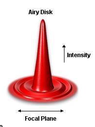
7 Principles of STORM


8 Principles of STORM Conventional fluorescence Raw images STORM Image Localisation Stochastic Optical Reconstruction Microscopy = STORM Rust, Bates & Zhuang, Nat. Methods, 2006 Bates, Huang, Dempsey & Zhuang, Science, 2007
9 History of acronyms PALM GSDIM ipalm STORM dstorm DNA-PAINT qpaint sptpalm SAME Blinking fluorescence Multiple raw frames Localise & reconstruct DIFFERENT Labelling strategies Laser sequences
10 N-STORM Super-Resolution System
11 N-STORM Acquisition Sequence

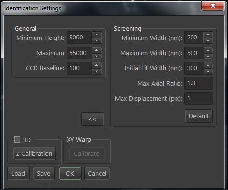
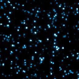
12 Identify Molecules

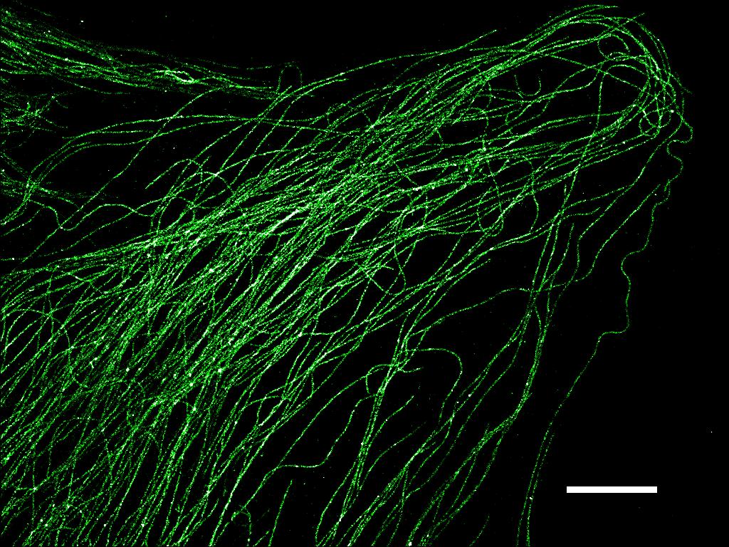
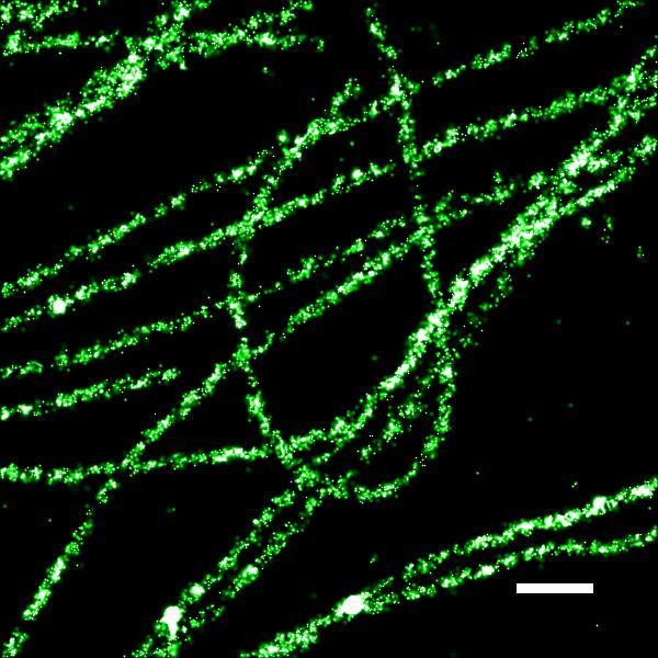
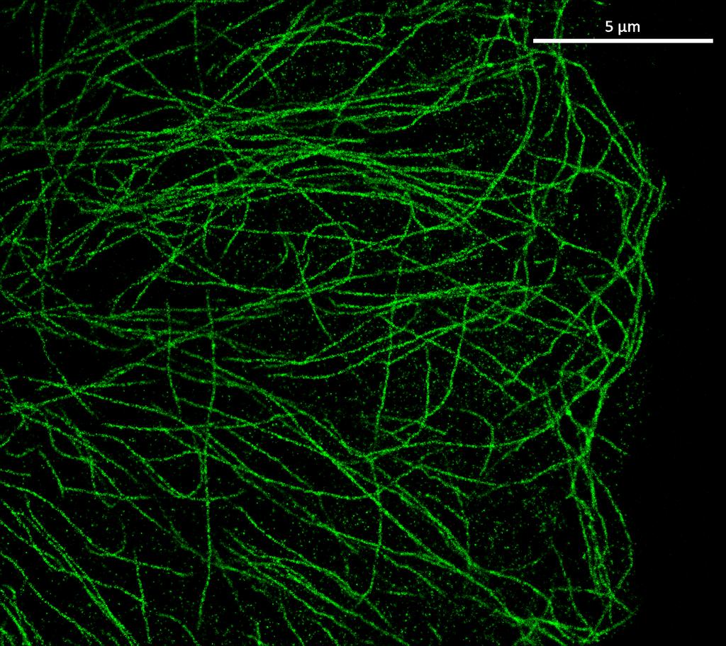
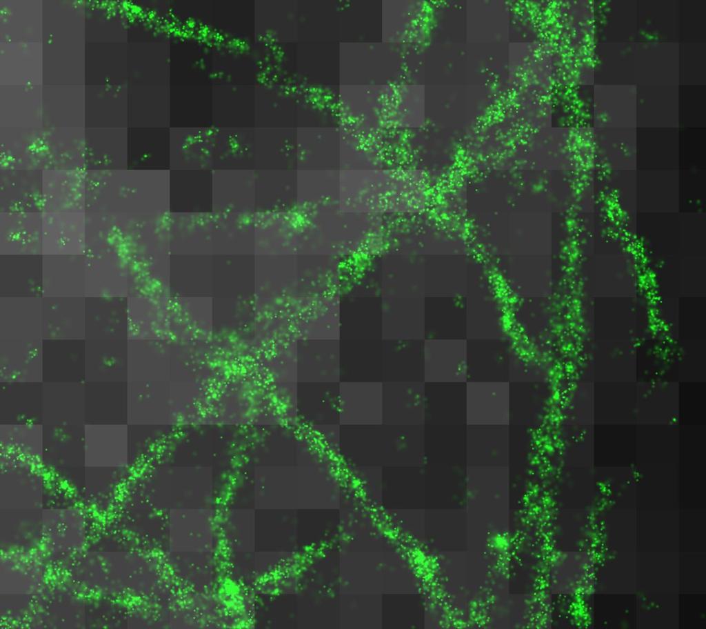
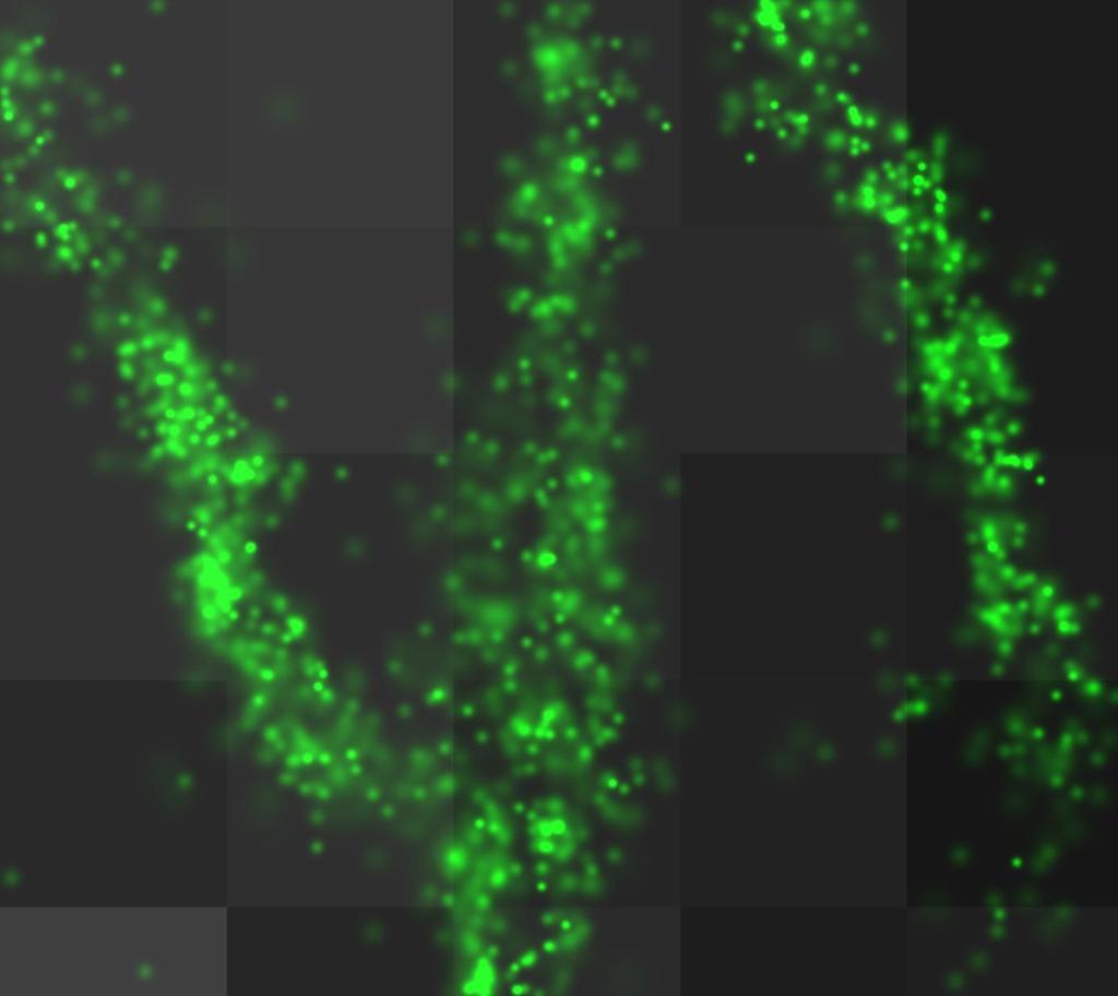
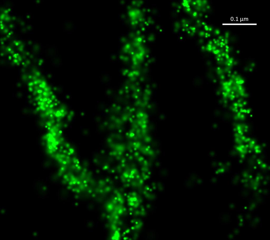
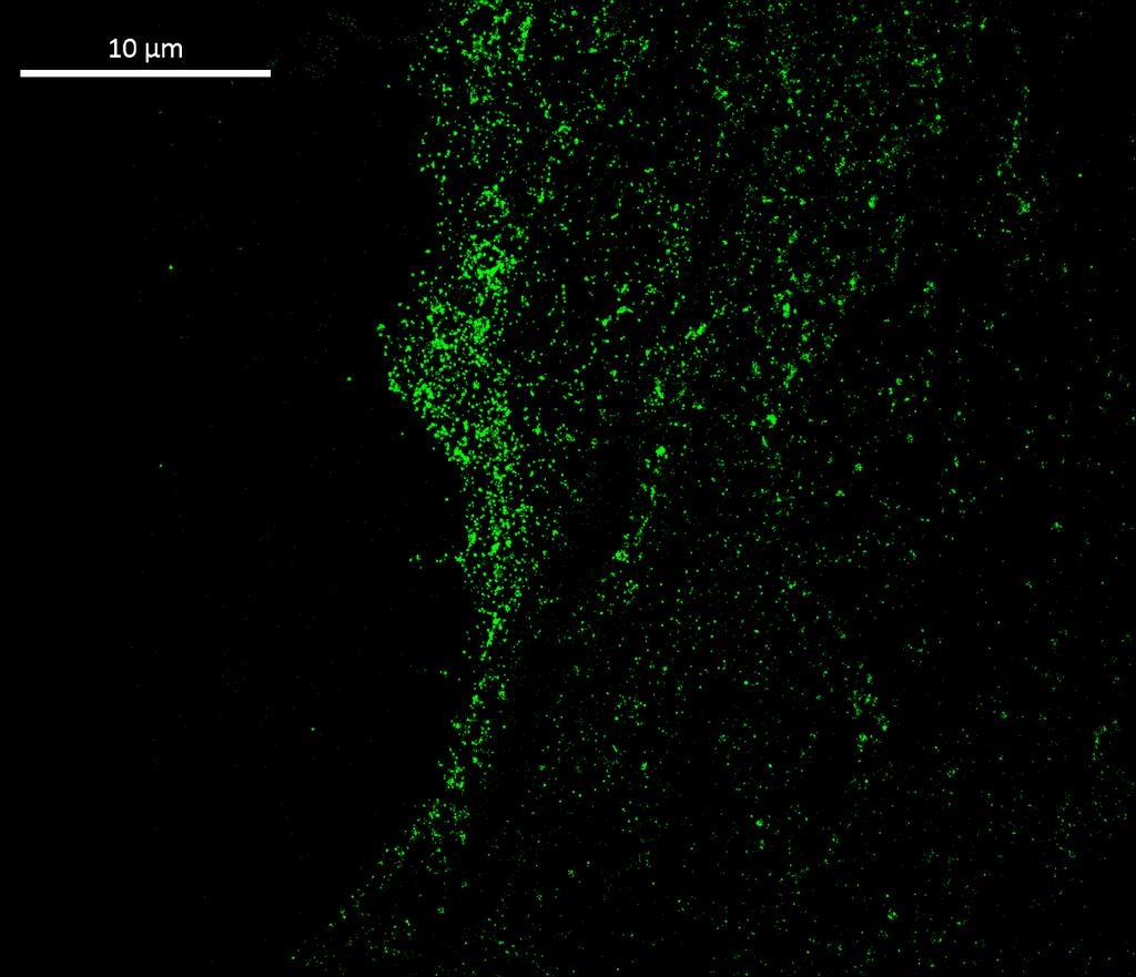
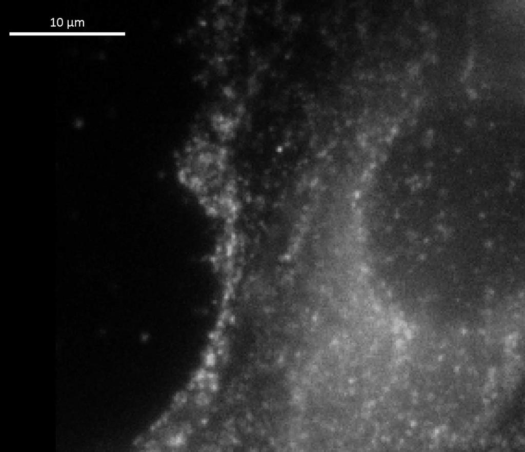
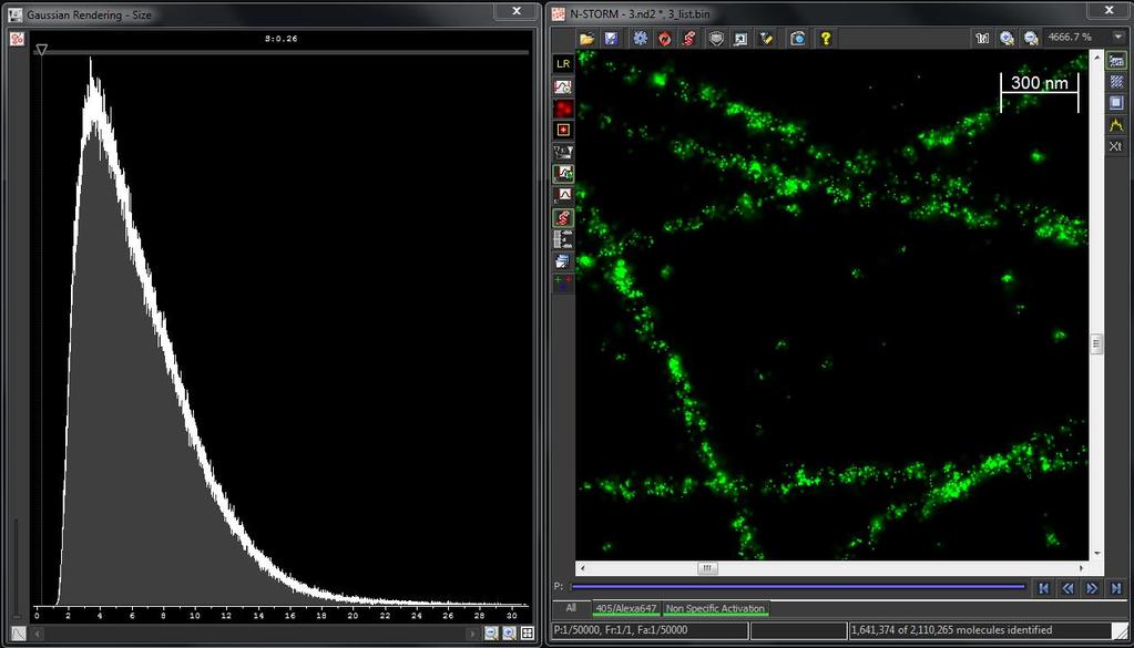
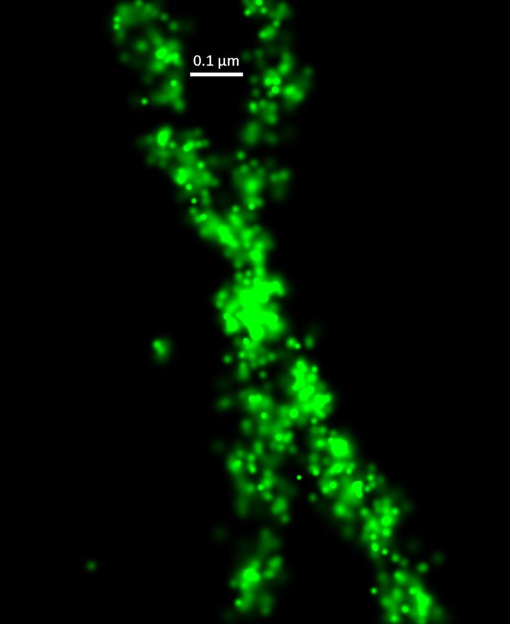
13 40,000 frames, 1,502,569 localization points 500 nm 5 μm Microtubules

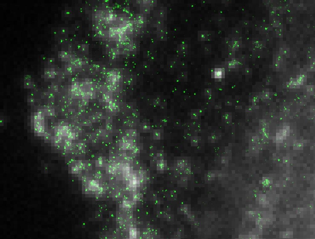
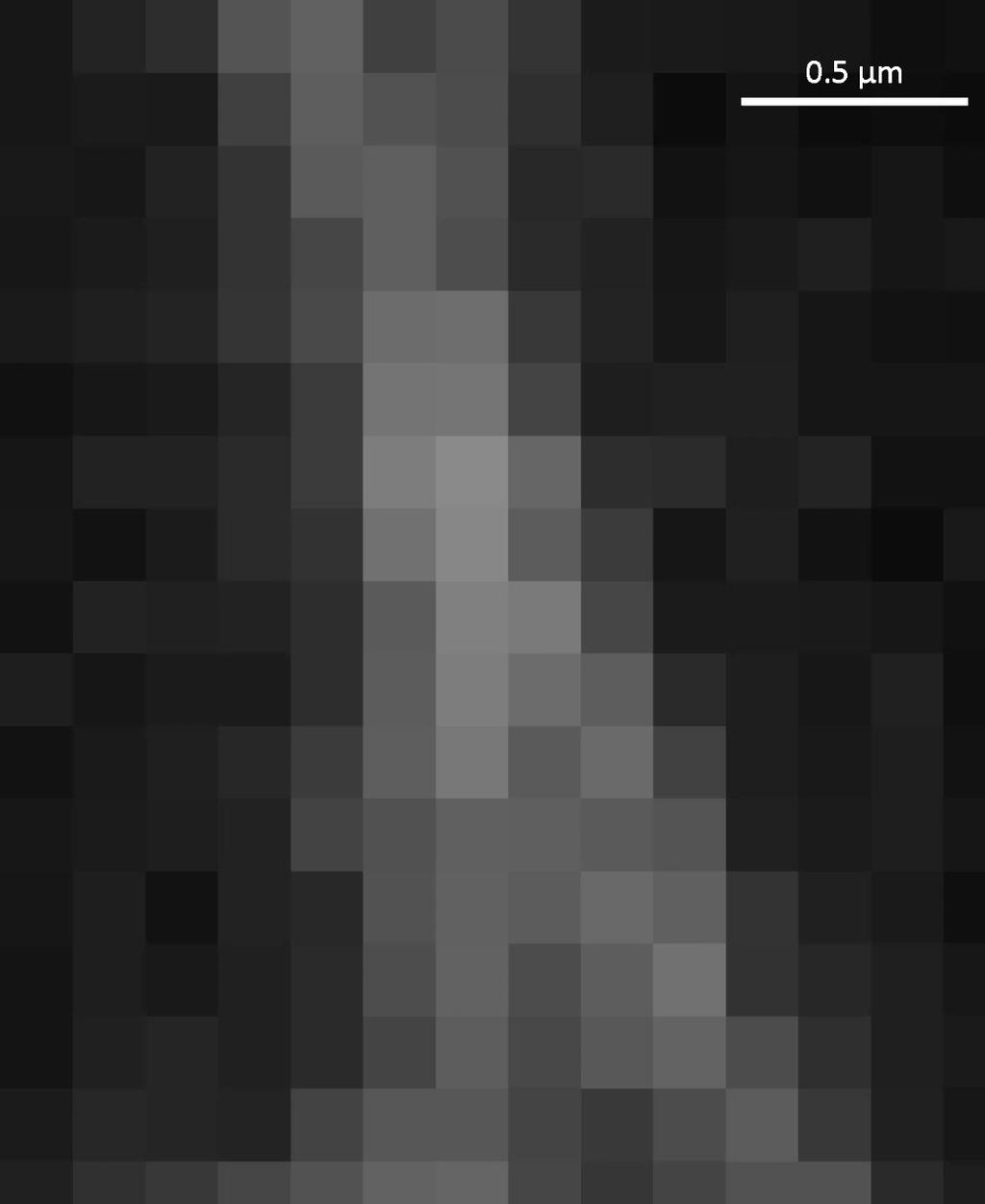
14 Widefield image Low laser power Overlays - Widefield

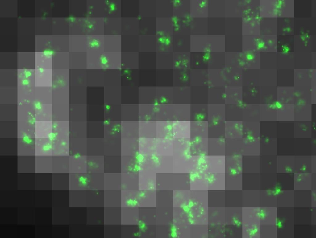
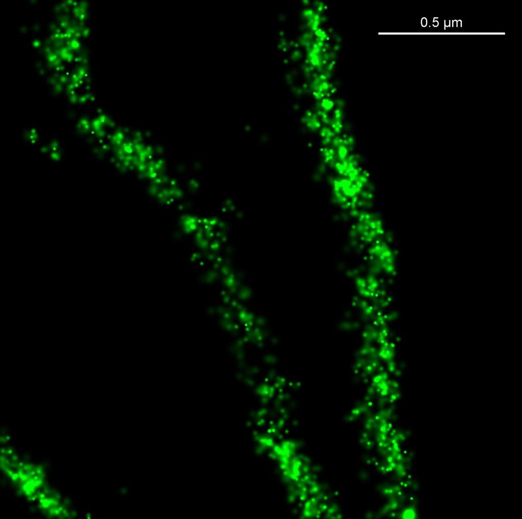
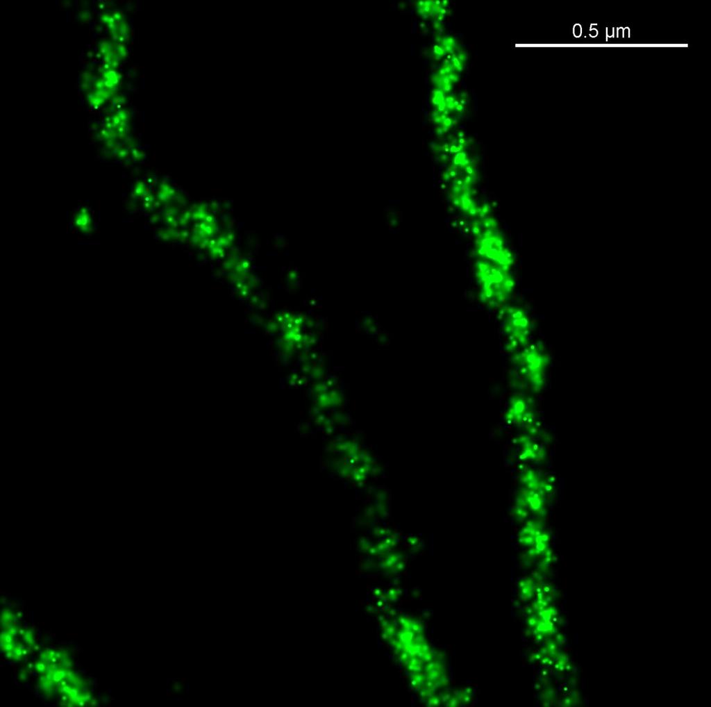
15 STORM acquisition 100 fold increase in laser power Overlays - Blinks


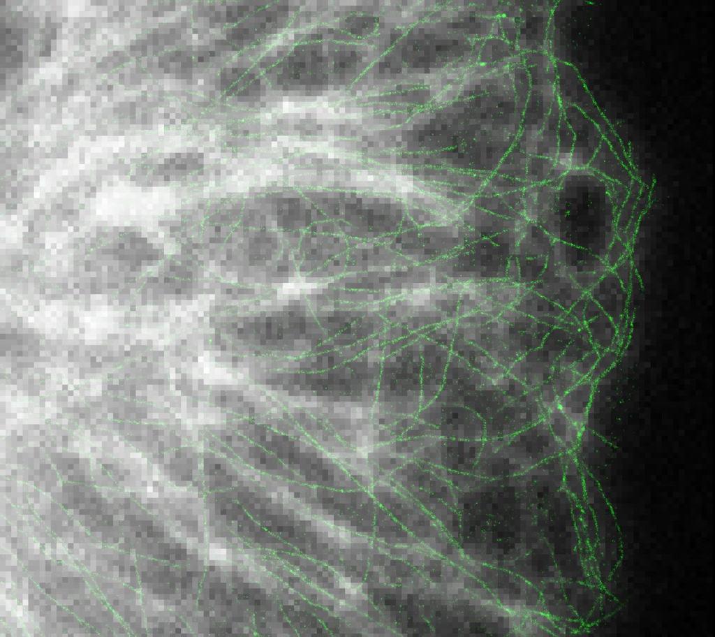

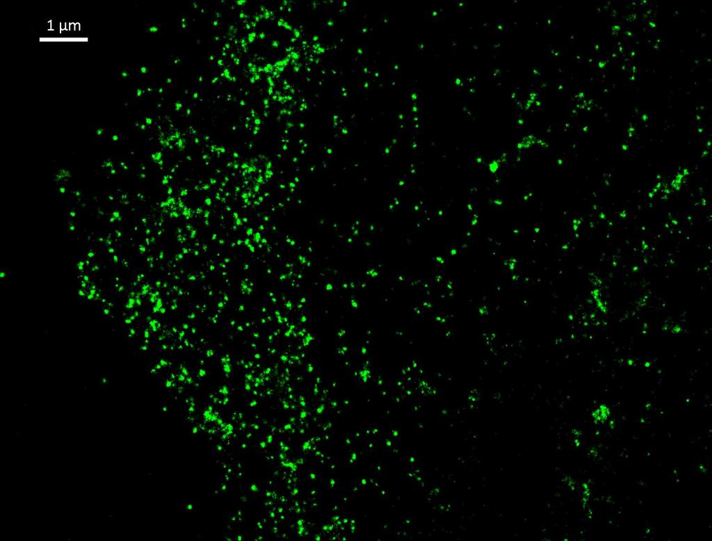
16 Overlays - STORM STORM image
17 Microtubules Widefield image STORM image
18 Clathrin
19 Clathrin
20 Clathrin
21 Outline Introduction Principles of STORM Applications N-STORM overview
22 Selected publication 1

23 Comparison of SIM and epi

24 Organisation of integrin - SIM

25 Organisation of integrin - STORM

26 Organisation of integrin - STORM
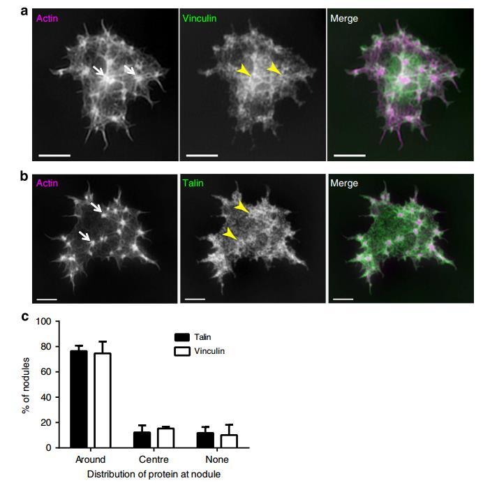
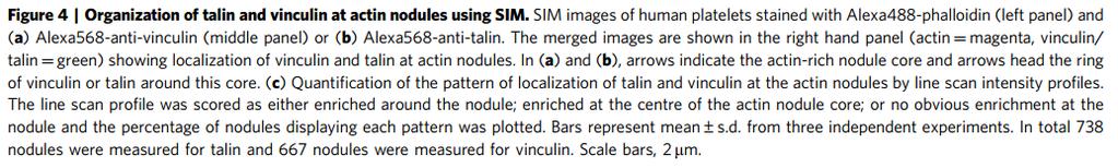
27 Organisation of talin & vinculin
28 Phosphoyrlation
29 Selected publication 2
30 qpaint (a) In DNA-PAINT, fluorescently labeled 'imager' strands (P*) transiently bind from solution to complementary 'docking' strands (P) attached to a target. Intensity vs. time traces show characteristic fluorescence on- and off-times
 of a protein residing in small so-called nuclear pore complexes that permit shuttling of various")
31 qpaint image With super resolution microscopy and qpaint analysis, researchers will be able to quantify individual molecules at specific locations in the cell. These images show varying copy numbers (shown are three, four, five, and six in the bottom images) of a protein residing in small so-called nuclear pore complexes that permit shuttling of various molecules in and out of the cell's nucleus. The arrows indicate pores with only a single protein that serve to calibrate the counting method. Credit: Wyss Institute at Harvard University.
32 Selected publication 3

33 Nuclear pore complexes Fig. 1.dSTORM of the NPC integral membrane protein gp210. (A) Comparison of widefield fluorescence (upper left corner) anddstorm image (lower right corner). gp210 proteins in nuclear envelopes isolated from Xenopus laevis oocytes were labeled by indirect immunofluorescence using the primary antibody X222 directed against an epitope located in the lumen of the nuclear envelope bordering the pore wall (Gajewski et al., 1996) and Alexa647 secondary antibodies. (B D) Higher magnifications of fluorescent circular structures to highlight the eightfold symmetrical arrangement of gp210 proteins in NPCs. (E) NPCs are generally seen as eightfold symmetrical ring structures in electron microscopy using negative staining. Scale bars: 1 μm (A), 250 nm (B), 150 nm (C E).
 The integral membrane protein gp210 surrounding the NPC labeled by immunofluorescence with Alexa647. The cross-section profile (below) of the left ring structure yields a ring diameter of 146 nm.")
34 NPC diameters Distribution of gp210 and WGA in NPCs as revealed by dstorm. In each case two representative examples are shown to emphasize differences in labeling efficiency. (A) The integral membrane protein gp210 surrounding the NPC labeled by immunofluorescence with Alexa647. The cross-section profile (below) of the left ring structure yields a ring diameter of 146 nm. (B) Nucleoporins of the central channel labeled with WGA Alexa647. The cross-section profile of the left image yields a channel diameter of 40 nm. (C) Diameters were confirmed by double staining of gp210 and WGA-binding nucleoporins located in the central channel of the NPC. Here diameters of 152 and 35 nm were calculated from a cross-section profile of the left image for the outer and the inner ring, respectively. (D) Average values of outer ring and central channel diameters as calculated from 50 different NPC ring structures. The diameter of the gp210 ring structure was determined as 161±17 nm, and the diameter of the central channel to be 38±5 nm. In addition, the distribution of FWHM values extracted from the cross-sectional profiles for gp210 and WGA showed the FWHM to be 29±7 nm for gp210 and 15±4 nm for WGA. Scale bars: 150 nm (A,C) 50 nm (B).
 For gp210, 426 individual rings containing ~160,000 localizations were combined to yield an average diameter of 164±7 nm for the gp210 ring surrounding the NPC.")
35 NPC diameters Fig. 3.Image analysis of accumulated NPC dstorm data. (A) For gp210, 426 individual rings containing ~160,000 localizations were combined to yield an average diameter of 164±7 nm for the gp210 ring surrounding the NPC. (B) For WGA, 621 rings containing ~40,000 localizations were combined and a diameter of 41±7 nm was determined for the central channel of the NPC. (C) Superimposed image of both structures. Scale bars: 100 nm.
 Comparison of conventional widefield fluorescence image (lower left corner) anddstorm image (upper right corner).")
36 Multicolour NPC imaging Fig. 4.Two-color dstorm images of NPCs using WGA-ATTO520 and Alexa647-labeled secondary antibodies directed against an epitope of gp210 on the luminal side. (A) Comparison of conventional widefield fluorescence image (lower left corner) anddstorm image (upper right corner). (B D) Higher magnification reveals the typical eightfold symmetrical ring structure of gp210 proteins (violet) surrounding the NPC and N-acetyl glucosamine-containing nucleoporins in the central channel labeled with WGA-ATTO520 (green). Scale bars: 2.5 μm (A), 100 nm (B D).
37 Outline Introduction Principles of STORM Applications N-STORM overview
38 N-STORM Super-Resolution System Hardware 3 colour STORM 2D & 3D capability TIRF/HILO/Epi illumination EMCCD camera Software Low and high density algorithms Gaussian & cross visualisation Drift correction 3D and chromatic calibrations Molecule statistics
39 N-STORM overview Resolution dyes & labelling Depth & 3D strategies Astigmatism & HILO Multi-colour strategies N-STORM dye pairs Analysis Interpreting data
40 N-STORM overview Resolution dyes & labelling Depth & 3D strategies Astigmatism & HILO Multi-colour strategies N-STORM dye pairs Analysis Interpreting data
41 Sample Preparation - Dyes Dempsey et al., Nature Methods, 2011
42 Sample Preparation - Glass
43 Accuracy of Localisation
44 Microtubules Widefield image STORM image
45 Localisation Precisions


46 Data Quality Good localisation precision + Large number of molecules identified
47 Microtubules Widefield image STORM image


48 Sample Preparation - labelling Antibodies Fab fragments Nanobodies Direct conjugation Optimisation tips Use smaller labels Use dyes at 1:1 label
49 Drift Correction
50 N-STORM overview Resolution dyes & labelling Depth & 3D strategies Astigmatism & HILO Multi-colour strategies N-STORM dye pairs Analysis Interpreting data

 Molecules localized in Z")

51 Above Focus z (nm) 3D STORM (x, y, z) Above Focus In Focus 0 In Focus Below Focus Below Focus 2γ Tube Lens Cylindrical Lens DU-897 EMCCD Huang, Zhuang et al, Science (2008) Molecules localized in Z Molecules above focus maintain symmetry in Y Molecules below focus maintain symmetry in X Fitted to Gaussians similar to XY
 300 0")


52 3D STORM 600 z (nm) nm 5 μm Huang, Wang, Bates and Zhuang, Science, 2008
53 Image Acquisition
54 N-STORM overview Resolution dyes & labelling Depth & 3D strategies Astigmatism & HILO Multi-colour strategies N-STORM dye pairs Analysis Interpreting data

55 Option 1 find 2 or more dyes Dempsey et al., Nature Methods, 2011
56 Option 1 recommendations Channel nm laser Alexa 647 Cy5 Channel nm laser Alexa 568 Alexa 555 CF Biotium 555 meos2 Channel nm laser Not recommended GLOX buffer + MEA-HCL


57 Option 1 - Chromatic Warp Uncorrected Corrected
58 Option 2 - N-STORM Dye Pairs Composition Activator Reporter Antibody
59 Photo-Switchable Dyes Pairs Alexa405 Alexa647 CY2 Alexa647 CY3 Alexa647 3 kinds of Activator and 1 kind of Reporter are available
60 N-STORM Sequence Step 1: Excite Activator molecule to energize the Reporter to a ready state 561nm CY3 Alexa647 Target sample (molecule)
61 N-STORM Sequence Step 2: Excite Reporter and emitted light is captured by the camera. 647nm 670nm CY3 Alexa647 Target sample (molecule)


62 2 Colour STORM Clathrin Microtubules 1 μm Bates, Huang, Dempsey and Zhuang, Science, 2007

63 2 Colour STORM 200 nm Image by Harvard Univ.

64 200 nm 2 Color 3D STORM Clathrin Formin Wu et al., Nature Cell Biology, 2010
65 Multi-colour summary Option 1 different dyes Option 2 dye pairs Easy sample prep 2 colour imaging Channel 2 (561 nm) poorer quality Chromatic correction required Harder sample prep High quality 3 colour imaging No chromatic correction required Longer image acquisition Better resolution
66 N-STORM overview Resolution dyes & labelling Depth & 3D strategies Astigmatism & HILO Multi-colour strategies N-STORM dye pairs Analysis Interpreting data

67 ROI Statistics Molecule lists


68 Overlapping Peak Fitting Original data Detected molecules with conventional fitting Detected molecules with Fit Overlapping Peaks Denser samples &/or wider range of fluorophores


69 Overlapping Peak Fitting 351,560 mol. 1,065,088 mol.
70 Summary Introduction Principles of STORM Applications N-STORM overview
STORM/PALM. Super Resolution Microscopy 10/31/2011. Looking into microscopic world of life
 Super Resolution Microscopy STORM/PALM Bo Huang Department of Pharmaceutical Chemistry, UCSF CSHL Quantitative Microscopy, 1/31/211 Looking into microscopic world of life 1 µm 1 µm 1 nm 1 nm 1 nm 1 Å Naked
Localization Microscopy
 Localization Microscopy Theory, Sample Prep & Practical Considerations Patrina Pellett & Ann McEvoy Applications Scientist GE Healthcare, Cell Technologies May 27 th, 2015 Localization Microscopy Talk
PALM/STORM, BALM, STED
 PALM/STORM, BALM, STED Last class 2-photon Intro to PALM/STORM Cyanine dyes/dronpa This class Finish localization super-res BALM STED Localization microscopy Intensity Bins = pixels xx 2 = ss2 + aa 2 /12
Fluorescence Nanoscopy
 Fluorescence Nanoscopy Keith A. Lidke University of New Mexico panda3.phys.unm.edu/~klidke/index.html Optical Microscopy http://en.wikipedia.org/wiki/k%c3%b6hler_illumination 30 µm Fluorescent Probes Michalet
Super Resolution Imaging Solution Provider. Imaging Future
 Super Resolution Imaging Solution Provider Imaging Future Imaging Solution More Than Equipment NanoBioImaging(NBI) is the Industrial Partner of HKUST Super Resolution Imaging Center (SRIC). NBI aims to
SIM SSIM. nanoscopy RESOLFT. Super-resolution STORM GSDIM. dstorm PALMIRA FPALM PALM PAINT SPRAIPAINT SOFI BALM CALM. Bo Huang
 STEDGSD STORM SOFI nanoscopy GSDIM PALMIRA SMACM BBB PAINT SPRAIPAINT CALM RESOLFT BALM SIM SSIM Super-resolution Bo Huang 2013.08.01 dstorm FPALM PALM 50 years to extend the resolution Confocal microscopy
Fast, three-dimensional super-resolution imaging of live cells
 Nature Methods Fast, three-dimensional super-resolution imaging of live cells Sara A Jones, Sang-Hee Shim, Jiang He & Xiaowei Zhuang Supplementary Figure 1 Supplementary Figure 2 Supplementary Figure 3
Super-resolution imaging: early days w/ Video-enhanced DIC, TIRF, PALM, STORM, etc.
 15/05/2012 Super-resolution imaging: early days w/ Video-enhanced DIC, TIRF, PALM, STORM, etc. Prof. Dr. Rainer Duden duden@bio.uni-luebeck.de 1 Using conventional light microscopy resolution is limited
PEER REVIEW FILE. Reviewers' comments: Reviewer #1 (Remarks to the Author):
:") PEER REVIEW FILE Reviewers' comments: Reviewer #1 (Remarks to the Author): General In its beginnings in the mid 1990s, localization microscopy based on optical isolation of fluorescent point targets was
PEER REVIEW FILE Reviewers' comments: Reviewer #1 (Remarks to the Author): General In its beginnings in the mid 1990s, localization microscopy based on optical isolation of fluorescent point targets was
Nature Methods: doi: /nmeth Supplementary Figure 1
 Supplementary Figure 1 File Hierarchy, Single Molecule Profiler software interface and datamining using Cell Profiler Analyst The Single Molecule Profiler (SMP) software automatically generates a database
Multiplexed imaging using same species primary antibodies with signal amplification
 Multiplexed imaging using same species primary antibodies with signal amplification Yu Wang 1,2, Wenxin Xie 1,2, Richie E. Kohman 1 and George M. Church 1,2 1. Wyss Institute for Biologically Inspired
Bi177 - Lecture 13 Microscopy Outside the Box. Fluorescence Nanoscopy TIRF 4-pi STED STORM/PALM
 Bi177 - Lecture 13 Microscopy Outside the Box Fluorescence Nanoscopy TIRF 4-pi STED STORM/PALM The diffraction limit: Abbe s law The Problem Diffraction limit 100x larger than molecular scale! Green Fluorescent
Super-resolution Microscopy
 Semr oc kwhi t epaperser i es : 1. Introduction Super-resolution Microscopy Fluorescence microscopy has revolutionized the study of biological samples. Ever since the invention of fluorescence microscopy
Tracking sub-microscopic protein organisation at the plasma membrane of live cells using triple-colour superresolution
 Tracking sub-microscopic protein organisation at the plasma membrane of live cells using triple-colour superresolution microscopy Jacob Piehler Division of Biophysics, University of Osnabrück, Germany
More on fluorescence
 More on fluorescence Last class Fluorescence Absorption emission Jablonski diagrams This class More on fluorescence Common fluorophores Jablonski diagrams to spectra Properties of fluorophores Excitation
Measure of surface protein mobility with u-paint technique
 Measure of surface protein mobility with u-paint technique How dynamic image can solve the situation? Random distribution or cluster? Why live super-resolution microscopy can solve the situation With mobility
Fluorescence Light Microscopy for Cell Biology
 Fluorescence Light Microscopy for Cell Biology Why use light microscopy? Traditional questions that light microscopy has addressed: Structure within a cell Locations of specific molecules within a cell
SUPPLEMENTARY INFORMATION
 doi: 10.1038/nature06147 SUPPLEMENTARY INFORMATION Figure S1 The genomic and domain structure of Dscam. The Dscam gene comprises 24 exons, encoding a signal peptide (SP), 10 IgSF domains, 6 fibronectin
SUPER-RESOLUTION MICROSCOPY. Dr. Nathalie Garin
 SUPER-RESOLUTION MICROSCOPY Dr. Nathalie Garin Content Motivation for superresolution Superresolution, nanoscopy, : definition Structured Illumination Microscopy (SIM) Localization microscopy STimulated
BIO 315 Lab Exam I. Section #: Name:
 Section #: Name: Also provide this information on the computer grid sheet given to you. (Section # in special code box) BIO 315 Lab Exam I 1. In labeling the parts of a standard compound light microscope
3D Multicolor Super-Resolution Imaging Offers Improved Accuracy in Neuron Tracing
 3D Multicolor Super-Resolution Imaging Offers Improved Accuracy in Neuron Tracing The Harvard community has made this article openly available. Please share how this access benefits you. Your story matters
Nature Methods: doi: /nmeth Supplementary Figure 1. Retention of RNA with LabelX.
 Supplementary Figure 1 Retention of RNA with LabelX. (a) Epi-fluorescence image of single molecule FISH (smfish) against GAPDH on HeLa cells expanded without LabelX treatment. (b) Epi-fluorescence image
BIO 315 Lab Exam I. Section #: Name:
 Section #: Name: Also provide this information on the computer grid sheet given to you. (Section # in special code box) BIO 315 Lab Exam I 1. In labeling the parts of a standard compound light microscope
Acetylated Microtubules Are Preferentially Bundled Leading to Enhanced
 Biophysical Journal, Volume 113 Supplemental Information Acetylated Microtubules Are Preferentially Bundled Leading to Enhanced Kinesin-1 Motility Linda Balabanian, Christopher L. Berger, and Adam G. Hendricks
Widefield Microscopy Bleed-Through
 In widefield microscopy the excitation wavelengths which illuminate the sample, and the emission wavelengths which reach the CCD camera are selected throughout a filter cube. A filter cube consists of
Visualisation, Sizing and Counting of Fluorescent and Fluorescently-Labelled Nanoparticles
 Visualisation, Sizing and Counting of Fluorescent and Fluorescently-Labelled Nanoparticles Introduction Fluorescent molecules have long been used to specifically label particular structures and features
DNA Microarray Technology
 2 DNA Microarray Technology 2.1 Overview DNA microarrays are assays for quantifying the types and amounts of mrna transcripts present in a collection of cells. The number of mrna molecules derived from
Post-expansion antibody delivery, after epitope-preserving homogenization.
 Supplementary Figure 1 Post-expansion antibody delivery, after epitope-preserving homogenization. (a, b) Wide-field fluorescence images of Thy1-YFP-expressing mouse brain hemisphere slice before expansion
A Brief History of Light Microscopy And How It Transformed Biomedical Research
 A Brief History of Light Microscopy And How It Transformed Biomedical Research Suewei Lin Office: Interdisciplinary Research Building 8A08 Email: sueweilin@gate.sinica.edu.tw TEL: 2789-9315 Microscope
ALP (alkaline phosphatase) calibrators were analyzed manually in microtiter plates to find the linearity range by following this protocol:
 calibrators were analyzed manually in microtiter plates to find the linearity range by following this protocol:") Exam Mol 3008 May 2009 Subject 1 (15p) ALP (alkaline phosphatase) calibrators were analyzed manually in microtiter plates to find the linearity range by following this protocol: Reaction solutions: 50
Exam Mol 3008 May 2009 Subject 1 (15p) ALP (alkaline phosphatase) calibrators were analyzed manually in microtiter plates to find the linearity range by following this protocol: Reaction solutions: 50
Visualizing Cells Molecular Biology of the Cell - Chapter 9
 Visualizing Cells Molecular Biology of the Cell - Chapter 9 Resolution, Detection Magnification Interaction of Light with matter: Absorbtion, Refraction, Reflection, Fluorescence Light Microscopy Absorbtion
Super-Resolution Localization Microscopy
 APPLICATION NOTE Super-Resolution Localization Microscopy Light microscopy techniques have been vital to our understanding of biological structures and systems since their invention in the late 16 th Century.
Fluorescence Microscopy. Terms and concepts to know: 10/11/2011. Visible spectrum (of light) and energy
 and energy") Fluorescence Microscopy Louisiana Tech University Ruston, Louisiana Microscopy Workshop Dr. Mark DeCoster Associate Professor Biomedical Engineering 1 Terms and concepts to know: Signal to Noise Excitation
Fluorescence Microscopy Louisiana Tech University Ruston, Louisiana Microscopy Workshop Dr. Mark DeCoster Associate Professor Biomedical Engineering 1 Terms and concepts to know: Signal to Noise Excitation
Stochastic Optical Reconstruction Microscopy (STORM): A Method for Superresolution Fluorescence Imaging
: A Method for Superresolution Fluorescence Imaging") Topic Introduction Stochastic Optical Reconstruction Microscopy (STORM): A Method for Superresolution Fluorescence Imaging Mark Bates, Sara A. Jones, and Xiaowei Zhuang The relatively low spatial resolution
Topic Introduction Stochastic Optical Reconstruction Microscopy (STORM): A Method for Superresolution Fluorescence Imaging Mark Bates, Sara A. Jones, and Xiaowei Zhuang The relatively low spatial resolution
Immunofluorescence Confocal Microscopy of 3D Cultures Grown on Alvetex
 Immunofluorescence Confocal Microscopy of 3D Cultures Grown on Alvetex 1.0. Introduction Immunofluorescence uses the recognition of cellular targets by fluorescent dyes or antigen-specific antibodies coupled
Cell Imaging. Cell Imaging 48
 Cell Imaging 48 bio-rad.com/zoe Cell Imaging Bio-Rad s suite of tools for fluorescence microscopy and cell imaging includes the ZOE fluorescent cell imager and nuclear dyes. See Also PureBlu Hoechst 33342
Dino-Lite knowledge & education. Fluorescence Microscopes
 Dino-Lite knowledge & education Fluorescence Microscopes Dino-Lite Fluorescence models Smallest fluorescence microscope in the world Revolution to biomedical and educational applications Flexible Easy
Lab 5: Optical trapping and single molecule fluorescence
 Lab 5: Optical trapping and single molecule fluorescence PI: Matt Lang Lab Instructor: Jorge Ferrer Summary Optical tweezers are an excellent experimental tool to study the biophysics of single molecule
Special Techniques 1. Mark Scott FILM Facility
 Special Techniques 1 Mark Scott FILM Facility SPECIAL TECHNIQUES Multi-photon microscopy Second Harmonic Generation FRAP FRET FLIM In-vivo imaging TWO-PHOTON MICROSCOPY Alternative to confocal and deconvolution
Multiplexed 3D FRET imaging in deep tissue of live embryos Ming Zhao, Xiaoyang Wan, Yu Li, Weibin Zhou and Leilei Peng
 Scientific Reports Multiplexed 3D FRET imaging in deep tissue of live embryos Ming Zhao, Xiaoyang Wan, Yu Li, Weibin Zhou and Leilei Peng 1 Supplementary figures and notes Supplementary Figure S1 Volumetric
Confocal Microscopy & Imaging Technology. Yan Wu
 Confocal Microscopy & Imaging Technology Yan Wu Dec. 05, 2014 Cells under the microscope What we use to see the details of the cell? Light and Electron Microscopy - Bright light / fluorescence microscopy
Evaluation of fluorophores for optimal performance in localization-based super-resolution imaging
 Evaluation of fluorophores for optimal performance in localization-based super-resolution imaging Graham T Dempsey 1,6, Joshua C Vaughan 2,3,6, Kok Hao Chen 3,6, Mark Bates 4 & Xiaowei Zhuang 2,3,5 211
Introduction to Computational Fluorescence Microscopy!
 Introduction to Computational Fluorescence Microscopy! EE367/CS448I: Computational Imaging and Display! stanford.edu/class/ee367! Lecture 13! Gordon Wetzstein! Stanford University! Midterm! Tuesday, Feb
F* techniques: FRAP, FLIP, FRET, FLIM,
 F* techniques: FRAP, FLIP, FRET, FLIM, FCS Antonia Göhler March 2015 Fluorescence explained in the Bohr model Absorption of light (blue) causes an electron to move to a higher energy orbit. After a particular
Basic Principles in Flow Cytometry. Flow Cytometry
 Basic Principles in Flow Cytometry Flow Cytometry» Flow Cytometry is the technological process that allows for the individual measurements of cell fluorescence and light scattering. This process is performed
Chapter 13. The Nucleus. The nucleus is the hallmark of eukaryotic cells; the very term eukaryotic means having a "true nucleus".
 Chapter 13 The Nucleus The nucleus is the hallmark of eukaryotic cells; the very term eukaryotic means having a "true nucleus". Fig.13.1. The EM of the Nucleus of a Eukaryotic Cell 13.1. The Nuclear Envelope
SUPPLEMENTARY INFORMATION
 DOI: 10.1038/NCHEM.1805 Visualization and Selective Chemical Targeting of RNA G-quadruplex Structures in the Cytoplasm of Human Cells Giulia Biffi 1, Marco Di Antonio 2, David Tannahill 1 and Shankar Balasubramanian
Q&A: Single-molecule localization microscopy for biological imaging
 Q U E S T I O N & ANSWER Q&A: Single-molecule localization microscopy for biological imaging Ann L McEvoy 1, Derek Greenfield 1,2,5, Mark Bates 3 and Jan Liphardt 1,2,4 * Open Access Why is it important
SURFACE ENHANCED RAMAN SCATTERING NANOPARTICLES AS AN ALTERNATIVE TO FLUORESCENT PROBES AN EVALUATION
 APPLICATION NOTE SURFACE ENHANCED RAMAN SCATTERING NANOPARTICLES AS AN ALTERNATIVE TO FLUORESCENT PROBES AN EVALUATION Summary: Interest in using nanoparticles specifically, Surface Enhanced Raman Scattering
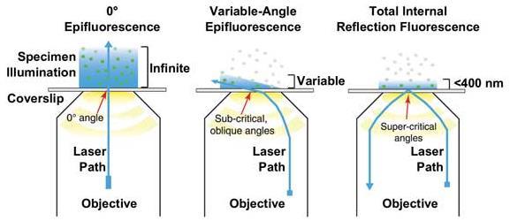
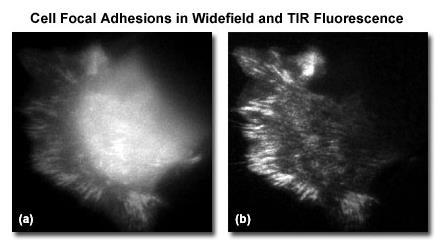
Total Internal Reflection Fluorescence Microscopy
 Total Internal Reflection Microscopy Nicole O Neil Indiana University October 24, 2005 Agenda Why use TIRFM? Theory behind TIR Snell s Law Instrumentation Evanescent Wave Excitation of Fluorophores Advantages/Disadvantages
Leica SR GSD Super-Resolution Microscopy with GSDIM
 Application NOTE 07/2012 Leica SR GSD Super-Resolution Microscopy with GSDIM Widefield GSD CONTENT The new world of resolution....... 3 The Leica SR GSD System for brilliant Super-Resolution Images.. 5
Fluorescence Imaging with One Nanometer Accuracy Lab
 I. Introduction. Fluorescence Imaging with One Nanometer Accuracy Lab Traditional light microscope is limited by the diffraction limit of light, typically around 250 nm. However, many biological processes
Single cell molecular profiling using Quantum Dots. Technical Journal Club Rahel Gerosa
 Single cell molecular profiling using Quantum Dots Technical Journal Club 01.10.2013 Rahel Gerosa Molecular Profiling Powerful technique to study complex molecular networks underlying physiological and
Biosensors. DNA Microarrays (for chemical analysis) Protein Sensors (for identifying viruses)
 Protein Sensors (for identifying viruses)") Biosensors DNA Microarrays (for chemical analysis) Protein Sensors (for identifying viruses) DNA Microarrays 40 000 detectors in parallel, each detecting a specific DNA sequence. Combinatorial Chemistry
Biosensors DNA Microarrays (for chemical analysis) Protein Sensors (for identifying viruses) DNA Microarrays 40 000 detectors in parallel, each detecting a specific DNA sequence. Combinatorial Chemistry
CHARACTERIZATION OF MOLECULAR ORIENTATION IN SUPER-RESOLUTION FLUORESCENCE MICROSCOPY
 Master Erasmus Mundus in Photonics Engineering, Nanophotonics and Biophotonics Europhotonics MASTER THESIS WORK CHARACTERIZATION OF MOLECULAR ORIENTATION IN SUPER-RESOLUTION FLUORESCENCE MICROSCOPY Yibing
Imaging of endocrine organs
 Imaging of endocrine organs Helen Christian Department of Physiology, Anatomy & Genetics St Anne s College, University of Oxford Diabetesforum, Stockholm 2017 Islets of Langerhan Pituitary gland Renin
Contents. SCHOOL of FLUORESCENCE. For more information, go to lifetechnologies.com/imagingbasics
 MPSF educator packet This packet contains illustrations and figures from the Molecular Probes School of Fluorescence website. They illustrate concepts from the basic physical properties that underlie fluorescence
MF-ChemiBIS. Today s most comprehensive solution for your bio-imaging needs and applications. Documenting Nature
 MF-ChemiBIS Today s most comprehensive solution for your bio-imaging needs and applications Documenting Nature MF-ChemiBIS Excellence in bio-imaging The DNR Advantage As pioneers in bio-imaging technologies
Real-Time PCR Principles and Applications
 Real-Time PCR Principles and Applications Dr Esam Ibraheem Azhar (BSc, MSc, Ph.D Molecular Medical Virology) Asst. Prof. Medical Laboratory Technology Department Objectives Real-Time PCR Principles and
Anti-MOUSE IgG (H&L) (GOAT) Antibody DyLight 488 Conjugated (Min X Bv Ch Gt GP Ham Hs Hu Rb Rt & Sh Serum Proteins)
 (GOAT) Antibody DyLight 488 Conjugated (Min X Bv Ch Gt GP Ham Hs Hu Rb Rt & Sh Serum Proteins)") Anti-MOUSE IgG (H&L) (GOAT) Antibody DyLight 488 Conjugated (Min X Bv Ch Gt GP Ham Hs Hu Rb Rt & Sh Serum Proteins) - 610-141-121 Code: 610-141-121 Size: 100 µg Product Description: Anti-MOUSE IgG (H&L)
Anti-MOUSE IgG (H&L) (GOAT) Antibody DyLight 488 Conjugated (Min X Bv Ch Gt GP Ham Hs Hu Rb Rt & Sh Serum Proteins) - 610-141-121 Code: 610-141-121 Size: 100 µg Product Description: Anti-MOUSE IgG (H&L)
Amnis ImageStream : Technical Reports & Applications
 Amnis ImageStream : Technical Reports & Applications ImageStream : Flow Cytometry and Microscopy in a Single Platform The ImageStream achieves true multispectral Imaging in Flow by combining microscopy
Characterizing Phenotypes of Bacteria by Staining Method
 Experiment 3 Laboratory to Biology III Diversity of Microorganisms / Wintersemester / page 1 Experiment 3 Characterizing Phenotypes of Bacteria by Staining Method Advisor NN Reading Chapters in BBOM 9
Characterizing Phenotypes of Bacteria by Staining Method
 Experiment 3 Laboratory to Biology III Diversity of Microorganisms / Wintersemester / page 1 Experiment Characterizing Phenotypes of Bacteria by Staining Method Advisor Reading NN Chapters 3.1, 3.7, 3.8,
Superresolution Pattern Recognition Reveals the Architectural Map of the
 Supplementary Information Superresolution Pattern Recognition Reveals the Architectural Map of the Ciliary Transition Zone T. Tony Yang a, Jimmy Su b, Won-Jing Wang c, Branch Craige d, George B. Witman
Chapter 10: Classification of Microorganisms
 Chapter 10: Classification of Microorganisms 1. The Taxonomic Hierarchy 2. Methods of Identification 1. The Taxonomic Hierarchy Phylogenetic Tree of the 3 Domains Taxonomic Hierarchy 8 successive taxa
T he recent widespread uptake of new super resolution techniques has revolutionized and invigorated molecular
 OPEN SUBJECT AREAS: SUPER-RESOLUTION MICROSCOPY SINGLE-MOLECULE BIOPHYSICS Received 16 October 2014 Accepted 11 December 2014 Published 21 January 2015 Correspondence and requests for materials should
SAPIENZA Università di Roma Laurea magistrale in Ingegneria delle Nanotecnologie A.A Biophotonics Laboratory Course
 SAPIENZA Università di Roma Laurea magistrale in Ingegneria delle Nanotecnologie A.A. 2016-2017 Biophotonics Laboratory Course Prof. Francesco Michelotti SAPIENZA Università di Roma Facoltà di Ingegneria
Single-molecule real-time detection of telomerase extension activity
 Supplementary Information Single-molecule real-time detection of telomerase extension activity Helen Hwang 1, Patricia Opresko 2, Sua Myong 1,3,4,5 1. Bioengineering Department, University of Illinois
LysoTracker Red DND-99 (Invitrogen) was used as a marker of lysosome or acidic
 was used as a marker of lysosome or acidic") information MATERIAL AND METHODS Lysosome staining LysoTracker Red DND-99 (Invitrogen) was used as a marker of lysosome or acidic compartments, according to the manufacturer s protocol. Plasmid independent
information MATERIAL AND METHODS Lysosome staining LysoTracker Red DND-99 (Invitrogen) was used as a marker of lysosome or acidic compartments, according to the manufacturer s protocol. Plasmid independent
Confocal Microscopy Analyzes Cells
 Choosing Filters for Fluorescence A Laurin Publication Photonic Solutions for Biotechnology and Medicine November 2002 Confocal Microscopy Analyzes Cells Reprinted from the November 2002 issue of Biophotonics
Visualizing mechanical tension across membrane receptors with a fluorescent sensor
 Nature Methods Visualizing mechanical tension across membrane receptors with a fluorescent sensor Daniel R. Stabley, Carol Jurchenko, Stephen S. Marshall, Khalid S. Salaita Supplementary Figure 1 Fabrication
Flow Cytometry - The Essentials
 Flow Cytometry - The Essentials Pocket Guide to Flow Cytometry: 1. Know your Cytometer 2. Understanding Fluorescence and Fluorophores 3. Gating Process 4. Controls 5. Optimization 6. Panel Building 7.
Principles of flow cytometry: overview of flow cytometry and its uses for cell analysis and sorting. Shoreline Community College BIOL 288
 Principles of flow cytometry: overview of flow cytometry and its uses for cell analysis and sorting Shoreline Community College BIOL 288 Flow Cytometry What is Flow Cytometry? Measurement of cells or particles
NEWTON 7.0 BIOLUMINESCENCE & FLUORESCENCE IMAGING IN VIVO - IN VITRO IMAGING
 NEWTON 7.0 BIOLUMINESCENCE & FLUORESCENCE IMAGING IN VIVO - IN VITRO IMAGING SMART IMAGING SYSTEM The NEWTON 7.0 system combines high sensitivity with advanced animal-handling features and userfriendly
New single-molecule imaging system ends prna debate over phi29 motor
 Page 1 of 5 January 30, 2007 New single-molecule imaging system ends prna debate over phi29 motor WEST LAFAYETTE, Ind. - Scientists are able to view active molecules within a biological motor of the nanometer
OCTOPLUS QPLEX FLUORESCENCE IMAGER. for fast & powerful fast 2D Gel image acquisition
 OCTOPLUS QPLEX FLUORESCENCE IMAGER for fast & powerful fast 2D Gel image acquisition Octoplus QPLEX Fluorescence Imager The new Octoplus QPLEX fluorescence imager sets a novel standard fluorescence 2D
Current ( pa) Current (pa) Voltage (mv) Voltage ( mv)
 Current (pa) Voltage (mv) Voltage ( mv)") Current ( pa) 3000 2000 1000 0-1000 -2000-3000 a -400-200 0 200 400 Voltage (mv) P1 P2 P3 P4 P5 P6 P7 P8 P9 P10 P11 P12 P13 P14 P15 P16 P17 P18 Average Current (pa) 4500 3000 1500 0-1500 -3000-4500 b -400-200
Current ( pa) 3000 2000 1000 0-1000 -2000-3000 a -400-200 0 200 400 Voltage (mv) P1 P2 P3 P4 P5 P6 P7 P8 P9 P10 P11 P12 P13 P14 P15 P16 P17 P18 Average Current (pa) 4500 3000 1500 0-1500 -3000-4500 b -400-200
Cell analysis and bioimaging technology illustrated
 Cell analysis and bioimaging technology illustrated The Cell Analysis Center Scientific Bulletin Part 1 Sysmex has been studying and exploring principles of automated haematology analysers, making full
Three Dimensional Orientation of Anisotropic. Plasmonic Aggregates at Intracellular Nuclear. Indentation Sites by Integrated Light Sheet Super-
 Supporting Information Three Dimensional Orientation of Anisotropic Plasmonic Aggregates at Intracellular Nuclear Indentation Sites y Integrated Light Sheet Super- Resolution Microscopy Suresh Kumar Chakkarapani,
Lecture 13. Motor Proteins I
 Lecture 13 Motor Proteins I Introduction: The study of motor proteins has become a major focus in cell and molecular biology. Motor proteins are very interesting because they do what no man-made engines
Supplementary Information. Arrays of Individual DNA Molecules on Nanopatterned Substrates
 Supplementary Information Arrays of Individual DNA Molecules on Nanopatterned Substrates Roland Hager, Alma Halilovic, Jonathan R. Burns, Friedrich Schäffler, Stefan Howorka S1 Figure S-1. Characterization
APPLICATION NOTE Rev. 7/2017, v4.0 Fluorescent Nanodiamonds: Bio-applications. Physical and Fluorescence Properties
 APPLICATION NOTE Rev. 7/2017, v4.0 Fluorescent Nanodiamonds: Bio-applications Fluorescent nanodiamonds (FNDs) offer a unique alternative to currently existing fluorescent biomarkers. With exceptional photo
Supplementary Figure 1. Design of linker truncation library between Nluc and either Venus or mneongreen
 1 2 3 Supplementary Figure 1. Design of linker truncation library between Nluc and either Venus or mneongreen 4 5 6 7 8 9 10 The cdna of C-terminally deleted FPs (mneongreen or Venus) mutants and N-terminally
ab CytoPainter ER Staining Kit Red Fluorescence
 ab139482 CytoPainter ER Staining Kit Red Fluorescence Instructions for Use Designed to detect Human endoplasmic reticulum by microscopy. This product is for research use only and is not intended for diagnostic
Fluorescence microscopy
 Fluorescence microscopy 1 Fluorescence microscopies basic fluorescence, fluorophores Deconvolution Confocal Two-photon/multi-photon 4Pi Light sheet Total internal reflection STED FRAP/FLIP/FCS FRET PALM/STORM/iPALM
Chapter 1. A Preview of the Cell. Lectures by Kathleen Fitzpatrick Simon Fraser University Pearson Education, Inc.
 Chapter 1 A Preview of the Cell Lectures by Kathleen Fitzpatrick Simon Fraser University The Cell Theory: A Brief History Robert Hooke (1665) observed compartments in cork, under a microscope, and first
7.1 Techniques for Producing and Analyzing DNA. SBI4U Ms. Ho-Lau
 7.1 Techniques for Producing and Analyzing DNA SBI4U Ms. Ho-Lau What is Biotechnology? From Merriam-Webster: the manipulation of living organisms or their components to produce useful usually commercial
Direct stochastic optical reconstruction microscopy with standard fluorescent probes
 Direct stochastic optical reconstruction microscopy with standard fluorescent probes Sebastian van de Linde, Anna Löschberger, Teresa Klein, Meike Heidbreder, Steve Wolter, Mike Heilemann & Markus Sauer
Resolution of Microscopes Visible light is nm Dry lens(0.5na), green(530nm light)=0.65µm=650nm for oil lens (1.4NA) UV light (300nm) = 0.13µm f
, green(530nm light)=0.65µm=650nm for oil lens (1.4NA) UV light (300nm) = 0.13µm f") Microscopes and Microscopy MCB 380 Good information sources: Alberts-Molecular Biology of the Cell http://micro.magnet.fsu.edu/primer/ http://www.microscopyu.com/ Approaches to Problems in Cell Biology
Microscopes and Microscopy MCB 380 Good information sources: Alberts-Molecular Biology of the Cell http://micro.magnet.fsu.edu/primer/ http://www.microscopyu.com/ Approaches to Problems in Cell Biology
THE JOURNAL OF CELL BIOLOGY
 Supplemental Material THE JOURNAL OF CELL BIOLOGY Yeung et al., http://www.jcb.org/cgi/content/full/jcb.200903020/dc1 Figure S1. Assessment of the surface charge of maturing phagosomes. (A C and F H) RAW
HYPERSPECTRAL MICROSCOPE PLATFORM FOR HIGHLY MULTIPLEX BIOLOGICAL IMAGING. Marc Verhaegen
 HYPERSPECTRAL MICROSCOPE PLATFORM FOR HIGHLY MULTIPLEX BIOLOGICAL IMAGING Marc Verhaegen CMCS, MONTREAL, MAY 11 th, 2017 OVERVIEW Hyperspectral Imaging Multiplex Biological Imaging Multiplex Single Particle
NOTES Gene Expression ACP Biology, NNHS
 Name Date Block NOTES Gene Expression ACP Biology, NNHS Model 1: Transcription the process of genes in DNA being copied into a messenger RNA 1. Where in the cell is DNA found? 2. Where in the cell does
N- SIM. Choose a primary antibody that localises strongly to the relevant structure and has low background fluorescence.
 Specifications: Resolution: approx. 100nm in XY and 300nm in Z 3D Axial Range: up to 20μm Speed: up to 0.6 sec/frame (2D- SIM/TIRF- SIM) or up to 1sec/frame (3D- SIM) Light sources: Lasers for N- SIM:
Super-Resolution Imaging System. Professionals serve professionals. Three-Dimensional Simultaneous Multi-Channel Super Resolution Imaging
 Professionals serve professionals Super-Resolution Imaging System Three-Dimensional Simultaneous Multi-Channel Super Resolution Imaging NanoBioImaging Ltd. Powerful Tool of Scientists Super-resolution
Nonspecific binding of 10 nm Cy5-labeled DinB on nine different surfaces, measured by the number of DinB spots over an imaging area of 2,500 µm 2.
 Supplementary Figure 1 Nonspecific binding of 10 nm Cy5-labeled DinB on nine different surfaces, measured by the number of DinB spots over an imaging area of 2,500 µm 2. Free DinB was washed out using
Meeting the growing needs of developmental biology. Solutions brochure
 Meeting the growing needs of developmental biology Solutions brochure Imaging for developmental biology Outstanding optical quality is at the heart of all Nikon s microscopic imaging systems and this,
Electronic Supplementary Information
 Electronic Supplementary Material (ESI) for Integrative Biology. This journal is The Royal Society of Chemistry 2015 Electronic Supplementary Information Table S1. Definition of quantitative cellular features
QImaging Camera Application Notes Multicolor Immunofluorescence Imaging
 QImaging Camera Application Notes Multicolor Immunofluorescence Imaging In order to image localization of intracellular proteins with high specificity, it is frequently necessary to multiplex antibody
FRET Sensitized Emission
 Page 1 of 8 FRET Sensitized Emission Function The FRET Sensitized Emission wizard is used for measuring the FRET efficiency. For this purpose, the fluorescence emission of the acceptor that results from
Exam MOL3007 Functional Genomics
 Faculty of Medicine Department of Cancer Research and Molecular Medicine Exam MOL3007 Functional Genomics Thursday December 20 th 9.00-13.00 ECTS credits: 7.5 Number of pages (included front-page): 5 Supporting