Introduction to Computational Fluorescence Microscopy!
|
|
|
- Willa Fleming
- 5 years ago
- Views:
Transcription

1 Introduction to Computational Fluorescence Microscopy! EE367/CS448I: Computational Imaging and Display! stanford.edu/class/ee367! Lecture 13! Gordon Wetzstein! Stanford University!
2 Midterm! Tuesday, Feb 27, 10:30-11:50am in Gates 03! In-class you need to be here! open book: use slides, internet, bring computer, whatever you like! can all be solved without programming, similar to theoretical questions of the assignments! only SCPD students can do remotely, we will be ing you!
 tadpole neurons (green); technique: confocal")
3 H. Rankin, transgenic xenopus laevis (african clawed toad) tadpole neurons (green); technique: confocal 10x!
; technique: super resolution")
4 M. Kandasamy, stained cells: actin (pink), DNA (yellow), mitochondria (green); technique: super resolution microscopy!
; technique: confocal 40X!")
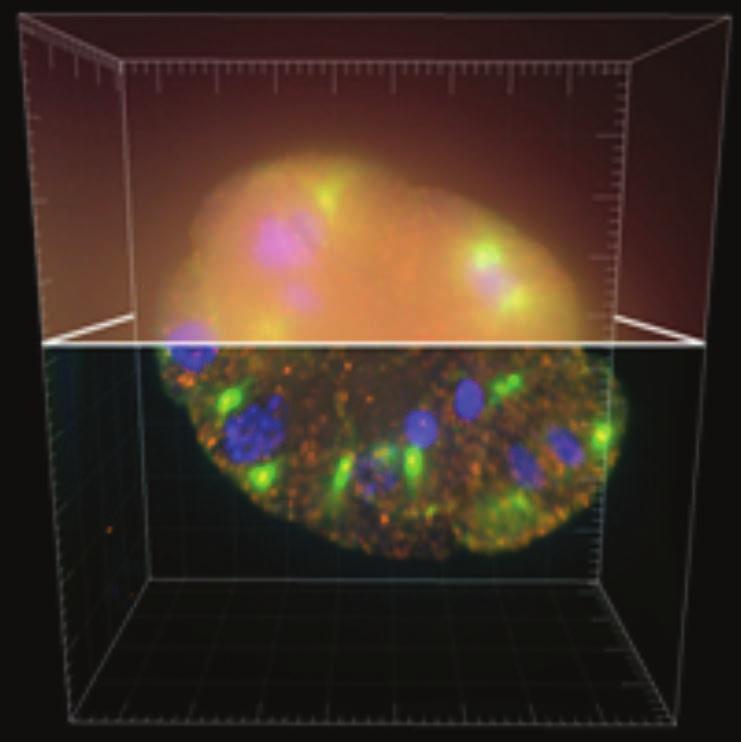
5 M. Boyle, larva of nephasoma pellucidum (peanut worm); technique: confocal 40X!
, DNA (blue); technique: structured illumination")
6 D. Burnette, osteosarcoma cell (bone cancer) showing actin (purple), mitochondria (yellow), DNA (blue); technique: structured illumination microscopy (SIM)!

7 T. Deerinck, HeLa cells with microtubules; technique: 2-photon microscopy 300X!
8 Nikon Small World Competition! annual photography competition, see showed only fluorescent samples (many others in the gallery)! this lecture: overview of fluorescence microscopy techniques!

9

10 source: white house & nature!
11 Brain Initiative! frontier of science (past frontiers: fly to moon, decode human genome)! two key factors: fluorescence microscopy & computational illumination!
12 Deisseroth Lab, Stanford; CLARITY; Nature 2013!

13 Widefield Microscopy! source: microscopyu!

14 Microscope Objective! source: Zeiss!


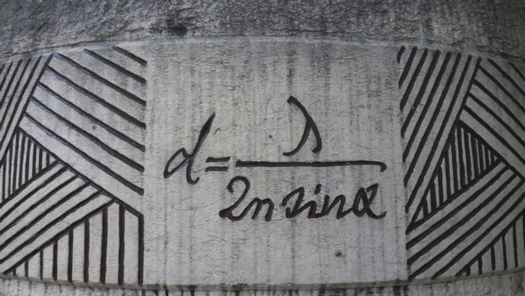
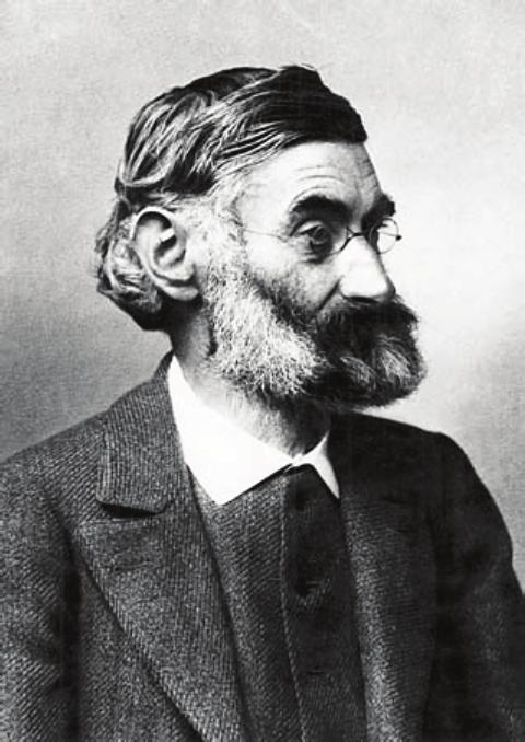
15 The Diffraction Limit! Ernst Abbe, 1905! source: wikipedia!
16 The Diffraction Limit! d =! 2nsin" =! 2NA!

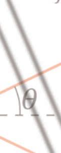


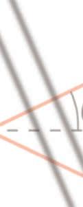

17 The Diffraction Limit! d =! 2nsin" =! 2NA!!
18 The Diffraction Limit! d =! 2nsin" =! 2NA!! Airy disk!


19 The Diffraction Limit! d =! 2nsin" =! 2NA Rayleigh Criterion! Airy disk! source: wikipedia!

20 Lateral and Axial Resolution & Missing Cone!

21 Fluorescence Microscopy!!!!! excitation and emission! coherence / incoherence! fluorescent labels! calcium imaging!
22 Fluorescence Microscopy (epi setup)! source: wikipedia!

23 Fluorescence Microscopy (epi setup)! source: Nikon MicroscopyU! source: wikipedia!

!")
24 Sensors used in Microscopy!! e.g., Andor ixon Ultra 897: cooled to -100 C or Hamamatsu Ocra Flash4.0 V2!! scientific CMOS & CCD (~20-50K)!! reduce pretty much all noise, except for photon or shot noise!
25 Fluorescence Microscopy - Challenges! inherently 2D need 3D for active brain imaging! higher-resolution in 2D and 3D! scattering! larger fields of view, bleaching! solution: engineer detection and illumination optics, algorithms, chemistry!!
26 ! Fluorescence Microscopy - Challenges!! inherently 2D need 3D for active brain imaging!! higher-resolution in 2D and 3D!!! scattering! larger fields of view, bleaching!! solution: engineer detection and illumination optics, algorithms, chemistry!
27 Superresolution Fluorescence Microscopy! stimulated emission-depletion (STED) microscopy! localization microscopy! 2D: STORM/PALM etc.! 3D: double helix PSF! localization algorithms! structured illumination microscopy (SIM)!
! Stefan Hell! (Max Planck Institute)! W. E. Moerner!")
28 2014 Nobel Price in Chemistry: super-resolved fluorescence microscopy! Eric Betzig! (Howard Hughes Institute)! Stefan Hell! (Max Planck Institute)! W. E. Moerner! (Stanford)!
 Microscopy!")

29 Stimulated Emission-Depletion (STED) Microscopy! excitation spot! de-excitation spot! emitted spot! source: wikipedia!

30 Stimulated Emission-Depletion (STED) Microscopy!

31 Localization Microscopy: PALM / STORM!

32 Structured Illumination Microscopy (SIM)!
33 3D Fluorescence Microscopy! confocal microscopy! 2 photon microscopy! light sheet microscopy! 3D deconvolution microscopy / focal stacks! others: spinning disk confocal, aperture correlation,!
34 1957! Confocal Microscopy!
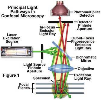
35 Confocal Microscopy!

36 Widefield vs Confocal Thin Sample! source:

37 Widefield vs Confocal Thick Sample! source:

38 2-Photon Microscopy! Denk et al. Two-photon laser scanning fluorescence microscopy, Science 1990; photo: microscopy.berkeley.edu!


39 2-Photon Microscopy! deep imaging! good scattering properties! Denk et al. Two-photon laser scanning fluorescence microscopy, Science 1990; photo: microscopy.berkeley.edu!
40 3D Deconvolution Microscopy!

41 3D Deconvolution Microscopy! whiteboard!

42 Light Sheet Microscopy!! invented by R. Zsigmondy, Nobel price in 1925!! Nature Method of the Year 2014! Huisken et al. Selective Plane Illumination Techniques in developmental biology, Development 2009!

43 Ahrens et al. Whole-brain functional imaging at cellular resolution! using light-sheet microscopy, Nature Methods 2013!

!")
44 Light Field Microscopy!!!! can do refocus, but more interesting: instantaneous 3D volume (for fluorescence)! diffraction becomes an issue! [Levoy et al. 2006]!

45 Levoy Group, Stanford! Light Field Microscopy!

46 Levoy Group, Stanford!

47 Levoy Group, Stanford! Light Field Microscopy!

48 3D Light Field Deconvolution!!! light field contains aliasing! use 3D deconvolution to get higher resolution! [Broxton et al. 2013]!

49 3D Light Field Deconvolution! lateral resolution is depth dependent! [Broxton et al. 2013]!


50 Functional 3D Brain Imaging! C. elegans! [Prevedel et al. 2014]!


51 Functional 3D Brain Imaging! optics design by Marc Levoy! Captured Light Field! [Prevedel et al. 2014]!


52 maximum intensity projection of volume! 350um x 350 um x 24 um at 50Hz! ~70 neurons in head region! [Prevedel et al. 2014]!

53 [Prevedel et al. 2014]!
A Brief History of Light Microscopy And How It Transformed Biomedical Research
 A Brief History of Light Microscopy And How It Transformed Biomedical Research Suewei Lin Office: Interdisciplinary Research Building 8A08 Email: sueweilin@gate.sinica.edu.tw TEL: 2789-9315 Microscope
Super Resolution Microscopy - Breaking the Diffraction Limit Radiological Research Accelerator Facility
 Super Resolution Microscopy - Breaking the Diffraction Limit Radiological Research Accelerator Facility Sabrina Campelo, Dr. Andrew Harken Outline Motivation Fluorescence Microscopy -Multiphoton Imaging
Bi177 - Lecture 13 Microscopy Outside the Box. Fluorescence Nanoscopy TIRF 4-pi STED STORM/PALM
 Bi177 - Lecture 13 Microscopy Outside the Box Fluorescence Nanoscopy TIRF 4-pi STED STORM/PALM The diffraction limit: Abbe s law The Problem Diffraction limit 100x larger than molecular scale! Green Fluorescent
Confocal Microscopy & Imaging Technology. Yan Wu
 Confocal Microscopy & Imaging Technology Yan Wu Dec. 05, 2014 Cells under the microscope What we use to see the details of the cell? Light and Electron Microscopy - Bright light / fluorescence microscopy
Super-resolution Microscopy
 Semr oc kwhi t epaperser i es : 1. Introduction Super-resolution Microscopy Fluorescence microscopy has revolutionized the study of biological samples. Ever since the invention of fluorescence microscopy
5/11/2015 MICROSCOPIC TECHNIQUES 2. Fluorescence microscopy SPECIAL TECHNIQUES BASED ON FLUORESCENCE MICROSCOPY
 UNIVERSITY OF PÉCS MEDICAL SCHOOL www.medchool.pte.hu MICROSCOPIC TECHNIQUES 2 SPECIAL TECHNIQUES BASED ON FLUORESCENCE MICROSCOPY BIOPHYSICS 2. 2015 25th March Dr. Beáta Bugyi Department of Biophyic Fluorecence
SUPER-RESOLUTION MICROSCOPY. Dr. Nathalie Garin
 SUPER-RESOLUTION MICROSCOPY Dr. Nathalie Garin Content Motivation for superresolution Superresolution, nanoscopy, : definition Structured Illumination Microscopy (SIM) Localization microscopy STimulated
Final Exam, 176 points PMB 185: Techniques in Light Microscopy
 Final Exam, 176 points Name PMB 185: Techniques in Light Microscopy Point value is in parentheses at the end of each question. 1) Order the steps in setting up Köhler illumination. It is not necessary
Fluorescence Nanoscopy
 Fluorescence Nanoscopy Keith A. Lidke University of New Mexico panda3.phys.unm.edu/~klidke/index.html Optical Microscopy http://en.wikipedia.org/wiki/k%c3%b6hler_illumination 30 µm Fluorescent Probes Michalet
PALM/STORM, BALM, STED
 PALM/STORM, BALM, STED Last class 2-photon Intro to PALM/STORM Cyanine dyes/dronpa This class Finish localization super-res BALM STED Localization microscopy Intensity Bins = pixels xx 2 = ss2 + aa 2 /12
Microscopy. CS/CME/BioE/Biophys/BMI 279 Nov. 2, 2017 Ron Dror
 Microscopy CS/CME/BioE/Biophys/BMI 279 Nov. 2, 2017 Ron Dror 1 Outline Microscopy: the basics Fluorescence microscopy Resolution limits The diffraction limit Beating the diffraction limit 2 Microscopy:
STORM/PALM. Super Resolution Microscopy 10/31/2011. Looking into microscopic world of life
 Super Resolution Microscopy STORM/PALM Bo Huang Department of Pharmaceutical Chemistry, UCSF CSHL Quantitative Microscopy, 1/31/211 Looking into microscopic world of life 1 µm 1 µm 1 nm 1 nm 1 nm 1 Å Naked
Confocal Microscopes. Evolution of Imaging
 Confocal Microscopes and Evolution of Imaging Judi Reilly Hans Richter Massachusetts Institute of Technology Environment, Health & Safety Office Radiation Protection What is Confocal? Pinhole diaphragm
High Throughput Whole Organ Imaging Based on Multifocal Multiphoton Microscope
 High Throughput Whole Organ Imaging Based on Multifocal Multiphoton Microscope LBRC researchers: Peter So, Jae Won Cha, Elijah Yew, Vijay Singh External technology collaborators: Prof. Hanry Yu (University
Sample region with fluorescent labeled molecules
 FLUORESCENCE IMAGING I. Fluorescence-imaging with diffraction limited spots The resolution in optical microscopy has been hampered by the smallest spot possible (~ λ/2) that can be achieved by conventional
Super-resolution imaging: early days w/ Video-enhanced DIC, TIRF, PALM, STORM, etc.
 15/05/2012 Super-resolution imaging: early days w/ Video-enhanced DIC, TIRF, PALM, STORM, etc. Prof. Dr. Rainer Duden duden@bio.uni-luebeck.de 1 Using conventional light microscopy resolution is limited
New developments in STED Microscopy
 New developments in STED Microscopy Arnold Giske*, Jochen Sieber, Hilmar Gugel, Marcus Dyba, Volker Seyfried, Dietmar Gnass Leica Microsystems CMS, Am Friedensplatz 3, 68126 Mannheim, Germany ABSTRACT
PEER REVIEW FILE. Reviewers' comments: Reviewer #1 (Remarks to the Author):
:") PEER REVIEW FILE Reviewers' comments: Reviewer #1 (Remarks to the Author): General In its beginnings in the mid 1990s, localization microscopy based on optical isolation of fluorescent point targets was
PEER REVIEW FILE Reviewers' comments: Reviewer #1 (Remarks to the Author): General In its beginnings in the mid 1990s, localization microscopy based on optical isolation of fluorescent point targets was
Visualizing Cells Molecular Biology of the Cell - Chapter 9
 Visualizing Cells Molecular Biology of the Cell - Chapter 9 Resolution, Detection Magnification Interaction of Light with matter: Absorbtion, Refraction, Reflection, Fluorescence Light Microscopy Absorbtion
Genetically targeted all-optical electrophysiology with a transgenic Credependent
 Genetically targeted all-optical electrophysiology with a transgenic Credependent Optopatch mouse Short title: Transgenic Optopatch mouse Shan Lou 1, Yoav Adam 1, Eli N. Weinstein 1,4, Erika Williams 2,
Wednesday, October 8. Today: Last Time: Changes in T & P Units Equilibrium calculations: some examples. Readings: Chang & Thoman:
 Wednesday, October 8 Last Time: Entropy of mixing Chemical potential and equilibrium The equilibrium constant Today: Changes in T & P Units Equilibrium calculations: some examples Readings: Chang & Thoman:
Super Resolution Imaging Solution Provider. Imaging Future
 Super Resolution Imaging Solution Provider Imaging Future Imaging Solution More Than Equipment NanoBioImaging(NBI) is the Industrial Partner of HKUST Super Resolution Imaging Center (SRIC). NBI aims to
Fluorescence microscopy
 Fluorescence microscopy 1 Fluorescence microscopies basic fluorescence, fluorophores Deconvolution Confocal Two-photon/multi-photon 4Pi Light sheet Total internal reflection STED FRAP/FLIP/FCS FRET PALM/STORM/iPALM
07.04 Topic: Detecting and avoiding bleed through, antibody staining and sample carriers
 Live Cell Imaging Facility Microscopy course Course #2870 (everything) Course #2871 (light and dark green) Public lectures (dark green). No registration required 07.04 Topic: Detecting and avoiding bleed
Nanoscale measurement examples in biology: Sub-diffractive imaging & the fly brain challenge
 Nanoscale measurement examples in biology: Sub-diffractive imaging & the fly brain challenge Harald Hess, HHMI Janelia Farm Introduction to HHMI Janelia Farms Sample: Nerves, Worm Brains, Fly Brains, Rodent
EuBI application for access
 * Title * First name * Last name * Email address * Institution/Company URL of the Institution/Company * Phone number Country code Phone * Street address * Zip Code * City * Country * Position * Not a Principal
Winter College on Micro and Nano Photonics for Life Sciences February General Overview
 1932-15 Winter College on Micro and Nano Photonics for Life Sciences 11-22 February 2008 General Overview Martina Havenith Ruhr University Bochum Bochum, Germany Microscopy- An Overview M. Havenith Ruhr-University
Fluorescence Light Microscopy for Cell Biology
 Fluorescence Light Microscopy for Cell Biology Why use light microscopy? Traditional questions that light microscopy has addressed: Structure within a cell Locations of specific molecules within a cell
Fluorescence Nanoscopy 高甫仁 ) Institute of Biophotonics, National Yang Ming University. Outline
 Institute of Biophotonics, National Yang Ming University. Outline") Fluorescence Nanoscopy 高甫仁 ) Fu-Jen Kao ( 高甫仁 Institute of Biophotonics, National Yang Ming University Outline The Abbe s (diffraction) limit and nanoscopy Fundamentals and opportunities of FLIM/FRET Visualizing
Fluorescence Nanoscopy 高甫仁 ) Fu-Jen Kao ( 高甫仁 Institute of Biophotonics, National Yang Ming University Outline The Abbe s (diffraction) limit and nanoscopy Fundamentals and opportunities of FLIM/FRET Visualizing
SIM SSIM. nanoscopy RESOLFT. Super-resolution STORM GSDIM. dstorm PALMIRA FPALM PALM PAINT SPRAIPAINT SOFI BALM CALM. Bo Huang
 STEDGSD STORM SOFI nanoscopy GSDIM PALMIRA SMACM BBB PAINT SPRAIPAINT CALM RESOLFT BALM SIM SSIM Super-resolution Bo Huang 2013.08.01 dstorm FPALM PALM 50 years to extend the resolution Confocal microscopy
8:00 pm David A. Agard, HHMI/University of California, San Francisco Welcome and introduction to the meeting
 Sunday, May 20 th 3:00 pm Check-in 6:00 pm Reception 7:00 pm Dinner 8:00 pm Session 1: Introduction 8:00 pm David A. Agard, HHMI/University of California, San Francisco Welcome and introduction to the
BIO 315 Lab Exam I. Section #: Name:
 Section #: Name: Also provide this information on the computer grid sheet given to you. (Section # in special code box) BIO 315 Lab Exam I 1. In labeling the parts of a standard compound light microscope
Introduction to N-STORM
 Introduction to N-STORM Dan Metcalf Advanced Imaging Manager Outline Introduction Principles of STORM Applications N-STORM overview Biological Scale Mitochondrion Microtubule Amino Acid 1Å Kinesin 1nm
Improvements on expansion microscopy: protein-retention expansion microscopy (proexm) and expansion FISH (exfish)
 and expansion FISH (exfish)") Improvements on expansion microscopy: protein-retention expansion microscopy (proexm) and expansion FISH (exfish) Technical Journal Club 19.07.2016. Orsolya Török Microscopy-history μικρός, mikrós, "small"
Improvements on expansion microscopy: protein-retention expansion microscopy (proexm) and expansion FISH (exfish) Technical Journal Club 19.07.2016. Orsolya Török Microscopy-history μικρός, mikrós, "small"
SAPIENZA Università di Roma Laurea magistrale in Ingegneria delle Nanotecnologie A.A Biophotonics Laboratory Course
 SAPIENZA Università di Roma Laurea magistrale in Ingegneria delle Nanotecnologie A.A. 2016-2017 Biophotonics Laboratory Course Prof. Francesco Michelotti SAPIENZA Università di Roma Facoltà di Ingegneria
Two-Photon Microscopy for Deep Tissue Imaging of Living Specimens
 for Deep Tissue Imaging of Living Specimens Tilman Franke* and Sebastian Rhode TILL Photonics GmbH, an FEI company, Lochhamer Schlag 21, D-82166 Gräfelfing, Germany *tilman.franke@fei.com Introduction
A legacy of innovation and discovery
 A legacy of innovation and discovery CellInsight CX7 LZR High Content Analysis Platform Quantifiably brilliant data Since the introduction of Thermo Scientific ArrayScan High Content Analysis (HCA) Readers
A NATIONAL RESEARCH INFRASTRUCTURE FOR BIOLOGICAL IMAGING
 A NATIONAL RESEARCH INFRASTRUCTURE FOR BIOLOGICAL IMAGING 1 FOUNDATION WP1-5 Project handling WP7 - Access to Innovative Technologies ALM WP6 - Advanced Light Microscopy (ALM) WP11 - Data Storage and Analysis
Mystery microscope images
 Mystery microscope images Equipment: 6X framed microscopy images 6X cards saying what each microscope image shows 6X cards with the different microscopy techniques written on them 6X cards with the different
Superresolution Pattern Recognition Reveals the Architectural Map of the
 Supplementary Information Superresolution Pattern Recognition Reveals the Architectural Map of the Ciliary Transition Zone T. Tony Yang a, Jimmy Su b, Won-Jing Wang c, Branch Craige d, George B. Witman
a) JOURNAL OF BIOLOGICAL CHEMISTRY b) PNAS c) NATURE
 JOURNAL OF BIOLOGICAL CHEMISTRY b) PNAS c) NATURE") a) JOURNAL OF BIOLOGICAL CHEMISTRY b) c) d) ........................ JOURNAL OF BIOLOGICAL CHEMISTRY MOLECULAR PHARMACOLOGY TRENDS IN PHARMACOLOGICAL S AMERICAN JOURNAL OF PHYSIOLOGY-HEART AND CIRCULATORY
a) JOURNAL OF BIOLOGICAL CHEMISTRY b) c) d) ........................ JOURNAL OF BIOLOGICAL CHEMISTRY MOLECULAR PHARMACOLOGY TRENDS IN PHARMACOLOGICAL S AMERICAN JOURNAL OF PHYSIOLOGY-HEART AND CIRCULATORY
Cellular imaging using Nano- Materials. A Case-Study based approach Arun Murali, Srivats V
 Cellular imaging using Nano- Materials A Case-Study based approach Arun Murali, Srivats V Agenda Discuss a few papers Explain a couple of new imaging techniques and their benefits over conventional imaging
High Power Diode Lasers and Multi Laser Engines, Expanding the Range of Biophotonics Applications. Konstantin Birngruber TOPTICA Photonics AG
 High Power Diode Lasers and Multi Laser Engines, Expanding the Range of Biophotonics Applications Konstantin Birngruber TOPTICA Photonics AG TOPTICA Photonics AG Company facts Founded 1998 180 employees
HHS Public Access Author manuscript Phys Rev Lett. Author manuscript; available in PMC 2014 March 18.
 Photo-imprint Photoacoustic Microscopy for Three-dimensional Label-free Sub-diffraction Imaging Junjie Yao, Lidai Wang, Chiye Li, Chi Zhang, and Lihong V. Wang * Optical Imaging Laboratory, Department
BIO 315 Lab Exam I. Section #: Name:
 Section #: Name: Also provide this information on the computer grid sheet given to you. (Section # in special code box) BIO 315 Lab Exam I 1. In labeling the parts of a standard compound light microscope
Imaging of endocrine organs
 Imaging of endocrine organs Helen Christian Department of Physiology, Anatomy & Genetics St Anne s College, University of Oxford Diabetesforum, Stockholm 2017 Islets of Langerhan Pituitary gland Renin
Multiphoton Microscopy: Seeing deeper and clearer
 Multiphoton Microscopy: Seeing deeper and clearer Since the invention of simple microscope by Leuwenhoek and Hooke in the 17th century, different types of light microscopy techniques (such as phase contrast,
CHARACTERIZATION OF MOLECULAR ORIENTATION IN SUPER-RESOLUTION FLUORESCENCE MICROSCOPY
 Master Erasmus Mundus in Photonics Engineering, Nanophotonics and Biophotonics Europhotonics MASTER THESIS WORK CHARACTERIZATION OF MOLECULAR ORIENTATION IN SUPER-RESOLUTION FLUORESCENCE MICROSCOPY Yibing
ADVANCED LIGHT MICROSCOPY TECHNOLOGY SPECIFIC REVIEW CRITERIA FOR EURO-BIOIMAGING NODE
 ADVANCED LIGHT MICROSCOPY TECHNOLOGY SPECIFIC REVIEW CRITERIA FOR EURO-BIOIMAGING NODE October 15, 2012 TABLE OF CONTENTS Introduction... 3 Common Technology Review Criteria for Advanced Light Microscopy
Shining the light into the brain
 Shining the light into the brain To cite this version:. Shining the light into the brain. Master. France. 2011. HAL Id: sfo-00658672 https://hal-sfo.ccsd.cnrs.fr/sfo-00658672 Submitted on
LASER SCANNING CONFOCAL MICROSCOPY
 LASER SCANNING CONFOCAL MICROSCOPY Nathan S. Claxton, Thomas J. Fellers, and Michael W. Davidson Department of Optical Microscopy and Digital Imaging, National High Magnetic Field Laboratory, The Florida
Monday: Y42 G53 Tuesday: Y42 G53 Wednesday: Y42 J11
 Locations: Irchel building 42, Level H and F Locations: Irchel building 42, Level H and F Self-study sessions: Monday: Y42 G53 Tuesday: Y42 G53 Wednesday: Y42 J11 1 Center for Microscopy and Image Analysis
SUMMER SCHOOL LABORATORY ACTIVITIES
 SUMMER SCHOOL LABORATORY ACTIVITIES ACTIVITIES Monday Tuesday Wednesday Thursday 18 th 19 th 20 th 21 st 1 and 2 A B C D 3 and 4 B C D A 5 and 6 C D A B 7 and 8 D A B C The students are divided into 4
Lasers for Microscopy: Major Trends
 Lasers for Microscopy: Major Trends Marco Arrigoni, Nigel Gallaher, Darryl McCoy, Volker Pfeufer and Matthias Schulze, Coherent Inc. Laser development for the microscopy market continues to be driven by
Measurement of point-spread function (PSF) for confocal fluorescence microscopy
 for confocal fluorescence microscopy") Measurement of point-spread function () for confocal fluorescence microscopy Inheon Song * a, HongKi Yoo a, Jaebum hoo b, Dae-Gab Gweon a a Department of Mechanical Engineering, Korea Advanced Institute
Measurement of point-spread function () for confocal fluorescence microscopy Inheon Song * a, HongKi Yoo a, Jaebum hoo b, Dae-Gab Gweon a a Department of Mechanical Engineering, Korea Advanced Institute
MicroTime 200 STED. Super-resolution add-on for the confocal time-resolved microscopy platform
 MicroTime 200 STED Super-resolution add-on for the confocal time-resolved microscopy platform confocal STED 2 Vision The MicroTime 200... The MicroTime 200 is a high-end confocal fluorescence lifetime
Resolution of Microscopes Visible light is nm Dry lens(0.5na), green(530nm light)=0.65µm=650nm for oil lens (1.4NA) UV light (300nm) = 0.13µm f
, green(530nm light)=0.65µm=650nm for oil lens (1.4NA) UV light (300nm) = 0.13µm f") Microscopes and Microscopy MCB 380 Good information sources: Alberts-Molecular Biology of the Cell http://micro.magnet.fsu.edu/primer/ http://www.microscopyu.com/ Approaches to Problems in Cell Biology
Microscopes and Microscopy MCB 380 Good information sources: Alberts-Molecular Biology of the Cell http://micro.magnet.fsu.edu/primer/ http://www.microscopyu.com/ Approaches to Problems in Cell Biology
Biophotonics I W. Petrich
 Biophotonics I W. Petrich Slides of lecture #6 November 20 th, 2017 http://www.kip.uni-heidelberg.de/biophotonik/teaching Lecture Biophotonics I will be credited with 2 CP subject to successfully passing
Optical Observation - Hyperspectral Characterization of Nano-scale Materials In-situ
 Optical Observation - Hyperspectral Characterization of Nano-scale Materials In-situ Research at the nanoscale is more effective, when research teams can quickly and easily observe and characterize a wide
Symposium 20 years of nano-optics April 6th, 2004 Auditorium, Institute of Physics, St.Johanns-Ring 25
 Symposium 20 years of nano-optics April 6th, 2004 Auditorium, Institute of Physics, St.Johanns-Ring 25 9:30 9:45 Coffee and Gipfeli 9:45 10:00 Welcome address and introduction B. Hecht Uni Basel H.-J.
Fluorescence Microscopy: A Biological Perspective
 Fluorescence Microscopy: A Biological Perspective From nanometre to metre: the scale of life Instrumentation and accessible scale limits the questions that can be addressed in biology Why are there limits?
Far-field fluorescence microscopy beyond the diffraction limit: Fluorescence imaging with ultrahigh resolution
 Far-field fluorescence microscopy beyond the diffraction limit: Fluorescence imaging with ultrahigh resolution James H. Rice School of Chemical Sciences and Pharmacy, University of East Anglia, Norwich
Satoshi Kawata. Near-Field Optic s and Surface Plasmon Polaritons
 Satoshi Kawata Near-Field Optic s and Surface Plasmon Polaritons Near-Field Optics and the Surface Plasmon Polariton Dieter W. Pohl 1 1. Introduction 1 2. Back to the Roots 1 2.1. Rayleigh and Mie Scattering
How to use the SP5 confocal microscope
 How to use the SP5 confocal microscope Mailfert Sébastien (mailfert@ciml.univ-mrs.fr) Tel : 9126 Imaging Immunity (ImagImm) photonic microscopy facility Centre d Immunologie de Marseille-Luminy 2016 /
Size Estimation of Protein Clusters in the Nanometer Range by Using Spatially Modulated Illumination Microscopy
 FORMATEX 2007 A. Méndez-Vilas and J. Díaz (Eds.) Size Estimation of Protein Clusters in the Nanometer Range by Using Spatially Modulated Illumination Microscopy U. J. Birk 1,2, I. Upmann 1, D. Toomre 3,
In spite of its long history, optical
 Major Trends Laser development for the microscopy market continues to be driven by key trends in applications, which currently include superresolution techniques, multiphoton applications in optogenetics
AURORA AIRY BEAM LIGHT SHEET IMAGING SYSTEM THE CUSTOM DEVELOPMENT PROGRAMME
 AURORA AIRY BEAM LIGHT SHEET IMAGING SYSTEM THE CUSTOM DEVELOPMENT PROGRAMME The Custom Development Programme Collaboration breeds innovation Our aim at M Squared Life, a new Biophotonics division within
Nature Methods: doi: /nmeth Supplementary Figure 1. Retention of RNA with LabelX.
 Supplementary Figure 1 Retention of RNA with LabelX. (a) Epi-fluorescence image of single molecule FISH (smfish) against GAPDH on HeLa cells expanded without LabelX treatment. (b) Epi-fluorescence image
Contents. The Objective. Exploring the Secrets of Cell Logistics: TIRF Microscopy Visualizes Intracellular Transport Paths. November.
 November 2007 No 6 Exploring the Secrets of Cell Logistics: TIRF Microscopy Visualizes Intracellular Transport Paths by Dominik Schneider and Prof. Ralf Jacob, Department of Cytobiology and Cytopathology
Molecular BioSystems, 3 (11):
:") Provided by the author(s) and University College Dublin Library in accordance with publisher policies. Please cite the published version when available. Title Beyond the diffraction limit : far-field fluorescence
Provided by the author(s) and University College Dublin Library in accordance with publisher policies. Please cite the published version when available. Title Beyond the diffraction limit : far-field fluorescence
Special Techniques 1. Mark Scott FILM Facility
 Special Techniques 1 Mark Scott FILM Facility SPECIAL TECHNIQUES Multi-photon microscopy Second Harmonic Generation FRAP FRET FLIM In-vivo imaging TWO-PHOTON MICROSCOPY Alternative to confocal and deconvolution
Multiplexed 3D FRET imaging in deep tissue of live embryos Ming Zhao, Xiaoyang Wan, Yu Li, Weibin Zhou and Leilei Peng
 Scientific Reports Multiplexed 3D FRET imaging in deep tissue of live embryos Ming Zhao, Xiaoyang Wan, Yu Li, Weibin Zhou and Leilei Peng 1 Supplementary figures and notes Supplementary Figure S1 Volumetric
Confocal Microscopy. Alberto Diaspro Mario Faretta Paolo Sapuppo
 Confocal Microscopy Alberto Diaspro Mario Faretta Paolo Sapuppo Confocal Book Alberto Diaspro LAMBS-MicroScoBio, Department of Physics, University of Genoa, Genoa, Italy Istituto FIRC di Oncologia Molecolare
Laboratory in Cell Biology
 BIOL 447 Laboratory in Cell Biology This is an in-depth lab course on the current and most utilized techniques in the study of cells. A weekly lecture covering the theory and/or practice of these techniques
Abstract. Introduction
 Abstract The investigation of interactions between engineered nanostructures and biological systems is a key component in the assessment of potential environmental and health implications due to the increasing
Laboratory in Cell Biology
 BIOL 447 Laboratory in Cell Biology This is an in-depth lab course on the current and most utilized techniques in the study of cells. A weekly lecture covering the theory and/or practice of these techniques
Confocal Microscopy Analyzes Cells
 Choosing Filters for Fluorescence A Laurin Publication Photonic Solutions for Biotechnology and Medicine November 2002 Confocal Microscopy Analyzes Cells Reprinted from the November 2002 issue of Biophotonics
Supplementary Figure 1
 Supplementary Figure 1 Line temporal focusing performance vs. widefield temporal focusing. (a) Imaging of 10μm fluorescent beads with the same laser power shows a much stronger fluorescence signal (x35)
 Page 1 of 9 Fundamentals and Applications in Multiphoton Excitation Microscopy Two-photon excitation microscopy (also referred to as non-linear, multiphoton, or two-photon laser scanning microscopy) is
Introduction CHAPTER 1
 CHAPTER 1 Introduction 1.1 Light Microscopy The light microscope is one of the significant inventions in the history of humankind that, along with the telescope, played a central role in the Scientific
Crystallographic Characterization of GaN Nanowires by Raman Spectral Image Mapping
 Crystallographic Characterization of GaN Nanowires by Raman Spectral Image Mapping Heerad Farkhoor, Adam Schwartzberg, Jeffrey Urban August 12, 2009 Abstract Obtaining structural information of nano-structured
Spectral Separation of Multifluorescence Labels with the LSM 510 META
 Microscopy from Carl Zeiss Spectral Separation of Multifluorescence Labels with the LSM 510 META Indians living in the South American rain forest can distinguish between almost 200 hues of green in their
BIOCHEMIST ALL IN ONE ARTICLE
 BIOCHEMIST ALL IN ONE ARTICLE Bringing ease-of-use to microscopy From the Philosopher s Stone to the Researcher s Dream Although naturally occurring luminescence has been observed for many centuries, the
Total Internal Reflection Fluorescence Microscopy
 Total Internal Reflection Microscopy Nicole O Neil Indiana University October 24, 2005 Agenda Why use TIRFM? Theory behind TIR Snell s Law Instrumentation Evanescent Wave Excitation of Fluorophores Advantages/Disadvantages
SUMMER SCHOOL LABORATORY ACTIVITIES
 SUMMER SCHOOL LABORATORY ACTIVITIES ACTIVITIES Monday Tuesday Wednesday Thursday 18 th 19 th 20 th 21 st 1 and 2 A B C D 3 and 4 B C D A 5 and 6 C D A B 7 and 8 D A B C The students are divided into 4
TOWARDS 3-D NEAR FIELD MICROSCOPY
 TOWARDS 3-D NEAR FIELD MICROSCOPY Gael Moneron, Alexandra Fragola, Florian Formanek, Laurent Williame, Arnaud Dubois, Lionel Aigouy, Yannick de Wilde, Samuel Grésillon and Claude Boccara Laboratoire d
Far-field Light Microscopy
 Far-field Light Microscopy Christoph G Cremer, University of Heidelberg, Heidelberg, Germany Far-field light microscopy has reemerged as one of the major tools for studying the structure and dynamics of
DCU Nano Research. Facility. Nano Research. Facility
 DCU Nano Research Facility Nano Research Facility 02 Introduction The Nano Research Facility at DCU aims to enhance and support research within academia and industry by providing open access to high end,
STED microscopy with single light source. TeodoraŞcheul
 STED microscopy with single light source TeodoraŞcheul Dr. Iréne Wang, Dr. Jean-Claude Vial LIPhy, Grenoble, France Summary I. Introduction to STED microscopy II. STED with one laser source 1. Two-photon
Imagerie et spectroscopie de fluorescence par excitation non radiative
 Imagerie et spectroscopie de fluorescence par excitation non radiative comment s affranchir de la limite de diffraction Rodolphe Jaffiol, Cyrille Vézy, Marcelina Cardoso Dos Santos LNIO, UTT, Troyes NanoBioPhotonics
Skull optical clearing window for in vivo imaging of the mouse cortex at synaptic resolution
 SUPPLEMENTARY INFORMATION for Skull optical clearing window for in vivo imaging of the mouse cortex at synaptic resolution Yanjie Zhao 1,2, Tingting Yu 1,2, Chao Zhang 1,2, Zhao Li 1,2, Qingming Luo 1,2,
Dark- Field Total Internal Reflection Microscopy for the Study of Kinesin Motor Proteins
 Dark- Field Total Internal Reflection Microscopy for the Study of Kinesin Motor Proteins Christopher Pfeiffer- Kelly Penn State College of Engineering Summer REU Program (CERI) Abstract: Kinesin motor
Super-Resolution Localization Microscopy
 APPLICATION NOTE Super-Resolution Localization Microscopy Light microscopy techniques have been vital to our understanding of biological structures and systems since their invention in the late 16 th Century.
Simultaneous multi-color, multiphoton fluorophore excitation using dual-color fiber lasers
 Multiphoton Microscopy / Fiber Laser Simultaneous multi-color, multiphoton fluorophore excitation using dual-color fiber lasers Matthias Handloser, Tim Paasch-Colberg, Bernhard Wolfring TOPTICA Photonics
CS262 Lecture 12 Notes Single Cell Sequencing Jan. 11, 2016
 CS262 Lecture 12 Notes Single Cell Sequencing Jan. 11, 2016 Background A typical human cell consists of ~6 billion base pairs of DNA and ~600 million bases of mrna. It is time-consuming and expensive to
Imaging & analysis with the LSM780 NLO Discover the secrets beyond the twilight zone
 Imaging & analysis with the LSM780 NLO Discover the secrets beyond the twilight zone Sven Terclavers LSM780 System overview The Scan Module - Core of the LSM 780 1 V/tunable PTC laser ports (405/440, cw/ps;
Live cell microscopy
 Live cell microscopy 1. Why do live cell microscopy? 2. Maintaining living cells on a microscope stage. 3. Considerations for imaging living cells. 4. Fluorescence labeling of living cells. 5. Imaging
Development and Application of Two-Photon Excitation Stimulated Emission Depletion Microscopy for Superresolution Fluorescence Imaging in Thick Tissue
 Development and Application of Two-Photon Excitation Stimulated Emission Depletion Microscopy for Superresolution Fluorescence Imaging in Thick Tissue The Harvard community has made this article openly
Euro-BioImaging European Research Infrastructure for Imaging Technologies in Biological and Biomedical Sciences
 Euro-BioImaging 262023 D6.3 Definition of technical standards Euro-BioImaging European Research Infrastructure for Imaging Technologies in Biological and Biomedical Sciences WP6 Advanced Light Microscopy
LASERS, LEDS AND OTHER LIGHTING SOURCES FOR LIFE SCIENCE APPLICATIONS. Wallace Latimer Coherent
 LASERS, LEDS AND OTHER LIGHTING SOURCES FOR LIFE SCIENCE APPLICATIONS Wallace Latimer Coherent IMAGING VS INTERACTION Lighting in Life Science divides into two categories Imaging UID Packaging Lab Automation