BmDCs were generated as described by a modified protocol of Inaba et al S1. Briefly, bone
|
|
|
- Griffin Sanders
- 5 years ago
- Views:
Transcription
1 Generation and culture of bone marrow-derived dendritic cells (bmdcs) BmDCs were generated as described by a modified protocol of Inaba et al S1. Briefly, bone marrow cells from murine tibias and femurs were passed through a nylon mesh to remove debris, and 3x1 6 cells were placed in 6-well plates (BD Biosciences, Germany) containing 5 ml DC-media (RPMI supplemented with 5% FCS, 1x nonessential amino acids, 2mM L- glutamine, 5nM 2-ME, 1U/ml penicillin/streptomycin, 2µg/ml gentamycin), 15 U/ml GM-CSF (R&D Systems, Germany) and 75 U/ml IL-4 (BD Biosciences, Germany). After 3 days, 7% of the media was exchanged. On day 6, 2x1 6 non-adherent cells were transferred into a new six-well plate with 3ml DC-media/well. On day 7, cells were stimulated for 48h with αcd4 (1C1 S2, a kind gift from S. Amigorena, Paris, France). After a total of 9 days of culture, mature bmdcs were harvested and used in all subsequent experiments of the manuscript. Quantitative RT-PCR Antigen-pulsed bmdcs and OT-II T cells were cocultivated for 24 hours in a 3-D collagen matrix. Following isolation of cells by gel digestion with collagenase, total RNA was extracted according to the manufacturer's instructions using TriReagent (Sigma-Aldrich, Deisenhofen, Germany) and reverse transcription was performed using the RevertAid First Strand cdna Synthesis Kit (Fermentas, Burlington, Canada). IL-2, Tbet, GATA3 and FoxP3 mrna were quantified by real time PCR using TaqMan Gene Expression Assays and TaqMan PCR Master Mix (both Applied Biosystems, Foster City, USA). A two-step PCR procedure (15 sec at 95 C, 6 sec at 6 C; 4 cycles) was performed using the ABI PRISM 73 sequence detection system (Applied Biosystems). RNA expression levels were normalised to expression of endogenous β-actin mrna.
2 Silencing of CYTIP with small interfering RNA (sirna) Downregulation of CYTIP expression by sirna transfection was done as described by Hofer et. al. S3 with some slight modifications. Briefly, 5x1 5 d7 bmdcs were transfected with 8pM sirna (ready to use 21-bp duplexes, selected from the Dharmacon database [Dharmacon, Lafayette, CO]) using GeneSilencerTM sirna Transfection Reagent (PeqLab, Erlangen, Germany) according to manufacturer's manual. After 4 hours cells were stimulated with αcd4 antibody and used after 24 hours for further experiments. CYTIP knockdown was controlled by immune fluorescence and quantified by RT-PCR. Therefore, total RNA was isolated from control sirna transfected or CYTIP sirna transfected bmdcs by Trizol Reagent (Invitrogen, Karlsruhe, Germany). Subsequently, cdna synthesis was performed using oligo-dt primers and cdna reversed transcriptase (Promega, Mannhein, Germany) and CYTIP knockdown was detected by quantitative RT-PCR (5`GAT GGA AGA CAA CCG AAG G`3 and 3 CC TTT TCG TCC TGT TAC CTT `5) using the ABI PRISM 7 cycler (Applied Biosystem, Vienna, Austria). RNA expression levels were normalised to expression of endogenous β-actin mrna. Immunofluorescence microscopy CYTIP knockdown was controlled by fluorescence microscopy using αcytip antibody (rat IgG1, clone 2F9; gift of Prof. W. Kolanus), an αcytohesin-1 antibody (C-19, goat polyclonal; Santa Cruz Biotechnology, Santa Cruz, USA) followed by an anti-rat-alexa Fluor 488nm and an anti-goat-alexa 647nm (both Invitrogen, Karlsruhe, Germany), respectively. Samples were analysed on a Leica LSM confocal microscope using a 63x lens. Background staining with secondary mabs was negligible.
3 References 1. Inaba K, Swiggard WJ, Steinman RM et al. Isolation of dendritic cells. Curr Protoc Immunol. 21 May;Chapter 3:Unit Barr TA, Heath AW. Functional activity of CD4 antibodies correlates to the position of binding relative to CD154. Immunology 21;12: Hofer S, Pfeil K, Niederegger H et al. Dendritic cells regulate T-cell deattachment through the integrin-interacting protein CYTIP. Blood 26;17:13-19.
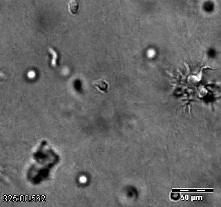
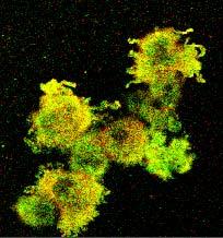
4 Figure S1 Cell surface activation markers are expressed to a similar extend in mature wildtype and bmdcs. Mean fluorescence intensity (MFI) of indicated cell surface markers on CD11c-positive mature bmdcs is depicted (n 6). Figure S2 Expression of active LFA-1 on dendritic cells leads to accumulation of antigen-specific T cells on the dendritic cell surface. Mature wildtype or bmdcs were cocultivated with naïve CD4 + OT-II T cells in a 3-D collagen gel and their interaction was visualised using time-lapse videomicroscopy. Two examples of DC/T cell interactions are shown demonstrating that in contrast to the profound T cell accumulation on DCs only few T cells associated with wildtype DCs. Figure S3 CYTIP silencing in CD18 deficient dendritic cells does not impair antigen-specific T cell proliferation. Mature bmdcs from wildtype and CD18-/- mice were transfected with control sirna or CYTIP specific sirna. Subsequently, naïve CD4 + OT-II T cells were cocultivated with DCs (ratio: 1 DC: 1 T cells) and T cell proliferation was assessed by incorporation of [ 3 H]-thymidine. Whereas, antigen-specific T cell proliferation induced by CYTIP silenced wildtype DCs was reduced, T cell proliferation upon coculture with CD18-/- DCs transfected with control sirna or CYTIP specific sirna was comparable. Assays were performed in triplicates. One representative assay out of two is shown. Figure S4 CYTIP and cytohesin-1 are co-expressed in mature dendritic cells. Expression of CYTIP and cytohesin-1 in mature wildtype bmdcs was analysed by immune fluorescence and is
5 demonstrated either as individual staining or as a merged picture. Leica LSM confocal, original magnification x63. Figure S5 CYTIP expression is induced upon maturation of dendritic cells. BmDCs were cultivated for, 3, 6, 8 and 9 days in conditions known to induce maturation (GM-CSF + IL-4). Subsequently, expression of CYTIP was analysed by immune fluorescence (A), western blot (α CYTIP mab clone 2F9) (B) and quantitative RT-PCR (C). CYTIP expression was found to be induced upon maturation of DCs. Additional stimulation of mature DCs (d9) with LPS did not further increase CYTIP expression.
6 Figure S1 MFI (x1) IA/IE none acd4 MFI (x1) CD54 4 CD8 4 CD86 MFI (x1) MFI (x1) CD4 1 CD MFI (x1) MFI (x1) 6 4 2

7 Figure S2
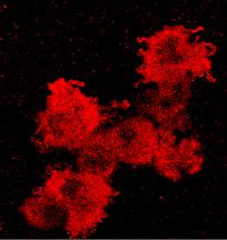
8 Figure S3 cytohesin-1 cytip Overlay 2 µm
9 Figure S4 6 5 p <.5 CD18 -/ control sirna cytip sirna control sirna cytip sirna cpm

 d d3 d6 d9")


 12 1")
10 Figure S5 A) d3 d6 d8 d9 d9 + LPS B) C) d d3 d6 d9 d9 +LPS x-fold increase (normalized against ß-actin) d d3 d6 d8 d8+acd4
HeLa cells stably transfected with an empty pcdna3 vector (HeLa Neo) or with a plasmid
 or with a plasmid") SUPPLEMENTAL MATERIALS AND METHODS Cell culture, transfection and treatments. HeLa cells stably transfected with an empty pcdna3 vector (HeLa Neo) or with a plasmid encoding vmia (HeLa vmia) 1 were cultured
SUPPLEMENTAL MATERIALS AND METHODS Cell culture, transfection and treatments. HeLa cells stably transfected with an empty pcdna3 vector (HeLa Neo) or with a plasmid encoding vmia (HeLa vmia) 1 were cultured
Establishing sirna assays in primary human peripheral blood lymphocytes
 page 1 of 7 Establishing sirna assays in primary human peripheral blood lymphocytes This protocol is designed to establish sirna assays in primary human peripheral blood lymphocytes using the Nucleofector
Isolation, culture, and transfection of primary mammary epithelial organoids
 Supplementary Experimental Procedures Isolation, culture, and transfection of primary mammary epithelial organoids Primary mammary epithelial organoids were prepared from 8-week-old CD1 mice (Charles River)
Table S1. Nucleotide sequences of synthesized oligonucleotides for quantitative
 Table S1. Nucleotide sequences of synthesized oligonucleotides for quantitative RT-PCR For CD8-1 transcript, forward primer CD8-1F 5 -TAGTAACCAGAGGCCGCAAGA-3 reverse primer CD8-1R 5 -TCTACTAAGGTGTCCCATAGCATGAT-3
Alpha or beta human chorionic gonadotropin knockdown decrease BeWo cell fusion by
 Alpha or beta human chorionic gonadotropin knockdown decrease BeWo cell fusion by down-regulating PKA and CREB activation Sudha Saryu Malhotra 1, Pankaj Suman 2 and Satish Kumar Gupta 1 * 1 Reproductive
Cell death analysis using the high content bioimager BD PathwayTM 855 instrument (BD
 Supplemental information Materials and Methods: Cell lines, reagents and antibodies: Wild type (A3) and caspase-8 -/- (I9.2) Jurkat cells were cultured in RPMI 164 medium (Life Technologies) supplemented
Supplementary Information (Ha, et. al) Supplementary Figures Supplementary Fig. S1
 Supplementary Figures Supplementary Fig. S1") Supplementary Information (Ha, et. al) Supplementary Figures Supplementary Fig. S1 a His-ORMDL3 ~ 17 His-ORMDL3 GST-ORMDL3 - + - + IPTG GST-ORMDL3 ~ b Integrated Density (ORMDL3/ -actin) 0.4 0.3 0.2 0.1
Supplementary Information (Ha, et. al) Supplementary Figures Supplementary Fig. S1 a His-ORMDL3 ~ 17 His-ORMDL3 GST-ORMDL3 - + - + IPTG GST-ORMDL3 ~ b Integrated Density (ORMDL3/ -actin) 0.4 0.3 0.2 0.1
Confocal immunofluorescence microscopy
 Confocal immunofluorescence microscopy HL-6 and cells were cultured and cytospun onto glass slides. The cells were double immunofluorescence stained for Mt NPM1 and fibrillarin (nucleolar marker). Briefly,
Supplementary Figure 1. Characterization of the POP2 transcriptional and post-transcriptional regulatory elements. (A) POP2 nucleotide sequence
 POP2 nucleotide sequence") 1 5 6 7 8 9 10 11 1 1 1 Supplementary Figure 1. Characterization of the POP transcriptional and post-transcriptional regulatory elements. (A) POP nucleotide sequence depicting the consensus sequence for
1 5 6 7 8 9 10 11 1 1 1 Supplementary Figure 1. Characterization of the POP transcriptional and post-transcriptional regulatory elements. (A) POP nucleotide sequence depicting the consensus sequence for
Percent survival. Supplementary fig. S3 A.
 Supplementary fig. S3 A. B. 100 Percent survival 80 60 40 20 Ml 0 0 100 C. Fig. S3 Comparison of leukaemia incidence rate in the triple targeted chimaeric mice and germline-transmission translocator mice
Gene Forward Primer Reverse Primer GAPDH ATCATCCCTGCCTCTACTGG GTCAGGTCCACCACTGACAC SSB1 AACTTCAGTGAGCCAAACCC GTTCTCAGAGGCTGGAGAGG
 Supplemental Data EXPERIMENTAL PROCEDURES Plasmids and Antibodies- Full length cdna of INT11 or INT12 were cloned into ps- Flag-SBP vector respectively. Anti-RNA pol II (RPB1) was purchased from Santa
Supplemental Methods Cell lines and culture
 Supplemental Methods Cell lines and culture AGS, CL5, BT549, and SKBR were propagated in RPMI 64 medium (Mediatech Inc., Manassas, VA) supplemented with % fetal bovine serum (FBS, Atlanta Biologicals,
To determine the effect of N1IC in the susceptibility of T cells to the tolerogenic effect of tumorassociated
 Supplementary Methods Tolerogenic effect of MDSC To determine the effect of N1IC in the susceptibility of T cells to the tolerogenic effect of tumorassociated MDSC in vivo, we used a model described previously
Supplemental Materials and Methods
 Supplemental Materials and Methods 125 I-CXCL12 binding assay KG1 cells (2 10 6 ) were preincubated on ice with cold CXCL12 (1.6µg/mL corresponding to 200nM), CXCL11 (1.66µg/mL corresponding to 200nM),
MeCP2. MeCP2/α-tubulin. GFP mir1-1 mir132
 Conservation Figure S1. Schematic showing 3 UTR (top; thick black line), mir132 MRE (arrow) and nucleotide sequence conservation (vertical black lines; http://genome.ucsc.edu). a GFP mir1-1 mir132 b GFP
Supplementary Material
 Supplementary Material Supplementary Methods Cell synchronization. For synchronized cell growth, thymidine was added to 30% confluent U2OS cells to a final concentration of 2.5mM. Cells were incubated
Grb2-Mediated Alteration in the Trafficking of AβPP: Insights from Grb2-AICD Interaction
 Journal of Alzheimer s Disease 20 (2010) 1 9 1 IOS Press Supplementary Material Grb2-Mediated Alteration in the Trafficking of AβPP: Insights from Grb2-AICD Interaction Mithu Raychaudhuri and Debashis
Add 5µl of 3N NaOH to DNA sample (final concentration 0.3N NaOH).
.") Bisulfite Treatment of DNA Dilute DNA sample to 2µg DNA in 50µl ddh 2 O. Add 5µl of 3N NaOH to DNA sample (final concentration 0.3N NaOH). Incubate in a 37ºC water bath for 30 minutes. To 55µl samples
Bisulfite Treatment of DNA Dilute DNA sample to 2µg DNA in 50µl ddh 2 O. Add 5µl of 3N NaOH to DNA sample (final concentration 0.3N NaOH). Incubate in a 37ºC water bath for 30 minutes. To 55µl samples
SUPPLEMENTARY INFORMATION
 doi:10.1038/nature12119 SUPPLEMENTARY FIGURES AND LEGENDS pre-let-7a- 1 +14U pre-let-7a- 1 Ddx3x Dhx30 Dis3l2 Elavl1 Ggt5 Hnrnph 2 Osbpl5 Puf60 Rnpc3 Rpl7 Sf3b3 Sf3b4 Tia1 Triobp U2af1 U2af2 1 6 2 4 3
Flow-cytometry assessment of parasitemia in cell parasite co-culture assay using hydroethidine staining Western blotting RT-PCR
 Flow-cytometry assessment of parasitemia in cell parasite co-culture assay using hydroethidine staining The capability of cells, either PBMC or purified T- cell lines, to inhibit parasite growth or merozoite
Supplemental material
 Supplemental material THE JOURNAL OF CELL BIOLOGY Taylor et al., http://www.jcb.org/cgi/content/full/jcb.201403021/dc1 Figure S1. Representative images of Cav 1a -YFP mutants with and without LMB treatment.
Antibodies used in this study Figure S1. Akt expression in neutrophils from WT and individual Akt isoform knockout mice
 ntibodies used in this study The anti β-actin monoclonal antibody (Sigma-ldrich, St. Louis, MO) was generated against a slightly modified human β-actin N-terminal peptide, c-sp-sp-sp-ile-la-la-leu-val-ile-
CD93 and dystroglycan cooperation in human endothelial cell adhesion and migration
 /, Supplementary Advance Publications Materials 2016 CD93 and dystroglycan cooperation in human endothelial cell adhesion and migration Supplementary Materials Supplementary Figure S1: In ECs CD93 silencing
Methods Western blot analysis of plg Quantification of plasminogen accumulation by ELISA Immunohistochemical analysis
 Methods Western blot analysis of plg Wild-type mice first received a standardized burn wound and then were intravenously administered 2 mg of human plg (Omnio AB, Umeå, Sweden). 24 hours after wounding
Human skin punch biopsies were obtained under informed consent from normal healthy
 SUPPLEMENTAL METHODS Acquisition of human skin specimens. Human skin punch biopsies were obtained under informed consent from normal healthy volunteers (n = 30) and psoriasis patients (n = 45) under a
Nature Immunology: doi: /ni Supplementary Figure 1
 Supplementary Figure 1 BALB/c LYVE1-deficient mice exhibited reduced lymphatic trafficking of all DC subsets after oxazolone-induced sensitization. (a) Schematic overview of the mouse skin oxazolone contact
Supplemental Materials and Methods
 Supplemental Materials and Methods In situ hybridization In situ hybridization analysis of HFE2 and genin mrna in rat liver tissues was performed as previously described (1). Briefly, the digoxigenin-labeled
Spironolactone ameliorates PIT1-dependent vascular osteoinduction in klotho-hypomorphic mice
 Spironolactone ameliorates PIT1-dependent vascular osteoinduction in klotho-hypomorphic mice Supplementary Material Supplementary Methods Materials Spironolactone, aldosterone and β-glycerophosphate were
Mayumi Egawa, Kaori Mukai, Soichiro Yoshikawa, Misako Iki, Naofumi Mukaida, Yohei Kawano, Yoshiyuki Minegishi, and Hajime Karasuyama
 Immunity, Volume 38 Supplemental Information Inflammatory Monocytes Recruited to Allergic Skin Acquire an Anti-inflammatory M2 Phenotype via Basophil-Derived Interleukin-4 Mayumi Egawa, Kaori Mukai, Soichiro
Supplementary Figure 1 - Characterization of rbag3 binding on macrophages cell surface.
 Supplementary Figure 1 - Characterization of rbag3 binding on macrophages cell surface. (a) Human PDAC cell lines were treated as indicated in Figure 1 panel F. Cells were analyzed for FITC-rBAG3 binding
Apoptosis assay: Apoptotic cells were identified by Annexin V-Alexa Fluor 488 and Propidium
 Apoptosis assay: Apoptotic cells were identified by Annexin V-Alexa Fluor 488 and Propidium Iodide (Invitrogen, Carlsbad, CA) staining. Briefly, 2x10 5 cells were washed once in cold PBS and resuspended
Emanuela Tumini, Sonia Barroso, Carmen Pérez Calero and Andrés Aguilera
 SUPPLEMENTARY INFORMATION Roles of human POLD1 and POLD3 in genome stability Emanuela Tumini, Sonia Barroso, Carmen Pérez Calero and Andrés Aguilera SUPPLEMENTARY METHODS Cell proliferation After sirna
Supporting Information
 Supporting Information Casson et al. 10.1073/pnas.1421699112 Sl Materials and Methods Macrophage Infections. In experiments where macrophages were primed with LPS, cells were pretreated with 0.5 μg/ml
T H E J O U R N A L O F C E L L B I O L O G Y
 T H E J O U R N A L O F C E L L B I O L O G Y Supplemental material Monteiro et al., http://www.jcb.org/cgi/content/full/jcb.201306162/dc1 Figure S1. 3D deconvolution microscopy analysis of WASH and exocyst
RNA was isolated using NucleoSpin RNA II (Macherey-Nagel, Bethlehem, PA) according to the
 according to the") Supplementary Methods RT-PCR and real-time PCR analysis RNA was isolated using NucleoSpin RNA II (Macherey-Nagel, Bethlehem, PA) according to the manufacturer s protocol and quantified by measuring the
Supplementary Methods RT-PCR and real-time PCR analysis RNA was isolated using NucleoSpin RNA II (Macherey-Nagel, Bethlehem, PA) according to the manufacturer s protocol and quantified by measuring the
Supplemental Material Igreja and Izaurralde 1. CUP promotes deadenylation and inhibits decapping of mrna targets. Catia Igreja and Elisa Izaurralde
 Supplemental Material Igreja and Izaurralde 1 CUP promotes deadenylation and inhibits decapping of mrna targets Catia Igreja and Elisa Izaurralde Supplemental Materials and methods Functional assays and
Anti-Pim-1 (Cat#3247), anti-met (Cat#3127), anti-ron (Cat#2654), Anti-EGFR
, anti-met (Cat#3127), anti-ron (Cat#2654), Anti-EGFR") Supplementary Methods Antibodies Anti-Pim-1 (Cat#3247), anti-met (Cat#3127), anti-ron (Cat#2654), Anti-EGFR (Cat#2646), anti-igf1r (Cat#3018), anti-insr (Cat#3020), anti-akt (pan, Cat#4691), anti-phospho-akt
Supplementary Methods Antibodies Anti-Pim-1 (Cat#3247), anti-met (Cat#3127), anti-ron (Cat#2654), Anti-EGFR (Cat#2646), anti-igf1r (Cat#3018), anti-insr (Cat#3020), anti-akt (pan, Cat#4691), anti-phospho-akt
Cytotoxicity of Botulinum Neurotoxins Reveals a Direct Role of
 Supplementary Information Cytotoxicity of Botulinum Neurotoxins Reveals a Direct Role of Syntaxin 1 and SNAP-25 in Neuron Survival Lisheng Peng, Huisheng Liu, Hongyu Ruan, William H. Tepp, William H. Stoothoff,
B. ADM: C. D. Apoptosis: 1.68% 2.99% 1.31% Figure.S1,Li et al. number. invaded cells. HuH7 BxPC-3 DLD-1.
 A. - Figure.S1,Li et al. B. : - + - + - + E-cadherin CK19 α-sma vimentin β -actin C. D. Apoptosis: 1.68% 2.99% 1.31% - : - + - + - + Apoptosis: 48.33% 45.32% 44.59% E. invaded cells number 400 300 200
FastLane Kits from Sample Direct to Result
 FastLane Kits from Sample Direct to Result New Sample & Assay Technologies Overview of FastLane technology Speed up and simplify your workflow FastLane Kits accelerate and streamline real-time RT-PCR analysis
SI Appendix. Tumor-specific CD8 + Tc9 cells are superior effector than Tc1 cells for. adoptive immunotherapy of cancers
 SI Appendix Tumor-specific CD8 + Tc9 cells are superior effector than Tc1 cells for adoptive immunotherapy of cancers Yong Lu, Bangxing Hong, Haiyan Li, Yuhuan Zheng, Mingjun Zhang, Siqing Wang, Jianfei
Supplementary Methods
 Supplementary Methods Microarray Data Analysis Gene expression data were obtained by hybridising a total of 24 samples from 6 experimental groups (n=4 per group) to Illumina HumanHT-12 Expression BeadChips.
Supplementary Figure 1. Localization of MST1 in RPE cells. Proliferating or ciliated HA- MST1 expressing RPE cells (see Fig. 5b for establishment of
 Supplementary Figure 1. Localization of MST1 in RPE cells. Proliferating or ciliated HA- MST1 expressing RPE cells (see Fig. 5b for establishment of the cell line) were immunostained for HA, acetylated
sirna Transfection Into Primary Neurons Using Fuse-It-siRNA
 sirna Transfection Into Primary Neurons Using Fuse-It-siRNA This Application Note describes a protocol for sirna transfection into sensitive, primary cortical neurons using Fuse-It-siRNA. This innovative
Supplementary Figure 1. Soft fibrin gels promote growth and organized mesodermal differentiation. Representative images of single OGTR1 ESCs cultured
 Supplementary Figure 1. Soft fibrin gels promote growth and organized mesodermal differentiation. Representative images of single OGTR1 ESCs cultured in 90-Pa 3D fibrin gels for 5 days in the presence
Generation of mouse BMDCs Vectors Short hairpin RNA constructs cdna constructs and generation of stable cell lines
 Generation of mouse BMDCs Mouse BMDCs were differentiated from BM cells isolated from femur and tibiae of the hind legs. Cell suspensions were filtered through a 40 µm cell strainer and incubated for 2
IKK is a therapeutic target in KRAS-induced lung cancer with disrupted p53 activity
 IKK is a therapeutic target in KRAS-induced lung cancer with disrupted p5 activity H6 5 5 H58 A59 H6 H58 A59 anti-ikkα anti-ikkβ anti-panras anti-gapdh anti-ikkα anti-ikkβ anti-panras anti-gapdh anti-ikkα
mmu-mir-200a-3p Real-time RT-PCR Detection and U6 Calibration Kit User Manual MyBioSource.com Catalog # MBS826230
 mmu-mir-200a-3p Real-time RT-PCR Detection and U6 Calibration Kit User Manual Catalog # MBS826230 For the detection and quantification of mirnas mmu-mir-200a-3p normalized by U6 snrna using Real-time RT-PCR
Four different active promoter genes were chosen, ATXN7L2, PSRC1, CELSR2 and
 SUPPLEMENTARY MATERIALS AND METHODS Chromatin Immunoprecipitation for qpcr analysis Four different active promoter genes were chosen, ATXN7L2, PSRC1, CELSR2 and IL24, all located on chromosome 1. Primer
Measurement of peritoneal macrophage apoptosis by Celigo plate imaging cytometer
 SUPPLEMENTAL METHODS Measurement of peritoneal macrophage apoptosis by Celigo plate imaging cytometer For Celigo experiments, 0.1 ml containing 5 x 10 4 cells was seeded into 96 well plates for 30 min
Modulation of the anti-tumor immune response by complement. Maciej M. Markiewski, Robert A. DeAngelis, Fabian Benencia, Salome K.
 Modulation of the anti-tumor immune response by complement Maciej M. Markiewski, Robert A. DeAngelis, Fabian Benencia, Salome K. Ricklin- Lichtsteiner, Anna Koutoulaki, Craig Gerard, George Coukos & John
-RT RT RT -RT XBMPRII
 Base pairs 2000 1650 1000 850 600 500 400 Marker Primer set 1 Primer set 2 -RT RT RT -RT XBMPRII kda 100 75 50 37 25 20 XAC p-xac A 300 B Supplemental figure 1. PCR analysis of BMPRII expression and western
Supplemental Figure 1 Human REEP family of proteins can be divided into two distinct subfamilies. Residues (single letter amino acid code) identical
 identical") Supplemental Figure Human REEP family of proteins can be divided into two distinct subfamilies. Residues (single letter amino acid code) identical in all six REEPs are highlighted in green. Additional
Supplemental Figure Human REEP family of proteins can be divided into two distinct subfamilies. Residues (single letter amino acid code) identical in all six REEPs are highlighted in green. Additional
mmu-mir-34a Real-time RT-PCR Detection Kit User Manual
 mmu-mir-34a Real-time RT-PCR Detection Kit User Manual Catalog # CPK1272 For the detection and quantification of mirna mmu-mir-34a using Real-Time RT-PCR detection instruments. For research use only. Not
Supplementary Figure 1. Expressions of stem cell markers decreased in TRCs on 2D plastic. TRCs were cultured on plastic for 1, 3, 5, or 7 days,
 Supplementary Figure 1. Expressions of stem cell markers decreased in TRCs on 2D plastic. TRCs were cultured on plastic for 1, 3, 5, or 7 days, respectively, and their mrnas were quantified by real time
Supplemental Table S1. RT-PCR primers used in this study
 Supplemental Table S1. RT-PCR primers used in this study -----------------------------------------------------------------------------------------------------------------------------------------------
Supplementary Figure 1 a. d 0.8 CON LPS PAN. 2nd ab nephrin podocin CON LPS PAN. upar. -tubulin. upar. upar / -tubulin CON LPS PAN
 Supplementary Figure 1 a Efferent arteriole Podocytes Afferent arteriole FP Endothelium GBM Glomerular filtration barrier b 188kD HEK + GFP HEK + GFP-Nphs1 Differentiated Podocytes HEK + GFP HEK + GFP-Nphs2
Cell viability. Cell viability was examined by MTT assay (Sigma-Aldrich).
.") Supplementary Materials Supplementary materials and methods Cell culture. Primary human dermal fibroblasts (DFs) were isolated from full-thickness skin samples. Tissue samples were dissected into small
Supplementary Materials Supplementary materials and methods Cell culture. Primary human dermal fibroblasts (DFs) were isolated from full-thickness skin samples. Tissue samples were dissected into small
Supporting Information
 Supporting Information Tal et al. 10.1073/pnas.0807694106 SI Materials and Methods VSV Infection and Quantification. Infection was carried out by seeding 5 10 5 MEF cells per well in a 6-well plate and
Supplementary Figure 1. Co-localization of GLUT1 and DNAL4 in BeWo cells cultured
 Supplementary Figure 1. Co-localization of GLUT1 and DNAL4 in BeWo cells cultured under static conditions. Cells were seeded in the chamber area of the device and cultured overnight without medium perfusion.
Supplementary Table 1. Primers used to construct full-length or various truncated mutants of ISG12b2.
 Supplementary Table 1. Primers used to construct full-length or various truncated mutants of ISG12b2. Construct name ISG12b2 (No tag) HA-ISG12b2 (N-HA) ISG12b2-HA (C-HA; FL-HA) 94-283-HA (FL-GFP) 93-GFP
SUPPLEMENTARY INFORMATION
 DOI: 10.1038/ncb3240 Supplementary Figure 1 GBM cell lines display similar levels of p100 to p52 processing but respond differentially to TWEAK-induced TERT expression according to TERT promoter mutation
Supplementary Fig. 1. Multiple five micron sections of liver tissues of rats treated
 Supplementary Figure Legends Supplementary Fig. 1. Multiple five micron sections of liver tissues of rats treated with either vehicle (left; n=3) or CCl 4 (right; n=3) were co-immunostained for NRP-1 (green)
Supplementary Data. Supplementary Materials and Methods Quantification of delivered sirnas. Fluorescence microscopy analysis
 Supplementary Data Supplementary Materials and Methods Quantification of delivered sirnas After transfection, cells were washed in three times with phosphate buffered saline and total RNA was extracted
Figure S2. Response of mouse ES cells to GSK3 inhibition. Mentioned in discussion
 Stem Cell Reports, Volume 1 Supplemental Information Robust Self-Renewal of Rat Embryonic Stem Cells Requires Fine-Tuning of Glycogen Synthase Kinase-3 Inhibition Yaoyao Chen, Kathryn Blair, and Austin
Cell proliferation was measured with Cell Counting Kit-8 (Dojindo Laboratories, Kumamoto, Japan).
.") 1 2 3 4 5 6 7 8 Supplemental Materials and Methods Cell proliferation assay Cell proliferation was measured with Cell Counting Kit-8 (Dojindo Laboratories, Kumamoto, Japan). GCs were plated at 96-well
1 2 3 4 5 6 7 8 Supplemental Materials and Methods Cell proliferation assay Cell proliferation was measured with Cell Counting Kit-8 (Dojindo Laboratories, Kumamoto, Japan). GCs were plated at 96-well
Supplementary Information
 Supplementary Information Mutual reinforcement of inflammation and carcinogenesis by the Helicobacter pylori CagA oncoprotein Nobumi Suzuki, Naoko Murata-Kamiya, Kohei Yanagiya, Wataru Suda, Masahira Hattori,
Supporting Information
 Supporting Information Patel et al. 10.1073/pnas. SI Materials and Methods Cells and Reagents. Hepatocyte-derived H35 cells were grown in high glucose DMEM (Gibco) media, supplemented with 10% FBS, 2%
Hes6. PPARα. PPARγ HNF4 CD36
 SUPPLEMENTARY INFORMATION Supplementary Table Positions and Sequences of ChIP primers -63 AGGTCACTGCCA -79 AGGTCTGCTGTG Hes6-0067 GGGCAaAGTTCA ACOT -395 GGGGCAgAGTTCA PPARα -309 GGCTCAaAGTTCAaGTTCA CPTa
Electrophoretic Mobility Shift Assay (EMSA). Nuclear extracts were. oligonucleotide spanning the NF-kB site (5 -GATCC-
. Nuclear extracts were. oligonucleotide spanning the NF-kB site (5 -GATCC-") SUPPLEMENTARY MATERIALS AND METHODS Electrophoretic Mobility Shift Assay (EMSA). Nuclear extracts were prepared as previously described. (1) A [ 32 P] datp-labeled doublestranded oligonucleotide spanning
SUPPLEMENTARY MATERIALS AND METHODS Electrophoretic Mobility Shift Assay (EMSA). Nuclear extracts were prepared as previously described. (1) A [ 32 P] datp-labeled doublestranded oligonucleotide spanning
The RT-qPCR analysis of selected type-i IFNs related genes, IRF7 and Oas3. The
 SUPPLEMENTARY MATERIALS AND METHODS Real time quantitative PCR The RT-qPCR analysis of selected type-i IFNs related genes, IRF7 and Oas3. The RT-qPCR was performed on the Applied Biosystems StepOne TM
Supplementary Figure 1
 Supplementary Figure 1 Virus infection induces RNF128 expression. (a,b) RT-PCR analysis of Rnf128 (RNF128) mrna expression in mouse peritoneal macrophages (a) and THP-1 cells (b) upon stimulation with
Flow cytometry Stained cells were analyzed and sorted by SORP FACS Aria (BD Biosciences).
.") Mice C57BL/6-Ly5.1 or -Ly5.2 congenic mice were used for LSK transduction and competitive repopulation assays. Animal care was in accordance with the guidelines of Keio University for animal and recombinant
Mice C57BL/6-Ly5.1 or -Ly5.2 congenic mice were used for LSK transduction and competitive repopulation assays. Animal care was in accordance with the guidelines of Keio University for animal and recombinant
Supplemental Information. Materials and methods.
 Supplemental Information Materials and methods. Cell culture. hmscs were isolated from bone marrow of 3 male donors, undergoing orthopedic surgery (mean age 69.7). Cells were cultured in high glucose DMEM
Supplement 1: Sequences of Capture Probes. Capture probes were /5AmMC6/CTG TAG GTG CGG GTG GAC GTA GTC
 Supplementary Appendixes Supplement 1: Sequences of Capture Probes. Capture probes were /5AmMC6/CTG TAG GTG CGG GTG GAC GTA GTC ACG TAG CTC CGG CTG GA-3 for vimentin, /5AmMC6/TCC CTC GCG CGT GGC TTC CGC
Mice TRAMP mice were maintained in a C57BL/6J background. Syngeneic UBI-GFP/BL6 mice were used for bone marrow engraftment. 2
 Antibodies Chicken IgY polyclonal α-gfp antibodies were purchased from Abcam (Cambridge, MA) and were detected using α-chicken IgY-FITC or α-chicken-hrp (also purchased from Abcam). The CD31-PE, CD11b-PE,
SUPPORTING ONLINE MATERIAL
 SUPPORTING ONLINE MATERIAL SUPPLEMENTAL EXPERIMENTAL PROCEDURES Primers for qpcr and semiquantitative PCR and conditions for semiquantitative PCR G6Pase 5 -ttgtggcagaagcatttgag-3, 5 -atatccttgcactggcaacc-3.
(A-B) P2ry14 expression was assessed by (A) genotyping (upper arrow: WT; lower
 P2ry14 expression was assessed by (A) genotyping (upper arrow: WT; lower") Supplementary Figures S1. (A-B) P2ry14 expression was assessed by (A) genotyping (upper arrow: ; lower arrow: KO) and (B) q-pcr analysis with Lin- cells, The white vertical line in panel A indicates that
Supplementary Figures S1. (A-B) P2ry14 expression was assessed by (A) genotyping (upper arrow: ; lower arrow: KO) and (B) q-pcr analysis with Lin- cells, The white vertical line in panel A indicates that
Supplementary Protocol. sirna transfection methodology and performance
 Supplementary Protocol sirna transfection methodology and performance sirna oligonucleotides, DNA construct and cell line. Chemically synthesized 21 nt RNA duplexes were obtained from Ambion Europe, Ltd.
Online Supplementary Information
 Online Supplementary Information NLRP4 negatively regulates type I interferon signaling by targeting TBK1 for degradation via E3 ubiquitin ligase DTX4 Jun Cui 1,4,6,7, Yinyin Li 1,5,6,7, Liang Zhu 1, Dan
Figure legend. The expression of BAFF and its receptors in macrophages was detected at lesion areas of atherosclerosis and arthritis.
 Data supplement #1 Figure legend. The expression of BAFF and its receptors in macrophages was detected at lesion areas of atherosclerosis and arthritis. A. Consecutive sections of an atherosclerotic plaque
PCR analysis was performed to show the presence and the integrity of the var1csa and var-
 Supplementary information: Methods: Table S1: Primer Name Nucleotide sequence (5-3 ) DBL3-F tcc ccg cgg agt gaa aca tca tgt gac tg DBL3-R gac tag ttt ctt tca ata aat cac tcg c DBL5-F cgc cct agg tgc ttc
Autocrine Complement Inhibits IL10-Dependent T-Cell Mediated. Antitumor Immunity to Promote Tumor Progression
 Supplemental Information Autocrine Complement Inhibits IL10-Dependent T-Cell Mediated Antitumor Immunity to Promote Tumor Progression Yu Wang, Sheng-Nan Sun, Qing Liu, Yang-Yang Yu, Jian Guo, Kun Wang,
Supplementary Materials for
 advances.sciencemag.org/cgi/content/full/4/9/eaat5401/dc1 Supplementary Materials for GLK-IKKβ signaling induces dimerization and translocation of the AhR-RORγt complex in IL-17A induction and autoimmune
Electronic Supplementary Information
 Electronic Supplementary Material (ESI) for Molecular BioSystems. This journal is The Royal Society of Chemistry 2017 Electronic Supplementary Information Dissecting binding of a β-barrel outer membrane
SANTA CRUZ BIOTECHNOLOGY, INC.
 TECHNICAL SERVICE GUIDE: Western Blotting 2. What size bands were expected and what size bands were detected? 3. Was the blot blank or was a dark background or non-specific bands seen? 4. Did this same
Supplementary Table 1. PCR amplification conditions for each primer pair. Primer sequence
 - 1 - Supplementary Tables Supplementary Table 1. PCR amplification conditions for each primer pair Primer sequence FN1 S - CAAAGCAAGCCCGGTTGT AS - CGCTCCCACTGTTGATTTATCTG ITGα2 S - TTAGGTTACTCTGTGGCTGCAATT
Supplemental Figure 1
 mrna level (fold induction) Supplemental Figure 1 a-flag pmx-rank WT pmx-rank Mt 0 1 3 6 0 1 3 6 (h) c-fos b-actin 5 4 3 2 1 0 0 1 3 6 0 1 3 6 (h) pmx-rank WT pmx-rank Mt TRAP+ Mononuclear pre-ocs/well
TRIM31 is recruited to mitochondria after infection with SeV.
 Supplementary Figure 1 TRIM31 is recruited to mitochondria after infection with SeV. (a) Confocal microscopy of TRIM31-GFP transfected into HEK293T cells for 24 h followed with SeV infection for 6 h. MitoTracker
Supplementary Information. A novel human endogenous retroviral protein inhibits cell-cell fusion. Supplementary Figures:
 Supplementary Information A novel human endogenous retroviral protein inhibits cell-cell fusion Jun Sugimoto, Makiko Sugimoto, Helene Bernstein, Yoshihiro Jinno and Danny J. Schust Supplementary Figures:
Supporting Information
 Supporting Information Stavru et al. 0.073/pnas.357840 SI Materials and Methods Immunofluorescence. For immunofluorescence, cells were fixed for 0 min in 4% (wt/vol) paraformaldehyde (Electron Microscopy
STING-Dependent Cytosolic DNA Sensing Promotes Radiation-Induced Type I Interferon-Dependent Antitumor Immunity in Immunogenic Tumors
 Immunity, Volume 41 Supplemental Information STING-Dependent Cytosolic DNA Sensing Promotes Radiation-Induced Type I Interferon-Dependent Antitumor Immunity in Immunogenic Tumors Liufu Deng, Hua Liang,
Supplementary Figure 1 PARP1 is involved in regulating the stability of mrnas from pro-inflammatory cytokine/chemokine mediators.
 Supplementary Figure 1 PARP1 is involved in regulating the stability of mrnas from pro-inflammatory cytokine/chemokine mediators. (a) A graphic depiction of the approach to determining the stability of
Accelerating skin wound healing by M-CSF through generating SSEA-1 and -3 stem cells. in the injured sites
 Accelerating skin wound healing by M-CSF through generating SSEA-1 and -3 stem cells in the injured sites Yunyuan Li, Reza Baradar Jalili, Aziz Ghahary Department of Surgery, University of British Columbia,
CD1a-autoreactive T cells are a normal component of the human αβ T cell repertoire
 CDa-autoreactive T cells are a normal component of the human αβ T cell repertoire Annemieke de Jong, Victor Peña-Cruz, Tan-Yun Cheng, Rachael A. Clark, Ildiko Van Rhijn,, D. Branch Moody Division of Rheumatology,
gacgacgaggagaccaccgctttg aggcacattgaaggtctcaaacatg
 Supplementary information Supplementary table 1: primers for cloning and sequencing cloning for E- Ras ggg aat tcc ctt gag ctg ctg ggg aat ggc ttt gcc ggt cta gag tat aaa gga agc ttt gaa tcc Tpbp Oct3/4
Thermo Scientific Dharmacon SMARTvector 2.0 Lentiviral shrna Particles
 Thermo Scientific Dharmacon SMARTvector 2.0 Lentiviral shrna Particles Long-term gene silencing shrna-specific design algorithm High titer, purified particles Thermo Scientific Dharmacon SMARTvector shrna
immunofluorescence. Name of antibodies Manufacturer Catalog Number Rabbit anti-pdyn Rabbit anti-kor-1
 Supplemental Tables Table S1. List of primary antibodies used for immunohistochemistry, FACS, and immunofluorescence. Name of antibodies Manufacturer Catalog Number Rabbit anti-pdyn Bioss USA bs-13041r
Supplementary Figure Legends
 Supplementary Figure Legends Figure S1 gene targeting strategy for disruption of chicken gene, related to Figure 1 (f)-(i). (a) The locus and the targeting constructs showing HpaI restriction sites. The
hnrnp C promotes APP translation by competing with FMRP for APP mrna recruitment to P bodies
 hnrnp C promotes APP translation by competing with for APP mrna recruitment to P bodies Eun Kyung Lee 1, Hyeon Ho Kim 1, Yuki Kuwano 1, Kotb Abdelmohsen 1, Subramanya Srikantan 1, Sarah S. Subaran 2, Marc