Fluorescence Microscopy: A Biological Perspective
|
|
|
- Dorcas Young
- 5 years ago
- Views:
Transcription
1 Fluorescence Microscopy: A Biological Perspective


2 From nanometre to metre: the scale of life Instrumentation and accessible scale limits the questions that can be addressed in biology Why are there limits?
 and numerical aperture (NA) of the objective. Limit: R = 0.61λ/NA so at 488 nm R ~ 0.2 µm.")

3 Resolution and microscopy Resolution: ability to distinguish two adjacent objects. Depends on the wavelength (λ) and numerical aperture (NA) of the objective. Limit: R = 0.61λ/NA so at 488 nm R ~ 0.2 µm. Small structures in cells can have a size close to or below this limit, may be imaged but as depicted in the image will appear larger than the real structure. Co-localizations:things may appear to co localize but are separate. NA = n x sin α where n is the refractive index
4 Five Dimensional Imaging: Goal of Fluorescence Microscopy in Biomedical Sciences 3 spatial dimensions, X, Y and Z Ability to follow multiple probes/targets Following processess in real time

5 Typical wide field fluorescence microscope Note key elements Objectives delivers exciting light and collects emmission signal Dichroic mirror; reflects excitation wavelength light but transmits emission wavelength. Filters: Filtering of different wavelengths. Different cuts off wavelenths and band passess Dichroic mirror Reflects the green light in excitation light but transmits the emitted red light from the fluorescent probe in sample Excitation light
6 Light Sources for fluorescent microscopy Mercury lamps ( W) & Excite HbO lamps variable intensities. LEDs long lasting, bright, use multiple wavelengths, variable power. Lasers (gas/solid state) high-intensity monochromatic light sources that are coherent and highly collimated to form a tight beam with a very low rate of expansion. Limited number of wavelengths available
 Size: 6.45 µm")

 monochrome FVII: 1376 x 1032 x 6.")
7 Detector systems: CCD cameras Axiocam HRc, colour camera resolution 1.4 megapixels (1388 x 1040) Size: 6.45 µm x 6.45 µm. Frame rate: 5/sec (20 ms exposure) at full resolution. DP71: 1.45 million pixel CCD with a Bayer color filter. This CCD is coupled to pixel-shifting technology; resulting in images with an ultrahigh resolution of 4080 x 3072 pixels. (high resolution) monochrome FVII: 1376 x 1032 x 6.45µm square pixel CCD monochrome imaging sensor. High frame rate, up to 22 fps in binning mode. colour High speed up 30 FPS with binning.

8 Electron multiplying CCD (EMCCD) technology EMCCDs operate by amplifying weak signal events (down to single photons) to a signal level that is well clear of the read noise floor of the camera, at any readout speed. cooling ~ -90 C in deep vacuum. ixon EM +885 Camera: high resolution with high QE. 32 FPS at full resolution. ixon EM +897 Camera: ultimate in sensitivity back illuminated EMCCD has single photon detection capability without an image intensifier, combined with greater than 90% QE of a back-illuminated sensor. Containing a 512 x 512 Frame. 16 µm square. Combines high sensitivity with high speed readout. EMCCD technology has revolutionized high speed imaging of live cells Solves problem of low light intensities and high speed acquisition


9 The Zeiss Axiovert 100TV Microscope
10 Visualization of multiple targets: Multi-channel imaging


11 Commonly used synthetic fluorophores Key concepts Selection: avoid spectral overlap. Try to separate emmission maxia. Sensitivity: high quantum yield: strong signals Stable: bleeching or quenching of signal A wide range of Alexa fluorophores available from Molecular Probes that meet the three requirements
12 Immunolocalization experiments in cells Fixation of cells (paraformaldehyde) Attachment of cells to slide Permeabilization & Blocking Probe with primary antibody. Probe with secondary antibodies with fluorophores (dyes) attached.. Washing. Viewing Y Y Y Fluor label Secondary ab Primary ab


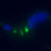
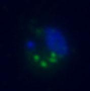
13 Example of immunolocalization of a flagellar associated protein in fixed Trypanosomes MERGE of all fluorescence channels MERGE of all channels Multichannel imaging shows DAPI (blue)staining dsdna in nucleus/kinetoplast Alexa 488/staining target protein using antibody Phase merge on wide field


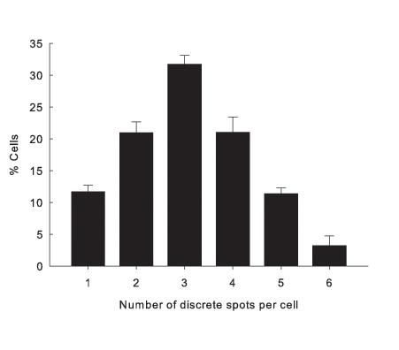
14 Quantitative microscopy Analysis of cell populations reveals a dynamic aspect to distribution of a myosin motor in trypanosomes Discrete spots per cell 1-6

.")
15 Morphology: molecular analysis HeLa Cells stained with anti-tubulin (green) and PI (red). Control Drug treated


16 Olympus IX81 with long focal length objectives Major advantage: direct viewing of cells in culture flasks Limitation: longer focal length objectives have lower NA values Long focal length objectives Dual camera system DP 71 and FVII Dual condenser system Excite HbO system Phase and DIC


17 Video imaging for analysis of cell motility Flagellar mutants
18 trackit software analysis gives quantitative data Detached flagellum cell Object ID Distance Length Extent Direction Speed Start X Start Y End X End Y Lost in fra µm µm µm µm/s µm µm µm µm Count Minimum Maximum Range Mean Std.-Dev Normal cell

19 Organelle Membrane-Specific Probes: DiOC6(3), NBD ceramide, Rhodamine These dyes stain specifically the organelle membranes, such as the endoplasmic reticulum membrane, Golgi membrane, and mitochondrial membrane. Organelle specific Fluorescent Probes for use in cells Organelle specific probes based on features of the comparment: Other probes accumulate specifically into organelles such lysosomes (weak bases) and mitochondria (weak acids or positively changed dyes). The nucleus can be stained with a variety of dyes that bind to dsdna e.g. DAPI, Hoest stain etc Membrane Potential-Sensible Probes: WW 781, RH-155, and Di-8-ANEPPS - These dyes are incorporated into the cell membrane. Their absorption or fluorescence intensity varies depending on the membrane potential. Fast-type dyes with response rates on the order of milliseconds should be used. Live cells usually incubated with the probe and then viewed directly fixed for viewing in combination with other probes

20 Visualization of single mitochondrion in live procyclic form trypanosomes


21 Fluorescent proteins: A vital tool in imaging in live cells Osamu Shimomura and Frank Johnson, a protein that lacked was able to produce green fluorescence when illuminated with ultraviolet light. The protein was eventually christened with the unceremonious name of green fluorescent protein (GFP). The GFP gene has been isolated and improved to obtain different color variants. These proteins are used to construct fluorescent chimeric proteins that can be expressed in living cells, tissues, and entire organisms, after transfection with the engineered vectors. The fluorescent protein technique avoids the problem of purifying, tagging, and introducing labeled proteins into cells or the task of producing specific antibodies for surface or internal antigens. Used widely in live cell imaging


22 Fluorescent proteins in cells, tissues and animals


23 Can also be used to make novelty pets
24 Problems with use of Fluorescent proteins 1. In the natural state most are oligomeric: can cause problems in vivo 2. Quantum yield of some variants is low. 3. Red proteins. Restricted choice in the red far red region. 4. Because of size (~20-30 kda) insertion of tag may affect location/function of tagged target protein.
25 The Z dimension: Confocal Microscopy
26 Why use Confocal Microscopy? High resolution images of your cells or sample, beyond that of a normal fluorescent microscope Localisation of a particle of interest within a cell Co-localisation of different particles We can take 2-D sections through 3-D cells & tissues and reconstruct an extended-focus series Use of 3 or 4 different fluorescent probes simultaneously We can combine fluorescence contrast with phase contrast and differential interference contrast (DIC) We can control the laser to do dynamic experiments
27 1. Single beam laser scanning confocal microscope Uses point illumination and point detection Uses a pinhole Both of these specifications restrict image information to the plane of focus Laser beam scans the specimen from left to right and is rapidly transported back to the start point in a process termed flyback


28

29 From Spot to image
30 Scanning Speed Field of View 512x 512 pixels = 262,144 points Build up entire field of view in 1 second Laser must dwell on each point for 3.8 µsec Need a PMT to read each sampling pixel
31 Advantages of Confocal Ability to serially produce thin ( µm) optical sections through fluorescent specimens up to 100 µm thick, non-invasively Contrast & definition dramatically improved cf widefield microscopy Reduction of out-of-focus fluorescence due to the presence of a pinhole Better lateral and axial resolution than wide-field Improved signal to noise ratio (SNR) Can un-mix two spectrally close fluors and image simultaneously without cross-talk Magnification zoom can be adjusted electronically As well as imaging in 3D (XYZ) one can image in 4D (XYZT)-live cell imaging with optical sectioning


32 Olympus FV1000 Point-Scanning Confocal microscope

33

34 Cells & Tissues are 3-dimensional3


35 Z-stacks

36 Multi colour labelling, Z-stack

37 Multi-colour labelling, single slice


38 Wide field imaging of a myosin motor TbMyo I reveal localization with various elements of the endocytic pathway TbMyo I Flagellar pocket/ Early endosomes TbMyo I Late endosomes TbMyo I Lysosome TbMyo I does not locate specifically with any individual compartment
, lysosome (red) and Myosin B (green) in")
39 Imaris Image analysis software Iso-surface Model showing the nucleus, kinetoplast (blue), lysosome (red) and Myosin B (green) in T. brucei
 and DNA stain-dapi")
40 Iso-surface model of fluorescently stained trypanosome Bloodstream forms of Trypanosoma brucei stained with antimyosin antibody(green) and DNA stain-dapi (blue)
41 Filter cube sets for DAPI, FITC & rhodamine chromophores Laser System contains: Argon laser, multi-line 458/ 488/ 515 nm, Green Helium-Neon laser 543 nm, Red Helium-Neon laser 633 nm, Near-Violet Laser diode 405 nm 10x, 20x & 40x dry and 40x & 60x oil objectives all of standard focal length 2 Stages available, one for standard slides and the other a heated stage for 35 mm or 50 mm glass bottom petri dishes Incubator for live cell experiments FluoView software has 3D & 4D capabilities spectral unmixing ; detection, spectral separation and visualization of multiple fluorescent labels with overlapping emission spectra such as CFP, GFP and YFP, which cannot be separated using conventional methods. The SIM scanning system can synchronize laser light stimulation and imaging to capture rapid changes in living cells immediately following fluorophore excitation e.g. photoactivation & FRAP
42 Live cell analysis and use of Multi-beam laser-scanning confocal microscope Uses point illumination and point detection Uses many pinholes in a Nipkow disc Very Fast
43

44 Basics of Spinning Disk Confocal


45 Andor Revolution XD Spinning Disc Confocal Microscope


46 Live Cell Microscopy 35/50 mm glass bottom petri dishes from Willco/MatTek LabTek chamber coverglass slides from Nunc
47 FEATURES CoolLED light source with wavelength peaks at 445nm, 490nm & 565nm. Filter cube sets for CFP, GFP, rhodamine & a triple filter cube to view all three flours simultaneously. Laser combiner comprises four solid state lasers, 445 nm, 488 nm, 514 nm, 561 nm The FRAPPA unit allows user defined ROI laser scanning for FRAP and Photo-activation. Yokogawa CSU-X1 Spinning Disk Unit, motorised version with brightfield bypass option. 5000rpm disk spin speed giving scan rate of 1000 scans per second. 2 interchangeable high-res cameras available for acquisition of images, using ixon +Ultra sensitive EMCCD technology
48 Advantages of Spinning Disk Confocal Can use CCD camera ( instant optical sectioning) rather than a PMT Parallel beam goes much faster-higher frame rate-limited only by the CCD camera read out rate Uses lower intensity laser illumination than single point scanning


49 Uptake of alum particles by dendritic cells

50 Cytoplasmic GFP-protein in live bloodstream forms of T. brucei, Spinning disk Confocal image
51 Specialized applications of Confocal Microscopy FRAP - photo-bleaching studies FRET Florescence Resonance Energy Transfer FLIM-Fluorescence Lifetime Imaging FLAP-Fluorescence localisation after photobleaching
52 FRET: useful for protein interaction studies

53 Using lasers on region of interest Looking for fluorescent tag to move back into area that is bleached. Used as a measure of mobility of tagged protein or organelle.
54 FRAP in bloodstream forms of T. brucei Bleeching of a protein in the flagellar pocket and recovery of signal due to trafficking of new protein to surface.
55 For multidimensional analysis What to use? Use Confocal for thick samples: up to 100 µm Use wide-field for thin samples: less than 2µm Use multi-beam confocal for moving or photo sensitive samples All can be used for multichannel imaging
56 Useful Links: Good Java Tutorials: Microscopy Primer website: Info on Fluorescent excitation & emission spectra
Fluorescence Light Microscopy for Cell Biology
 Fluorescence Light Microscopy for Cell Biology Why use light microscopy? Traditional questions that light microscopy has addressed: Structure within a cell Locations of specific molecules within a cell
Special Techniques 1. Mark Scott FILM Facility
 Special Techniques 1 Mark Scott FILM Facility SPECIAL TECHNIQUES Multi-photon microscopy Second Harmonic Generation FRAP FRET FLIM In-vivo imaging TWO-PHOTON MICROSCOPY Alternative to confocal and deconvolution
A Brief History of Light Microscopy And How It Transformed Biomedical Research
 A Brief History of Light Microscopy And How It Transformed Biomedical Research Suewei Lin Office: Interdisciplinary Research Building 8A08 Email: sueweilin@gate.sinica.edu.tw TEL: 2789-9315 Microscope
Confocal Microscopy & Imaging Technology. Yan Wu
 Confocal Microscopy & Imaging Technology Yan Wu Dec. 05, 2014 Cells under the microscope What we use to see the details of the cell? Light and Electron Microscopy - Bright light / fluorescence microscopy
Widefield Microscopy Bleed-Through
 In widefield microscopy the excitation wavelengths which illuminate the sample, and the emission wavelengths which reach the CCD camera are selected throughout a filter cube. A filter cube consists of
Imaging facilities at WUR
 Imaging facilities at WUR Advanced light microscopy facilities at Wageningen UR Programme Thursday 13 June 2013 Lunch meeting organized by Cat-Agro Food 12.00 Welcome and sandwich lunch 12.10 Introduction
Dino-Lite knowledge & education. Fluorescence Microscopes
 Dino-Lite knowledge & education Fluorescence Microscopes Dino-Lite Fluorescence models Smallest fluorescence microscope in the world Revolution to biomedical and educational applications Flexible Easy
Resolution of Microscopes Visible light is nm Dry lens(0.5na), green(530nm light)=0.65µm=650nm for oil lens (1.4NA) UV light (300nm) = 0.13µm f
, green(530nm light)=0.65µm=650nm for oil lens (1.4NA) UV light (300nm) = 0.13µm f") Microscopes and Microscopy MCB 380 Good information sources: Alberts-Molecular Biology of the Cell http://micro.magnet.fsu.edu/primer/ http://www.microscopyu.com/ Approaches to Problems in Cell Biology
Microscopes and Microscopy MCB 380 Good information sources: Alberts-Molecular Biology of the Cell http://micro.magnet.fsu.edu/primer/ http://www.microscopyu.com/ Approaches to Problems in Cell Biology
Fluorescence Microscopy
 Fluorescence Microscopy Dr. Arne Seitz Swiss Institute of Technology (EPFL) Faculty of Life Sciences Head of BIOIMAGING AND OPTICS BIOP arne.seitz@epfl.ch Fluorescence Microscopy Why do we need fluorescence
Spectral Separation of Multifluorescence Labels with the LSM 510 META
 Microscopy from Carl Zeiss Spectral Separation of Multifluorescence Labels with the LSM 510 META Indians living in the South American rain forest can distinguish between almost 200 hues of green in their
F* techniques: FRAP, FLIP, FRET, FLIM,
 F* techniques: FRAP, FLIP, FRET, FLIM, FCS Antonia Göhler March 2015 Fluorescence explained in the Bohr model Absorption of light (blue) causes an electron to move to a higher energy orbit. After a particular
Lab 5: Optical trapping and single molecule fluorescence
 Lab 5: Optical trapping and single molecule fluorescence PI: Matt Lang Lab Instructor: Jorge Ferrer Summary Optical tweezers are an excellent experimental tool to study the biophysics of single molecule
Fluorescence Microscopy
 Fluorescence Microscopy Dr. Arne Seitz Swiss Institute of Technology (EPFL) Faculty of Life Sciences Head of BIOIMAGING AND OPTICS BIOP arne.seitz@epfl.ch Fluorescence Microscopy Why do we need fluorescence
Visualizing Cells Molecular Biology of the Cell - Chapter 9
 Visualizing Cells Molecular Biology of the Cell - Chapter 9 Resolution, Detection Magnification Interaction of Light with matter: Absorbtion, Refraction, Reflection, Fluorescence Light Microscopy Absorbtion
Confocal Microscopes. Evolution of Imaging
 Confocal Microscopes and Evolution of Imaging Judi Reilly Hans Richter Massachusetts Institute of Technology Environment, Health & Safety Office Radiation Protection What is Confocal? Pinhole diaphragm
MICROSCOPY. "micro" (small) "scopeo" (to watch)
 scopeo (to watch)") MICROSCOPY "micro" (small) "scopeo" (to watch) THE RELATIVE SIZES OF MOLECULES, CELLS AND ORGANISMS THE RELATIVE SIZES OF MOLECULES, CELLS AND ORGANISMS MICROSCOPY 1590 2012 MICROSCOPY THE LIGHT Light:
MICROSCOPY "micro" (small) "scopeo" (to watch) THE RELATIVE SIZES OF MOLECULES, CELLS AND ORGANISMS THE RELATIVE SIZES OF MOLECULES, CELLS AND ORGANISMS MICROSCOPY 1590 2012 MICROSCOPY THE LIGHT Light:
FLUORESCENCE. Matyas Molnar and Dirk Pacholsky
 FLUORESCENCE Matyas Molnar and Dirk Pacholsky 1 Information This lecture contains images and information from the following internet homepages http://micro.magnet.fsu.edu/primer/index.html http://www.microscopyu.com/
Live cell microscopy
 Live cell microscopy 1. Why do live cell microscopy? 2. Maintaining living cells on a microscope stage. 3. Considerations for imaging living cells. 4. Fluorescence labeling of living cells. 5. Imaging
Multiplexed 3D FRET imaging in deep tissue of live embryos Ming Zhao, Xiaoyang Wan, Yu Li, Weibin Zhou and Leilei Peng
 Scientific Reports Multiplexed 3D FRET imaging in deep tissue of live embryos Ming Zhao, Xiaoyang Wan, Yu Li, Weibin Zhou and Leilei Peng 1 Supplementary figures and notes Supplementary Figure S1 Volumetric
Confocal Microscopy Analyzes Cells
 Choosing Filters for Fluorescence A Laurin Publication Photonic Solutions for Biotechnology and Medicine November 2002 Confocal Microscopy Analyzes Cells Reprinted from the November 2002 issue of Biophotonics
Automated Digital Microscopy
 A p p l i c a t i o n G u i d e Peter Banks, Ph.D. and Peter J. Brescia, Applications Department, BioTek Instruments, Inc., Winooski, VT Table of Contents Introduction ----------------------------------------------------------------------------------------------------------------------
Partha Roy
 Fluorescence microscopy http://micro.magnet.fsu.edu/primer/index.html Partha Roy 1 Lecture Outline Definition of fluorescence Common fluorescent reagents Construction ti of a fluorescence microscope Optical
Simultaneous multi-color, multiphoton fluorophore excitation using dual-color fiber lasers
 Multiphoton Microscopy / Fiber Laser Simultaneous multi-color, multiphoton fluorophore excitation using dual-color fiber lasers Matthias Handloser, Tim Paasch-Colberg, Bernhard Wolfring TOPTICA Photonics
Fluorescence Microscopy. Terms and concepts to know: 10/11/2011. Visible spectrum (of light) and energy
 and energy") Fluorescence Microscopy Louisiana Tech University Ruston, Louisiana Microscopy Workshop Dr. Mark DeCoster Associate Professor Biomedical Engineering 1 Terms and concepts to know: Signal to Noise Excitation
Fluorescence Microscopy Louisiana Tech University Ruston, Louisiana Microscopy Workshop Dr. Mark DeCoster Associate Professor Biomedical Engineering 1 Terms and concepts to know: Signal to Noise Excitation
Super Resolution Imaging Solution Provider. Imaging Future
 Super Resolution Imaging Solution Provider Imaging Future Imaging Solution More Than Equipment NanoBioImaging(NBI) is the Industrial Partner of HKUST Super Resolution Imaging Center (SRIC). NBI aims to
SUPPLEMENTARY INFORMATION
 a 14 12 Densitometry (AU) 1 8 6 4 2 t b 16 NMHC-IIA GAPDH NMHC-IIB Densitometry (AU) 14 12 1 8 6 4 2 1 nm 1 nm 1 nm 1 nm sirna 1 nm 1 nm Figure S1 S4 Quantification of protein levels. (a) The microtubule
ATFM 2015 ATFM Sample preparation guide
 ATFM 2016 Sample preparation guide The workshop is organized in the framework of the Czech-BioImaging research infrastructure supported by MEYS (LM2015062) 1 Table of Contents General recommendations...
cell and tissue imaging by fluorescence microscopy
 cell and tissue imaging by fluorescence microscopy Steven NEDELLEC Plateforme Micropicell SFR Santé François Bonamy Nantes 1 A matter of size Limit of resolution 0.15mm aims: building the image of an object
Super-resolution Microscopy
 Semr oc kwhi t epaperser i es : 1. Introduction Super-resolution Microscopy Fluorescence microscopy has revolutionized the study of biological samples. Ever since the invention of fluorescence microscopy
Workshop advanced light microscopy
 Workshop advanced light microscopy Multi-mode confocal laser scanning microscope Jan Willem Borst Laboratory of Biochemistry Biomolecular Networks www.bic.wur.nl MicroSpectroscopy Centre Wageningen Microspectroscopy
Rice/TCU REU on Computational Neuroscience. Fundamentals of Molecular Imaging
 Rice/TCU REU on Computational Neuroscience Fundamentals of Molecular Imaging June 2, 2009 Neal Waxham 713-500-5621 m.n.waxham@uth.tmc.edu Objectives Introduction to resolution in light microscopy Brief
How to use the SP5 confocal microscope
 How to use the SP5 confocal microscope Mailfert Sébastien (mailfert@ciml.univ-mrs.fr) Tel : 9126 Imaging Immunity (ImagImm) photonic microscopy facility Centre d Immunologie de Marseille-Luminy 2016 /
Nodes of regulation in cellular systems
 Nodes of regulation in cellular systems cell membrane signal transduction ligands receptors oligomerization transport signal transduction modified protein Golgi transcription factor transport ER transport
ab CytoPainter ER Staining Kit Red Fluorescence
 ab139482 CytoPainter ER Staining Kit Red Fluorescence Instructions for Use Designed to detect Human endoplasmic reticulum by microscopy. This product is for research use only and is not intended for diagnostic
BIOCHEMIST ALL IN ONE ARTICLE
 BIOCHEMIST ALL IN ONE ARTICLE Bringing ease-of-use to microscopy From the Philosopher s Stone to the Researcher s Dream Although naturally occurring luminescence has been observed for many centuries, the
Sapphire. Biomolecular Imager THE NEXT GENERATION OF LASER-BASED IMAGING
 Sapphire Biomolecular Imager THE NEXT GENERATION OF LASER-BASED IMAGING Breakthrough image capture and analysis The Sapphire Biomolecular Imager is a next generation laser scanning system that provides
The analysis of fluorescence microscopy images for FRET detection
 The analysis of fluorescence microscopy images for FRET detection Ela Claridge, Dale J. Powner and Michael J.O. Wakelam School of Computer Science, The University of Birmingham B5 2TT Institute for Cancer
Final Exam, 176 points PMB 185: Techniques in Light Microscopy
 Final Exam, 176 points Name PMB 185: Techniques in Light Microscopy Point value is in parentheses at the end of each question. 1) Order the steps in setting up Köhler illumination. It is not necessary
Contents. SCHOOL of FLUORESCENCE. For more information, go to lifetechnologies.com/imagingbasics
 MPSF educator packet This packet contains illustrations and figures from the Molecular Probes School of Fluorescence website. They illustrate concepts from the basic physical properties that underlie fluorescence
ab CytoPainter ER Staining Kit Red Fluorescence
 ab139482 CytoPainter ER Staining Kit Red Fluorescence Instructions for Use Designed to detect Human endoplasmic reticulum by microscopy. This product is for research use only and is not intended for diagnostic
Direct visualization, sizing and concentration measurement of fluorescently labeled nanoparticles using NTA
 Direct visualization, sizing and concentration measurement of fluorescently labeled nanoparticles using NTA NANOSIGHT RANGE Visualize and Measure Nanoparticle Size and Concentration PARTICLE SIZE PARTICLE
Imaging & analysis with the LSM780 NLO Discover the secrets beyond the twilight zone
 Imaging & analysis with the LSM780 NLO Discover the secrets beyond the twilight zone Sven Terclavers LSM780 System overview The Scan Module - Core of the LSM 780 1 V/tunable PTC laser ports (405/440, cw/ps;
Two-Photon Microscopy for Deep Tissue Imaging of Living Specimens
 for Deep Tissue Imaging of Living Specimens Tilman Franke* and Sebastian Rhode TILL Photonics GmbH, an FEI company, Lochhamer Schlag 21, D-82166 Gräfelfing, Germany *tilman.franke@fei.com Introduction
Supporting Information
 Supporting Information Stavru et al. 0.073/pnas.357840 SI Materials and Methods Immunofluorescence. For immunofluorescence, cells were fixed for 0 min in 4% (wt/vol) paraformaldehyde (Electron Microscopy
Introduction to Computational Fluorescence Microscopy!
 Introduction to Computational Fluorescence Microscopy! EE367/CS448I: Computational Imaging and Display! stanford.edu/class/ee367! Lecture 13! Gordon Wetzstein! Stanford University! Midterm! Tuesday, Feb
CytoPainter Golgi Staining Kit Green Fluorescence
 ab139483 CytoPainter Golgi Staining Kit Green Fluorescence Instructions for Use Designed for the detection of Golgi bodies by microscopy This product is for research use only and is not intended for diagnostic
HYPERSPECTRAL MICROSCOPE PLATFORM FOR HIGHLY MULTIPLEX BIOLOGICAL IMAGING. Marc Verhaegen
 HYPERSPECTRAL MICROSCOPE PLATFORM FOR HIGHLY MULTIPLEX BIOLOGICAL IMAGING Marc Verhaegen CMCS, MONTREAL, MAY 11 th, 2017 OVERVIEW Hyperspectral Imaging Multiplex Biological Imaging Multiplex Single Particle
Immunofluorescence Confocal Microscopy of 3D Cultures Grown on Alvetex
 Immunofluorescence Confocal Microscopy of 3D Cultures Grown on Alvetex 1.0. Introduction Immunofluorescence uses the recognition of cellular targets by fluorescent dyes or antigen-specific antibodies coupled
Practical light microscopy: an introduction
 Practical light microscopy: an introduction Dr. Mark Leake, Oxford University www.physics.ox.ac.uk/users/leake Aim of today s talk: Explanation of the very (very) basics of how a light microscope works
FLIM Fluorescence Lifetime IMaging
 FLIM Fluorescence Lifetime IMaging Fluorescence lifetime t I(t) = F0 exp( ) τ 1 τ = k f + k nr k nr = k IC + k ISC + k bl Batiaens et al, Trends in Cell Biology, 1999 τ τ = fluorescence lifetime (~ns to
How to perform-control immunostaining experiment - microscopist subjective point of view. Pawel Pasierbek
 How to perform-control immunostaining experiment - microscopist subjective point of view. Pawel Pasierbek Immunolabeling and fluorescent detection became such a standard procedure in the biomedical research
A legacy of innovation and discovery
 A legacy of innovation and discovery CellInsight CX7 LZR High Content Analysis Platform Quantifiably brilliant data Since the introduction of Thermo Scientific ArrayScan High Content Analysis (HCA) Readers
Fluorescence Nanoscopy
 Fluorescence Nanoscopy Keith A. Lidke University of New Mexico panda3.phys.unm.edu/~klidke/index.html Optical Microscopy http://en.wikipedia.org/wiki/k%c3%b6hler_illumination 30 µm Fluorescent Probes Michalet
QUICK GUIDE TO STED SAMPLE PREPARATION
 Page 1 13 Author Wernher Fouquet Date 2014-01-23 Quick Guide to STED Sample Preparation This document provides you with information about sample preparation for STED microscopy. It contains some tips and
LASER SCANNING CONFOCAL MICROSCOPY
 LASER SCANNING CONFOCAL MICROSCOPY Nathan S. Claxton, Thomas J. Fellers, and Michael W. Davidson Department of Optical Microscopy and Digital Imaging, National High Magnetic Field Laboratory, The Florida
Contact Details. Dr Alexander Galkin. Office: MBC Room 186. Tel: (028) Frequency and wavelength.
 Frequency and wavelength.") Contact Details The electromagnetic spectrum Biological Spectroscopy Dr Alexander Galkin Email: a.galkin@qub.ac.uk Dr Alexander Galkin MSc Biomolecular Function - BBC8045 Office: MBC Room 186 Tel: (028)
Contact Details The electromagnetic spectrum Biological Spectroscopy Dr Alexander Galkin Email: a.galkin@qub.ac.uk Dr Alexander Galkin MSc Biomolecular Function - BBC8045 Office: MBC Room 186 Tel: (028)
Selected Topics in Electrical Engineering: Flow Cytometry Data Analysis
 Selected Topics in Electrical Engineering: Flow Cytometry Data Analysis Bilge Karaçalı, PhD Department of Electrical and Electronics Engineering Izmir Institute of Technology Outline Experimental design
NEWTON 7.0 BIOLUMINESCENCE & FLUORESCENCE IMAGING IN VIVO - IN VITRO IMAGING
 NEWTON 7.0 BIOLUMINESCENCE & FLUORESCENCE IMAGING IN VIVO - IN VITRO IMAGING SMART IMAGING SYSTEM The NEWTON 7.0 system combines high sensitivity with advanced animal-handling features and userfriendly
Biochemistry. Biochemical Techniques. 18 Spectrofluorimetry
 Description of Module Subject Name Paper Name 12 Module Name/Title 1. Objectives 1.1 To understand technique of Spectrofluorimetry. 1.2 To explain instrumentation design 1.3 What are applications of Spectrofluorimetry?
Sapphire. Biomolecular Imager THE NEXT GENERATION OF LASER-BASED IMAGING
 Sapphire Biomolecular Imager THE NEXT GENERATION OF LASER-BASED IMAGING Breakthrough image capture and analysis The Sapphire Biomolecular Imager is a next generation laser scanning system that provides
Con-focal and Multi-photon Microscope Experiment Fundamental. Qian Hu, Lab of Laser Scanning Confocal & Two-Photon Microscopy, ION, CAS
 Con-focal and Multi-photon Microscope Experiment Fundamental Qian Hu, Lab of Laser Scanning Confocal & Two-Photon Microscopy, ION, CAS 1. Light is Electromagnetic Wave ν = c / λ 2. Image of a Point Source
PALM/STORM, BALM, STED
 PALM/STORM, BALM, STED Last class 2-photon Intro to PALM/STORM Cyanine dyes/dronpa This class Finish localization super-res BALM STED Localization microscopy Intensity Bins = pixels xx 2 = ss2 + aa 2 /12
The new LSM 700 from Carl Zeiss
 The new LSM 00 from Carl Zeiss Olaf Selchow, Bernhard Goetze To cite this version: Olaf Selchow, Bernhard Goetze. The new LSM 00 from Carl Zeiss. Biotechnology Journal, Wiley- VCH Verlag, 0, (), pp.. .
Chapter 10: Classification of Microorganisms
 Chapter 10: Classification of Microorganisms 1. The Taxonomic Hierarchy 2. Methods of Identification 1. The Taxonomic Hierarchy Phylogenetic Tree of the 3 Domains Taxonomic Hierarchy 8 successive taxa
Cell Structure and Function
 Cell Structure and Function Dead White Men Who Discovered (and were made of) Cells: Anton Van Leeuwenhoek Robert Hooke Where the Magic Happened Schleiden Cell Theory All plants are made of cells Schwann
Microscopy BIU Equipment Center course for M.Sc. Students
 Microscopy BIU Equipment Center course for M.Sc. Students Avi Jacob, Ph.D. Head of Light Microscopy Fluorescence Some of the slides in this course were taken with permission from Paul Robinson J. Paul
NEWTON 7.0 BIOLUMINESCENCE & FLUORESCENCE IMAGING IN VIVO - IN VITRO IMAGING
 NEWTON 7.0 BIOLUMINESCENCE & FLUORESCENCE IMAGING IN VIVO - IN VITRO IMAGING The NEWTON s protocol driven image acquisition is as quick as it is intuitive: adjust your exposure, save, print or quantify.
SURFACE ENHANCED RAMAN SCATTERING NANOPARTICLES AS AN ALTERNATIVE TO FLUORESCENT PROBES AN EVALUATION
 APPLICATION NOTE SURFACE ENHANCED RAMAN SCATTERING NANOPARTICLES AS AN ALTERNATIVE TO FLUORESCENT PROBES AN EVALUATION Summary: Interest in using nanoparticles specifically, Surface Enhanced Raman Scattering
Localization Microscopy
 Localization Microscopy Theory, Sample Prep & Practical Considerations Patrina Pellett & Ann McEvoy Applications Scientist GE Healthcare, Cell Technologies May 27 th, 2015 Localization Microscopy Talk
Real-Time Multiplex Kinase Phosphorylation Sensors in Living Cells
 Real-Time Multiplex Kinase Phosphorylation Sensors in Living Cells Nur P. Damayanti 1,2,5, Kevin Buno 3, Yi Cui 1,2,6, Sherry L. Voytik-Harbin 2-4, Roberto Pili 5, Jennifer Freeman 7 Joseph M. K. Irudayaraj
Bi177 - Lecture 13 Microscopy Outside the Box. Fluorescence Nanoscopy TIRF 4-pi STED STORM/PALM
 Bi177 - Lecture 13 Microscopy Outside the Box Fluorescence Nanoscopy TIRF 4-pi STED STORM/PALM The diffraction limit: Abbe s law The Problem Diffraction limit 100x larger than molecular scale! Green Fluorescent
Introduction to N-STORM
 Introduction to N-STORM Dan Metcalf Advanced Imaging Manager Outline Introduction Principles of STORM Applications N-STORM overview Biological Scale Mitochondrion Microtubule Amino Acid 1Å Kinesin 1nm
Super Resolution Microscopy - Breaking the Diffraction Limit Radiological Research Accelerator Facility
 Super Resolution Microscopy - Breaking the Diffraction Limit Radiological Research Accelerator Facility Sabrina Campelo, Dr. Andrew Harken Outline Motivation Fluorescence Microscopy -Multiphoton Imaging
Concept review: Fluorescence
 16 Concept review: Fluorescence Some definitions: Chromophore. The structural feature of a molecule responsible for the absorption of UV or visible light. Fluorophore. A chromophore that remits an absorbed
Tracking Cellular Protein Localization and Movement in Cells with a Flexible Fluorescent Labeling Technology. Chad Zimprich January 2015
 Tracking Cellular Protein Localization and Movement in Cells with a Flexible Fluorescent Labeling Technology Chad Zimprich January 2015 Presentation verview HaloTag Fusion Technology Design Functionality
Welcome! openmicberkeley.wordpress.com. Open Berkeley
 Welcome! openmicberkeley.wordpress.com Agenda Jen Lee: Introduction to FRET Marla Feller: Using FRET sensors to look at time resolved measurements Becky Lamason: Using FRET to determine if a bacterial
QImaging Camera Application Notes Multicolor Immunofluorescence Imaging
 QImaging Camera Application Notes Multicolor Immunofluorescence Imaging In order to image localization of intracellular proteins with high specificity, it is frequently necessary to multiplex antibody
Amnis ImageStream : Technical Reports & Applications
 Amnis ImageStream : Technical Reports & Applications ImageStream : Flow Cytometry and Microscopy in a Single Platform The ImageStream achieves true multispectral Imaging in Flow by combining microscopy
D e c N o. 2 8
 D e c. 2 0 0 7 N o. 2 8 CONFOCAL APPLICATION LETTER resolution FRET Acceptor Photobleaching LAS AF Application Wizard FRET with Leica TCS SP5 LAS AF Version 1.7.0 Introduction Fluorescence Resonance Energy
Think outside the dot.
 Think outside the dot. Insight & Analysis INNOVATIve Welcome to a Revolution in Cell Analysis: Imaging Flow Cytometry amnis The ImageStream system advances your science by combining quantitative cellular
Confocal Microscopy of Electronic Devices. James Saczuk. Consumer Optical Electronics EE594 02/22/2000
 Confocal Microscopy of Electronic Devices James Saczuk Consumer Optical Electronics EE594 02/22/2000 Introduction! Review of confocal principles! Why is CM used to examine electronics?! Several methods
Experts in Femtosecond Laser Technology. DermaInspect. Non-invasive multiphoton tomography of human skin
 Experts in Femtosecond Laser Technology DermaInspect Non-invasive multiphoton tomography of human skin In vivo optical biopsies with subcellular spatial resolution based on near infrared femtosecond laser
Imaging of BacMam Transfected U-2 OS Cells
 A p p l i c a t i o n N o t e Imaging of BacMam Transfected U-2 OS Cells Optimization of Transfection Conditions Using the Cytation 3 Multi- Mode Reader and Gen5 Data Analysis Software Paul Held Ph. D.
T H E J O U R N A L O F C E L L B I O L O G Y
 Supplemental material Prashar et al., http://www.jcb.org/cgi/content/full/jcb.201304095/dc1 T H E J O U R N A L O F C E L L B I O L O G Y Figure S1. FBT phagocytosis in cells expressing PM-GFP. (A) T-PC
11/19/2013. Janine Zankl FACS Core Facility 13. November Cellular Parameters. Cellular Parameters. Monocytes. Granulocytes.
 DEPARTEMENT BIOZENTRUM Janine Zankl FACS Core Facility 13. November 2013 Cellular Parameters Granulocytes Monocytes Basophils Neutrophils Lymphocytes Eosinophils Cellular Parameters 1 What Is Flow Cytometry?
Single cell molecular profiling using Quantum Dots. Technical Journal Club Rahel Gerosa
 Single cell molecular profiling using Quantum Dots Technical Journal Club 01.10.2013 Rahel Gerosa Molecular Profiling Powerful technique to study complex molecular networks underlying physiological and
Quantum Dot applications in Fluorescence Imaging for Calibration and Molecular Imaging
 Quantum Dot applications in Fluorescence Imaging for Calibration and Molecular Imaging Introduction In this application note, we will discuss the application of quantum dots in fluorescence imaging, both
More on fluorescence
 More on fluorescence Last class Fluorescence Absorption emission Jablonski diagrams This class More on fluorescence Common fluorophores Jablonski diagrams to spectra Properties of fluorophores Excitation
Direct Imaging of APP Proteolysis in Living Cells
 Direct Imaging of APP Proteolysis in Living Cells Niccoló Parenti, Ambra Del Grosso, Claudia Antoni, Marco Cecchini, Renato Corradetti, Francesco S. Pavone, Martino Calamai Supplementary Information Supplementary
Supplementary Figure 1. CryoTEM images of the barcoded nanoparticles (a) and
 and") a b Supplementary Figure 1. CryoTEM images of the barcoded nanoparticles (a) and size measurements of the particles (b). Liposomes were loaded with DNA barcodes and were imaged using cryo-tem and measured
a b Supplementary Figure 1. CryoTEM images of the barcoded nanoparticles (a) and size measurements of the particles (b). Liposomes were loaded with DNA barcodes and were imaged using cryo-tem and measured
Fluorescence microscopy
 Fluorescence microscopy 1 Fluorescence microscopies basic fluorescence, fluorophores Deconvolution Confocal Two-photon/multi-photon 4Pi Light sheet Total internal reflection STED FRAP/FLIP/FCS FRET PALM/STORM/iPALM
STORM/PALM. Super Resolution Microscopy 10/31/2011. Looking into microscopic world of life
 Super Resolution Microscopy STORM/PALM Bo Huang Department of Pharmaceutical Chemistry, UCSF CSHL Quantitative Microscopy, 1/31/211 Looking into microscopic world of life 1 µm 1 µm 1 nm 1 nm 1 nm 1 Å Naked
Microscopy from Carl Zeiss. DirectFRAP. News from the Cell. The New Class of Laser Manipulation for the Analysis of Cell Dynamics
 Microscopy from Carl Zeiss DirectFRAP News from the Cell The New Class of Laser Manipulation for the Analysis of Cell Dynamics DirectFRAP. New Insights into Cell Dynamics. Fluorescence breaks new ground:
Biophotonics. Light Matter Interactions & Lasers. NPTEL Biophotonics 1
 Biophotonics Light Matter Interactions & Lasers NPTEL Biophotonics 1 Overview In this lecture you will learn, Light matter interactions: absorption, emission, stimulated emission Lasers and some laser
Imaging of Cells using fluorescents dyes. By: Josué A. Benjamín Rivera September 27, 2018
 Imaging of Cells using fluorescents dyes By: Josué A. Benjamín Rivera September 27, 2018 1 History Sir William Henry Perkin BRITISH CHEMIST In 1856, at the age of 18, William Henry Perkin set out with
Supplementary Table 1. Components of an FCS setup (1PE and 2PE)
") Supplementary Table 1. Components of an FCS setup (1PE and 2PE) Component and function Laser source Excitation of fluorophores Microscope with xy-translation stage mounted on vibration isolated optical
Supplementary Table 1. Components of an FCS setup (1PE and 2PE) Component and function Laser source Excitation of fluorophores Microscope with xy-translation stage mounted on vibration isolated optical
COPYRIGHTED MATERIAL. Tissue Preparation and Microscopy. General Concepts. Chemical Fixation CHAPTER 1
 CHAPTER 1 Tissue Preparation and Microscopy General Concepts I. Biological tissues must undergo a series of treatments to be observed with light and electron microscopes. The process begins by stabilization
Absorption of an electromagnetic wave
 In vivo optical imaging?? Absorption of an electromagnetic wave Tissue absorption spectrum Extinction = Absorption + Scattering Absorption of an electromagnetic wave Scattering of an electromagnetic wave
Microscopy from Carl Zeiss
 Microscopy from Carl Zeiss LSM 710 In Tune with Your Application Enjoy new freedom in selecting fluorescent dyes with In Tune, the new laser system for the LSM 710. Whatever the wavelength, you can match
2004 Debye Lecture 4 C. B. Murray. Quantum Dot Applications: Sun Screen. Solar Cells. Bio-tagging. Solid State Lighting?
 2004 Debye Lecture 4 C. B. Murray Quantum Dot Applications: Sun Screen Solar Cells Bio-tagging Solid State Lighting? Quantum Dot Solar cells Nanocrystal Solar Cells Double-labeling of mitochondria