Fluorescence Microscopy
|
|
|
- Noel Taylor
- 5 years ago
- Views:
Transcription
1 Fluorescence Microscopy Dr. Arne Seitz Swiss Institute of Technology (EPFL) Faculty of Life Sciences Head of BIOIMAGING AND OPTICS BIOP
2 Fluorescence Microscopy Why do we need fluorescence microscopy Basics about fluorescence Fluorescent dyes and staining procedures Fluorescent microscopy Advanced applications

3 Purpose of fluorescence microscopy Cells are usually transparent and therefore study of dynamic processes is not always easily possible. Thus a staining procedure is needed.
 like e.")
 Fluorescent dyes: Sensitivity")
4 Different staining strategies Histological stain (Absorption) like e.g H&E staining (Hematoxilin and Eosin staining) Fluorescent dyes: Sensitivity (single molecule detection is possible)
![The term 'fluorescence' was coined Gabriel Stokes in his 1852 paper [1] ; the name was suggested "to denote the general appearance of a solution of sulphate of quinine and similar media". (Phil.](/docs-images/84/89720817/images/5-3.jpg "Trans. R. Soc. Lond. 1853 143, 385-396 [quote from page 387).")
5 The term 'fluorescence' was coined Gabriel Stokes in his 1852 paper [1] ; the name was suggested "to denote the general appearance of a solution of sulphate of quinine and similar media". (Phil. Trans. R. Soc. Lond , [quote from page 387). The name itself was derived from the mineral fluorite (calcium difluoride), some examples of which contain traces of divalent europium, which serves as the fluorescent activator to provide a blue fluorescent emission. The fluorite which provoked the observation originally, and which remains one of the most outstanding examples of the phenomenon, originated from the Weardale region, of northern England. (from Wikipedia) What is fluorescence?


6 The Atomic View 2 1 high energy low energy

7 Fluorescence energy diagram Jablonski Diagram (very simplified)


8 Absorbtion and Emission Spectra of Fluorophores
9 Excitation and emission spectra of fluorescent dyes Stokes Shifted => Scattered excitation light can be efficiently separated from fluorescence
10 Excitation and Emission Spectra Excitation and emission spectra are not discrete.
11 Excitation and Emission Spectra The profile of the emission spectra are independent of the excitation wavelength
12 Jablonski Diagram (simplified) 1. Excitation s 2. Internal conversion s 3. Solvent relaxation s 4. Fluorescence 10-9 s 5. Intersystem crossing 10-9 s 6. Phosphorescence 10-3 s Saturation of excited state possible 5 T1 6

13 Bleaching Bleaching is irreversible (=fluorophore is destroyed) Bleaching is dependent on the excitation power Bleaching can also cause photodamage bleached bleached
14 Some Features of a Useful Fluorophore High Absoption High quantum yield High stability, little photobleaching Compatibility with biological systems (labeling efficiency)
15 Fluorescence Microscopy Specificity (molecules can be specifically labelled) Sensitivity (single molecule detection is possible) Fluorescence can report on the environment of the labelled molecule
16 Organelle Specific Fluorescent Stains

17 Fluorescent Stains DAPI binds DNA at AT-rich streches in the minor groove DAPI
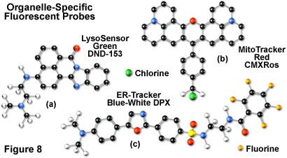
18 Fluorescent Stains Mitotracker LysoSensor
19 Fluorophore Labeled Proteins/Antibodies
20 Molecules can be specifically labelled Fluorescein Fluorescein isothiocyanate (FITC)
21 Molecules can be specifically labelled IgG labelled IgG IgG labelled IgG
22 Molecules can be specifically labelled (e.g. Immunofluorescence)

23 Protein of interest Production of a specific antibody Proteins can be specifically labelled Fluorescent labbeleing of the antibody Staining of cells, tissue etc. Alternative: Detection via a fluorescently labelled secondary antibody Major limitation: Targeting in live cells.

24 Quantum Dots conduction band Size quantization effect energy band gap Wannier exciton e - hν h + valence band Particle Size decreases Band gap increases Picture from: Chan WCW et al. Current Opinion in Biotechnology 2002, 13: 40-46


25 Quantum dots Advantages Quantum yield Similar, slightly lower as organic dyes Absorption Lager cross-section Reduced photo-bleaching-rate ZnS-capped CdSe QDs compared with Rhodamine 6G 20 time brighter times more stable

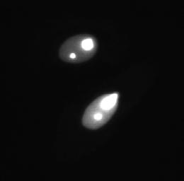
26 Sensitivity (Single Fluorophores)
27 Autofluorescent Proteins


28 Green Fluorescent Protein (GFP) 488 nm Aequorea victoria (Jellyfish) Chemistry Nobel price 2008 Osamu Shimomura Martin Chalfie Roger Y. Tsien

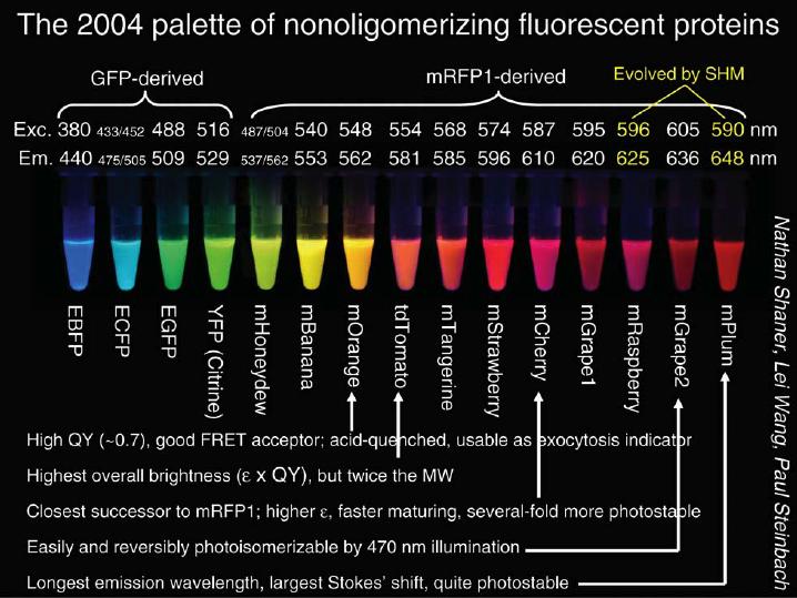
29 Applications of fluorescent proteins (FP) Two Most common applications of GFP variants From Chudakov et al, Trends Biotech., 2005
30
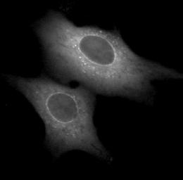
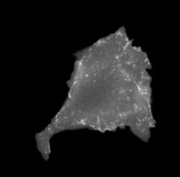
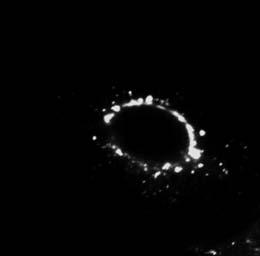
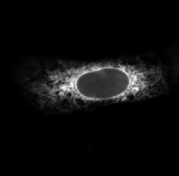
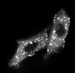
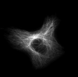
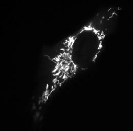
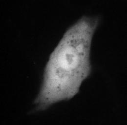
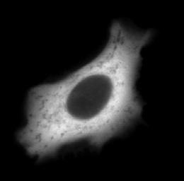

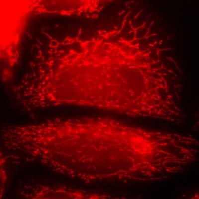
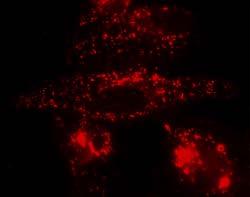
31 Protein Localization nucleus nucleolus nuclear envelope cytoplasm nucleus + cytoplasm mitochondria peroxisomes microtubules focal adhesions endoplasmic reticulum Golgi plasma membrane 10µm Dr. Arne Seitz
32 Summary Organelles and molecules can be labeled by: Organelle and protein specific fluorescent stains (e.g. Dapi). Labeling of antibodies/proteins with fluorophores. Autofluorescent proteins (e.g. GFPs). Live cell imaging: FP (Fluorescent proteins, e.g. GFP) are the method of choice to label proteins or organelles. Injection of labeled antibodies is possible. Organelle specific stains like e.g. DAPI can be toxic for the cell.
33 Fluorescent Microscopy Why do we need fluorescence microscopy Basics about fluorescence Fluorescent dyes and staining procedures Fluorescent microscopy Advanced applications
34 Fluorescence detection Excitation light (IE) Most excitation light Sample Fluorescent light (IFL) IE/IFL = 10 4 for strong fluorescence IE/IFL = for weak fluorescence (e.g. in situ hybrid.) In order to detect the fluorescence at 10% background the excitation light must be removed or attenuated by a factor up to 10 11
35 Epifluorescence Sample Objective Excitation Light
36 Epifluorescence Sample Back-scattered excitation light: IE/100 Objective Fluorescence
37 Epifluorescence Sample Objective Excitation Light Dichroic mirror (passes green but reflects blue light)
38 Epifluorescence Sample Back-scattered excitation light IE/100 Objective Dichroic mirror (passes green but reflects blue light) Fluorescence Detector
39 Epifluorescence (real world) Sample Back-scattered excitation light IE/100 Objective Dichroic mirror (passes green but reflects blue light) Back-scattered excitation light IE/10,000 Fluorescence Detector
40 Epifluorescence (real world) Sample Back-scattered excitation light IE/100 Objective Dichroic mirror (passes green but reflects blue light) Back-scattered excitation light IE/10,000 Back-scattered excitation light IE/10 11 Fluorescence Detector Emission filter (passes fluorescence but not back-scattered excitation light)
41 Typical Set-Up for Epifluorescence Sample Scattered light Objective HBO 488nm Dichroic mirror Alexa 488 Excitation Filterwheel Detector 520nm Emission Filterwheel
42 Set-Up for Green-Red Double Fluorescence Sample Scattered light Objective HBO 488nm Double dichroic mirror (λ1 = 505nm +λ2 = 560nm) Alexa 488 Excitation Filterwheel (Bandpass) Detector 520nm Emission Filterwheel (Bandpass)
43 Set-Up for Green-Red Double Fluorescence Sample Scattered light Objective HBO 550nm Double dichroic mirror Alexa 555 Excitation Filterwheel (Bandpass) Detector 590nm Emission Filterwheel (Bandpass, Longpass)

44 Implementation of Epifluorescence

45 Implementation of Epifluorescence

46 Implementation of Epifluorescence
47 Köhler illumination in Epifluorescence Transmission Focus on the specimen Close field diaphragm Focus condenser until field diaphragm is seen sharp Center field diaphragm Close field diaphragm up to % Remove eyepiece, look down to the aperture diaphragm Center (if possible) aperture diaphragm Open/Close aperture diaphragm up to % Fluorescence Focus on the specimen Swing in focusing aid (if available) Focus image of arc sharply Swing out focusing aid Close field diaphragm Center field diaphragm
48 Typical filter profiles Longpass Bandpass Shortpass
49 Typical Triple Bandpass Filter DAPI GFP TexasRed
50 Single Color Detection (e.g. GFP)

51 Single Color Detection (e.g. GFP) Use longpass filter in the emission!
52 Double Color Detection (e.g. GFP and TRITC)

53 GFP-TRITC Detection Filter Cubes GFP-detection TRITC-detection Bandpass emission filters are necessary in multicolor imaging
54 Triple Filter Cube
 Interference filters (high flexibility in")
55 Types of filters typically used Color glass filters (cheap, limited in wavelengths) Interference filters (high flexibility in wavelengths)
56 Light Sources Must fit the fluorescent dyes Must fit the Detectors
57 Light sources Halogen lamp Continuous spectrum: depends on temperature For 3400K maximum at 900 nm Lower intensity at shorter wavelengths Very strong in IR Mercury Lamp (HBO) Most of intensity in near UV Spectrum has a line structure Lines at 313, 334, 365, 406, 435, 546, and 578 nm Xenon lamp (XBO) Even intensity across the visible spectrum Has relatively low intensity in UV Strong in IR Metal halide lamp (Hg, I, Br) Stronger intensity between lines Stable output over short period of time Lifetime up to 5 times longer

58 Spectrum of a mercury arc lamp Dr. Arne Seitz Ideal for excitation of GFP2, CFP and DsRed imaging but less convenient for EGFP

59 Spectrum of a Xenon arc lamp
60 Summary Epifluorescence microscopy set-up is very sensitive. Bandpass detection filters are necessary for multicolor detection. Ideal excitation light sources should fit the dyes in use.
61 What is special about fluorescence microscopy? Specificity (molecules can be specifically labelled) Sensitivity (single molecule detection is possible) Fluorescence can report on the environment of the labelled molecule


62 Electron microscopy Light microscopy Molecular dynamics, molecular interactions Organelles Cells Worm Housefly Human 1 Å m 1 nm 10-9 m FRAP FCS LM FRET limit PALM,STORM 1 µm 10-6 m 1 mm 10-3 m 1 cm 10-2 m 1 m FRAP: Fluorescence recovery after photobleaching FCS: Fluorescence correlation spectroscopy FRET: Fluorescence resonance energy transfer

63 Quantum Yield Q = number of emitted versus absorbed photons Q = k f k f + k nr Lifetime τ = average time molecule spends in the excited state k= events/sec knr = k i τ = Q = 1 k f + k nr τ τ 0

64 Nonfluorescent relaxation can be due to: FRET k T Sensitized Emission Donor Acceptor
 1 = D")
65 Fluorescence Resonance Energy Transfer (FRET) Excitation Excitation Fluorescence Donor Fluorescence 1 τ D = k D k T Sensitized Emission Excitation Fluorescence Sensitized Emission Quenched Donor Fluorescence τ D ( k + k ) 1 = D T
66 Summary Fluorescence is dependant on the environment of the molecule. Parameters which can change due to the environment are: Intensity Fluorescence lifetime Frequency (=spectral shift) Fluorescence can be used as a reporter of the environment.
67 More about fluorescence microscopy 1. Lecture Biomicroscopy I + II, Prof. Theo Lasser, EPFL 2. Books a) Principle of fluorescence spectroscopy, Joseph R. Lackowicz, Springer 2 nd edition (1999) 3. Internet a) b) b) Web sites of microscope manufactures Leica Nikon Olympus Zeiss 4. BIOp EPFL, SV-AI 0241, SV-AI Dr. Arne Seitz -
Fluorescence Microscopy
 Fluorescence Microscopy Dr. Arne Seitz Swiss Institute of Technology (EPFL) Faculty of Life Sciences Head of BIOIMAGING AND OPTICS BIOP arne.seitz@epfl.ch Fluorescence Microscopy Why do we need fluorescence
BIOIMAGING AND OPTICS PLATFORM EPFL SV PTBIOP FLUORESCENCE MICROSCOPY
 FLUORESCENCE MICROSCOPY Internal course 2014 January 14 th FLUORESCENCE MICROSCOPY Why do we need it? - 2- UNSTAINED SPECIMEN Missing specificity 3 DIFFERENT STAINING STRATEGIES Histological stain (Absorption)
Partha Roy
 Fluorescence microscopy http://micro.magnet.fsu.edu/primer/index.html Partha Roy 1 Lecture Outline Definition of fluorescence Common fluorescent reagents Construction ti of a fluorescence microscope Optical
Dino-Lite knowledge & education. Fluorescence Microscopes
 Dino-Lite knowledge & education Fluorescence Microscopes Dino-Lite Fluorescence models Smallest fluorescence microscope in the world Revolution to biomedical and educational applications Flexible Easy
FLUORESCENCE. Matyas Molnar and Dirk Pacholsky
 FLUORESCENCE Matyas Molnar and Dirk Pacholsky 1 Information This lecture contains images and information from the following internet homepages http://micro.magnet.fsu.edu/primer/index.html http://www.microscopyu.com/
Concept review: Fluorescence
 16 Concept review: Fluorescence Some definitions: Chromophore. The structural feature of a molecule responsible for the absorption of UV or visible light. Fluorophore. A chromophore that remits an absorbed
Contact Details. Dr Alexander Galkin. Office: MBC Room 186. Tel: (028) Frequency and wavelength.
 Frequency and wavelength.") Contact Details The electromagnetic spectrum Biological Spectroscopy Dr Alexander Galkin Email: a.galkin@qub.ac.uk Dr Alexander Galkin MSc Biomolecular Function - BBC8045 Office: MBC Room 186 Tel: (028)
Contact Details The electromagnetic spectrum Biological Spectroscopy Dr Alexander Galkin Email: a.galkin@qub.ac.uk Dr Alexander Galkin MSc Biomolecular Function - BBC8045 Office: MBC Room 186 Tel: (028)
F* techniques: FRAP, FLIP, FRET, FLIM,
 F* techniques: FRAP, FLIP, FRET, FLIM, FCS Antonia Göhler March 2015 Fluorescence explained in the Bohr model Absorption of light (blue) causes an electron to move to a higher energy orbit. After a particular
Special Techniques 1. Mark Scott FILM Facility
 Special Techniques 1 Mark Scott FILM Facility SPECIAL TECHNIQUES Multi-photon microscopy Second Harmonic Generation FRAP FRET FLIM In-vivo imaging TWO-PHOTON MICROSCOPY Alternative to confocal and deconvolution
Fluorescence Light Microscopy for Cell Biology
 Fluorescence Light Microscopy for Cell Biology Why use light microscopy? Traditional questions that light microscopy has addressed: Structure within a cell Locations of specific molecules within a cell
A Brief History of Light Microscopy And How It Transformed Biomedical Research
 A Brief History of Light Microscopy And How It Transformed Biomedical Research Suewei Lin Office: Interdisciplinary Research Building 8A08 Email: sueweilin@gate.sinica.edu.tw TEL: 2789-9315 Microscope
More on fluorescence
 More on fluorescence Last class Fluorescence Absorption emission Jablonski diagrams This class More on fluorescence Common fluorophores Jablonski diagrams to spectra Properties of fluorophores Excitation
Fluorescence spectroscopy
 Fluorescence spectroscopy The light: electromagnetic wave Zoltán Ujfalusi Biophysics seminar Dept. of Biophysics, University of Pécs 14-16 February 2011 Luminescence: light is not generated by high temperatures!!!
Introduction to Fluorescence Jablonski Diagram
 ntroduction to Fluorescence Jablonski Diagram Excited Singlet Manifold S1 internal conversion S2 k -isc k isc Excited riplet Manifold 1 S0 k nr k k' f nr fluorescence k p phosphorescence Singlet round
MICROSCOPY. "micro" (small) "scopeo" (to watch)
 scopeo (to watch)") MICROSCOPY "micro" (small) "scopeo" (to watch) THE RELATIVE SIZES OF MOLECULES, CELLS AND ORGANISMS THE RELATIVE SIZES OF MOLECULES, CELLS AND ORGANISMS MICROSCOPY 1590 2012 MICROSCOPY THE LIGHT Light:
MICROSCOPY "micro" (small) "scopeo" (to watch) THE RELATIVE SIZES OF MOLECULES, CELLS AND ORGANISMS THE RELATIVE SIZES OF MOLECULES, CELLS AND ORGANISMS MICROSCOPY 1590 2012 MICROSCOPY THE LIGHT Light:
Visualizing Cells Molecular Biology of the Cell - Chapter 9
 Visualizing Cells Molecular Biology of the Cell - Chapter 9 Resolution, Detection Magnification Interaction of Light with matter: Absorbtion, Refraction, Reflection, Fluorescence Light Microscopy Absorbtion
FRET and FRET based Microscopy Techniques
 Big Question: We can see rafts in Model Membranes (GUVs or Supported Lipid Bilayers, LM), but how to study in cells? Do rafts really exist in cells? Are they static large structures? Are they small transient
Fluorescence spectroscopy
 Fluorescence spectroscopy The light: electromagnetic wave Tamás Huber Biophysics seminar Dept. of Biophysics, University of Pécs 05-07. February 2013. Luminescence: light emission of an excited system.
Imaging facilities at WUR
 Imaging facilities at WUR Advanced light microscopy facilities at Wageningen UR Programme Thursday 13 June 2013 Lunch meeting organized by Cat-Agro Food 12.00 Welcome and sandwich lunch 12.10 Introduction
Fluorescence Microscopy. Terms and concepts to know: 10/11/2011. Visible spectrum (of light) and energy
 and energy") Fluorescence Microscopy Louisiana Tech University Ruston, Louisiana Microscopy Workshop Dr. Mark DeCoster Associate Professor Biomedical Engineering 1 Terms and concepts to know: Signal to Noise Excitation
Fluorescence Microscopy Louisiana Tech University Ruston, Louisiana Microscopy Workshop Dr. Mark DeCoster Associate Professor Biomedical Engineering 1 Terms and concepts to know: Signal to Noise Excitation
Con-focal and Multi-photon Microscope Experiment Fundamental. Qian Hu, Lab of Laser Scanning Confocal & Two-Photon Microscopy, ION, CAS
 Con-focal and Multi-photon Microscope Experiment Fundamental Qian Hu, Lab of Laser Scanning Confocal & Two-Photon Microscopy, ION, CAS 1. Light is Electromagnetic Wave ν = c / λ 2. Image of a Point Source
Practical light microscopy: an introduction
 Practical light microscopy: an introduction Dr. Mark Leake, Oxford University www.physics.ox.ac.uk/users/leake Aim of today s talk: Explanation of the very (very) basics of how a light microscope works
Fluorescence Spectroscopy. Student: Marin Cristina Antonia Coordinator:S.l. Preda Liliana
 Fluorescence Spectroscopy Student: Marin Cristina Antonia Coordinator:S.l. Preda Liliana Fluorescence Electron in the ground state is excited to a higher energy state After loss of some energy in vibrational
Fluorescence spectroscopy
 Fluorescence spectroscopy The light: electromagnetic wave Tamás Huber Biophysics seminar Dept. of Biophysics, University of Pécs 05-06. February 2014. 1 Luminescence: light emission of an excited system.
Biochemistry. Biochemical Techniques. 18 Spectrofluorimetry
 Description of Module Subject Name Paper Name 12 Module Name/Title 1. Objectives 1.1 To understand technique of Spectrofluorimetry. 1.2 To explain instrumentation design 1.3 What are applications of Spectrofluorimetry?
Widefield Microscopy Bleed-Through
 In widefield microscopy the excitation wavelengths which illuminate the sample, and the emission wavelengths which reach the CCD camera are selected throughout a filter cube. A filter cube consists of
Fluorescence Microscopy: A Biological Perspective
 Fluorescence Microscopy: A Biological Perspective From nanometre to metre: the scale of life Instrumentation and accessible scale limits the questions that can be addressed in biology Why are there limits?
1. The fluorescence process.
 1. The fluorescence process. 1.1 introduction Fluorescence is the result of a three-stage process that occurs in certain molecules (generally polyaromatic hydrocarbons or heterocycles) called fluorophores
Reminder: absorption. OD = A = - log (I / I 0 ) = ε (λ) c x. I = I ε(λ) c x. Definitions. Fluorescence quenching and FRET.
 = ε (λ) c x. I = I ε(λ) c x. Definitions. Fluorescence quenching and FRET.") Reminder: absorption Special fluorescence applications I 0 I Fluorescence quenching and FRET Miklós Nyitrai; 24 th of Februry 2011. substance OD = A = - log (I / I 0 ) = ε (λ) c x optical density I = I
Reminder: absorption Special fluorescence applications I 0 I Fluorescence quenching and FRET Miklós Nyitrai; 24 th of Februry 2011. substance OD = A = - log (I / I 0 ) = ε (λ) c x optical density I = I
Imaging of Cells using fluorescents dyes. By: Josué A. Benjamín Rivera September 27, 2018
 Imaging of Cells using fluorescents dyes By: Josué A. Benjamín Rivera September 27, 2018 1 History Sir William Henry Perkin BRITISH CHEMIST In 1856, at the age of 18, William Henry Perkin set out with
What to look for in a fluorophore. What to do with a fluorophore. Types of fluorochromes
 What to do with a fluorophore Intracellular localization (ER, Golgi, PM, nuclear, lysosome, MT, actin,...) Dynamic processes (protein synthesis, trafficking, turnover, DNA replication, cytoskeletal remodeling,
cell and tissue imaging by fluorescence microscopy
 cell and tissue imaging by fluorescence microscopy Steven NEDELLEC Plateforme Micropicell SFR Santé François Bonamy Nantes 1 A matter of size Limit of resolution 0.15mm aims: building the image of an object
Advanced fluorescence microscopy techniques
 Practice-oriented, student-friendly modernization of the biomedical education for strengthening the international competitiveness of the rural Hungarian universities TÁMOP-4.1.1.C-13/1/KONV-2014-0001 Advanced
The Green Fluorescent Protein. w.chem.uwec.edu/chem412_s99/ppt/green.ppt
 The Green Fluorescent Protein w.chem.uwec.edu/chem412_s99/ppt/green.ppt www.chem.uwec.edu/chem412_s99/ppt/green.ppt Protein (gene) is from a jellyfish: Aequorea victoria www.chem.uwec.edu/chem412_s99/ppt/green.ppt
FLIM Fluorescence Lifetime IMaging
 FLIM Fluorescence Lifetime IMaging Fluorescence lifetime t I(t) = F0 exp( ) τ 1 τ = k f + k nr k nr = k IC + k ISC + k bl Batiaens et al, Trends in Cell Biology, 1999 τ τ = fluorescence lifetime (~ns to
Confocal Microscopes. Evolution of Imaging
 Confocal Microscopes and Evolution of Imaging Judi Reilly Hans Richter Massachusetts Institute of Technology Environment, Health & Safety Office Radiation Protection What is Confocal? Pinhole diaphragm
Advanced fluorescence microscopy techniques
 Practice-oriented, student-friendly modernization of the biomedical education for strengthening the international competitiveness of the rural Hungarian universities TÁMOP-4.1.1.C-13/1/KONV-2014-0001 Advanced
2004 Debye Lecture 4 C. B. Murray. Quantum Dot Applications: Sun Screen. Solar Cells. Bio-tagging. Solid State Lighting?
 2004 Debye Lecture 4 C. B. Murray Quantum Dot Applications: Sun Screen Solar Cells Bio-tagging Solid State Lighting? Quantum Dot Solar cells Nanocrystal Solar Cells Double-labeling of mitochondria
Janos Szabad Department of Biology University of Szeged 6720 Szeged, Somogyi str
 Janos Szabad Department of Biology University of Szeged 6720 Szeged, Somogyi str. 4. E-mail: szabad.janos@med.u-szeged.hu - Through the use of antibodies - against the protein - against a fusion partner
Lab 1: Ensemble Fluorescence Basics
 Lab 1: Ensemble Fluorescence Basics This laboratory module is divided into two sections. The first one is on organic fluorophores, and the second one is on ensemble measurement of FRET (Fluorescence Resonance
Intracellular localization and trafficking of proteins or How (and why) to find a needle in a haystack
 to find a needle in a haystack") Intracellular localization and trafficking of proteins or How (and why) to find a needle in a haystack :: The Structure of a Cell :: Relative sizes SUBUNITS 0.2 mm (200 μm) minimum resolvable by unaided
Intracellular localization and trafficking of proteins or How (and why) to find a needle in a haystack :: The Structure of a Cell :: Relative sizes SUBUNITS 0.2 mm (200 μm) minimum resolvable by unaided
BIO 315 Lab Exam I. Section #: Name:
 Section #: Name: Also provide this information on the computer grid sheet given to you. (Section # in special code box) BIO 315 Lab Exam I 1. In labeling the parts of a standard compound light microscope
Resolution of Microscopes Visible light is nm Dry lens(0.5na), green(530nm light)=0.65µm=650nm for oil lens (1.4NA) UV light (300nm) = 0.13µm f
, green(530nm light)=0.65µm=650nm for oil lens (1.4NA) UV light (300nm) = 0.13µm f") Microscopes and Microscopy MCB 380 Good information sources: Alberts-Molecular Biology of the Cell http://micro.magnet.fsu.edu/primer/ http://www.microscopyu.com/ Approaches to Problems in Cell Biology
Microscopes and Microscopy MCB 380 Good information sources: Alberts-Molecular Biology of the Cell http://micro.magnet.fsu.edu/primer/ http://www.microscopyu.com/ Approaches to Problems in Cell Biology
The most extensively used technique for tissue analysis is light microscopy.
 Fluorescence Theory Quantum yield Wavelength shift Ligand interactions Membrane interactions Using quenchning effects Fluorescence in-vivo Localization Distance measurements FRET The most extensively used
Absorption of an electromagnetic wave
 In vivo optical imaging?? Absorption of an electromagnetic wave Tissue absorption spectrum Extinction = Absorption + Scattering Absorption of an electromagnetic wave Scattering of an electromagnetic wave
Single cell molecular profiling using Quantum Dots. Technical Journal Club Rahel Gerosa
 Single cell molecular profiling using Quantum Dots Technical Journal Club 01.10.2013 Rahel Gerosa Molecular Profiling Powerful technique to study complex molecular networks underlying physiological and
BIO 315 Lab Exam I. Section #: Name:
 Section #: Name: Also provide this information on the computer grid sheet given to you. (Section # in special code box) BIO 315 Lab Exam I 1. In labeling the parts of a standard compound light microscope
Fluorescence quenching, Fluorescence anisotropy, Fluorescence resonance energy transfer (FRET)
") Fluorescence quenching, Fluorescence anisotropy, Fluorescence resonance energy transfer (FRET) Timescale of fluorescence processes The excited electron decay possibilities k f k ph k q k t k ic Biophysics
Fluorescence quenching, Fluorescence anisotropy, Fluorescence resonance energy transfer (FRET) Timescale of fluorescence processes The excited electron decay possibilities k f k ph k q k t k ic Biophysics
Nodes of regulation in cellular systems
 Nodes of regulation in cellular systems cell membrane signal transduction ligands receptors oligomerization transport signal transduction modified protein Golgi transcription factor transport ER transport
Live cell microscopy
 Live cell microscopy 1. Why do live cell microscopy? 2. Maintaining living cells on a microscope stage. 3. Considerations for imaging living cells. 4. Fluorescence labeling of living cells. 5. Imaging
Confocal Microscopy Analyzes Cells
 Choosing Filters for Fluorescence A Laurin Publication Photonic Solutions for Biotechnology and Medicine November 2002 Confocal Microscopy Analyzes Cells Reprinted from the November 2002 issue of Biophotonics
Rice/TCU REU on Computational Neuroscience. Fundamentals of Molecular Imaging
 Rice/TCU REU on Computational Neuroscience Fundamentals of Molecular Imaging June 2, 2009 Neal Waxham 713-500-5621 m.n.waxham@uth.tmc.edu Objectives Introduction to resolution in light microscopy Brief
Contents. SCHOOL of FLUORESCENCE. For more information, go to lifetechnologies.com/imagingbasics
 MPSF educator packet This packet contains illustrations and figures from the Molecular Probes School of Fluorescence website. They illustrate concepts from the basic physical properties that underlie fluorescence
1. INTRODUCTION 2. EXPERIMENTAL 3. REFERENCES
 1. INTRODUCTION 2. EXPERIMENTAL 3. REFERENCES 1 1. INTRODUCTION Fluorescence spectroscopy is one of the most widely used spectroscopic techniques in the fields of biochemistry and molecular biophysics
Each question may have MULTIPLE correct answers. Select all that are correct.
 Knowledge Assessment Flow Cytometry Workshop, Part 1 April 20, 2015 Each question may have MULTIPLE correct answers. Select all that are correct. 1. Tandem dyes are a. highly stable fluorophores after
The Basics of Flow Cytometry
 The Basics of Flow Cytometry F ACS C ore F acility Janine Bögli, Biozentrum, 29. January 2018 The functions of the FACS Core Facility Centralization of equipment and expertise Train users Sorter operation
Lesson Plan: Fluorescence
 Lesson Plan: Fluorescence Background Fluorescence is produced when a material or substance absorbs light of a given color and then gives off light of another color. The light that is given off, or emitted,
HYPERSPECTRAL MICROSCOPE PLATFORM FOR HIGHLY MULTIPLEX BIOLOGICAL IMAGING. Marc Verhaegen
 HYPERSPECTRAL MICROSCOPE PLATFORM FOR HIGHLY MULTIPLEX BIOLOGICAL IMAGING Marc Verhaegen CMCS, MONTREAL, MAY 11 th, 2017 OVERVIEW Hyperspectral Imaging Multiplex Biological Imaging Multiplex Single Particle
Monitoring and Optimizing the Lipopolysaccharides-plasmid DNA interaction by FLIM-FRET
 Transactions on Science and Technology Vol. 4, No. 3-3, 342-347, 2017 Monitoring and Optimizing the Lipopolysaccharides-plasmid DNA interaction by FLIM-FRET Nur Syahadatain Abdul Razak 1#, Clarence M.
ab CytoPainter ER Staining Kit Red Fluorescence
 ab139482 CytoPainter ER Staining Kit Red Fluorescence Instructions for Use Designed to detect Human endoplasmic reticulum by microscopy. This product is for research use only and is not intended for diagnostic
Time-resolved Measurements Using the Agilent Cary Eclipse Fluorescence Spectrophotometer A Versatile Instrument for Accurate Measurements
 Time-resolved Measurements Using the Agilent Cary Eclipse Fluorescence Spectrophotometer A Versatile Instrument for Accurate Measurements Technical Overview Authors Dr. Fabian Zieschang, Katherine MacNamara,
Welcome! openmicberkeley.wordpress.com. Open Berkeley
 Welcome! openmicberkeley.wordpress.com Agenda Jen Lee: Introduction to FRET Marla Feller: Using FRET sensors to look at time resolved measurements Becky Lamason: Using FRET to determine if a bacterial
ADVANCED PRACTICAL COURSE IN BIOPHYSICS: FRET
 : FRET 1 INTRODUCTION Fluorescence spectroscopy and fluorescence microscopy are essential tools in biology. Biological molecules can be labeled with fluorescent molecules and thus, their localization and
Compensation: Fundamental Principles
 Flow Cytometry Seminar Series 2017 : Fundamental Principles Spillover correction in multicolor flow cytometry 28.02.2017 http://www.cytometry.uzh.ch Contents Fluorescence and its detection Absorption and
Confocal Microscopy & Imaging Technology. Yan Wu
 Confocal Microscopy & Imaging Technology Yan Wu Dec. 05, 2014 Cells under the microscope What we use to see the details of the cell? Light and Electron Microscopy - Bright light / fluorescence microscopy
Spectral Separation of Multifluorescence Labels with the LSM 510 META
 Microscopy from Carl Zeiss Spectral Separation of Multifluorescence Labels with the LSM 510 META Indians living in the South American rain forest can distinguish between almost 200 hues of green in their
Fluorescence microscopy
 Fluorescence microscopy 1 Fluorescence microscopies basic fluorescence, fluorophores Deconvolution Confocal Two-photon/multi-photon 4Pi Light sheet Total internal reflection STED FRAP/FLIP/FCS FRET PALM/STORM/iPALM
Microscopy from Carl Zeiss
 Microscopy from Carl Zeiss LSM 710 In Tune with Your Application Enjoy new freedom in selecting fluorescent dyes with In Tune, the new laser system for the LSM 710. Whatever the wavelength, you can match
TARGETED IMAGING. Maureen Chan and Ruwani Mahathantila
 TARGETED IMAGING Maureen Chan and Ruwani Mahathantila Overview 2 Introduction to fluorescent imaging Fluorescent agents Quantum Dots Physical properties How QDs work In Vivo QD imaging Future Video What
Using Quantum Dots in Fluorescence Resonance Energy Transfer Studies
 p.1/31 Using Quantum Dots in Fluorescence Resonance Energy Transfer Studies Rajarshi Guha Pennsylvania State University p.2/31 Introduction Using organic fluorophores as labels A brief overview of fluorescence
Spectra Chacracterizations of Optical Nanoparticles
 THAI NGUYEN UNIVERSITY OF EDUCATION Spectra Chacracterizations of Optical Nanoparticles Chu Viet Ha Department of Physics 18/2018 1 THAI NGUYEN UNIVERSITY OF EDUCATION Address 20 Luong Ngoc Quyen Street,
Biophotonics?? Biophotonics. technology in biomedical engineering. Advantages of the lightwave
 Biophotonics - Imaging: X-ray, OCT, polarimetry, DOT, TIRF, photon migration, endoscopy, confocal microscopy, multiphoton microscopy, multispectral imaging - Biosensing: IR spectroscopy, fluorescence,
The analysis of fluorescence microscopy images for FRET detection
 The analysis of fluorescence microscopy images for FRET detection Ela Claridge, Dale J. Powner and Michael J.O. Wakelam School of Computer Science, The University of Birmingham B5 2TT Institute for Cancer
Single-Molecule Biophysics. Physical Cell Biology Guest lecture
 Single-Molecule Biophysics Physical Cell Biology Guest lecture Liviu Movileanu Syracuse University lmovilea@syr.edu Web: http://movileanulab.syr.edu Single-molecule versus bulk-phase measurements Bulk-phase
Workshop advanced light microscopy
 Workshop advanced light microscopy Multi-mode confocal laser scanning microscope Jan Willem Borst Laboratory of Biochemistry Biomolecular Networks www.bic.wur.nl MicroSpectroscopy Centre Wageningen Microspectroscopy
QuantaMaX and standard filters for fluorescence
 QuantaMaX and standard filters for fluorescence Our fluorescence filter product line is comprised of Stock QuantaMAX and Standard Vivid and Basic excitation, emission and dichroic interference filters,
Crystal Growth, Optical and Thermal Studies of 4-Nitroaniline 4- Aminobenzoic Acid: A Fluorescent Material
 Research Article Crystal Growth, Optical and Thermal Studies of 4-Nitroaniline 4- Aminobenzoic Acid: A Fluorescent Material A. Silambarasan, P. Rajesh * and P. Ramasamy Centre for Crystal Growth, SSN College
Fluorescence Resonance Energy Transfer (FRET) Microscopy
 Microscopy") Applications in Confocal Microscopy Fluorescence Resonance Energy Transfer (FRET) Microscopy Product Info Brochures Confocal Theory Java Tutorials Glossary Applications Image Gallery Resource Links The
Applications in Confocal Microscopy Fluorescence Resonance Energy Transfer (FRET) Microscopy Product Info Brochures Confocal Theory Java Tutorials Glossary Applications Image Gallery Resource Links The
11/19/2013. Janine Zankl FACS Core Facility 13. November Cellular Parameters. Cellular Parameters. Monocytes. Granulocytes.
 DEPARTEMENT BIOZENTRUM Janine Zankl FACS Core Facility 13. November 2013 Cellular Parameters Granulocytes Monocytes Basophils Neutrophils Lymphocytes Eosinophils Cellular Parameters 1 What Is Flow Cytometry?
Multiplexed 3D FRET imaging in deep tissue of live embryos Ming Zhao, Xiaoyang Wan, Yu Li, Weibin Zhou and Leilei Peng
 Scientific Reports Multiplexed 3D FRET imaging in deep tissue of live embryos Ming Zhao, Xiaoyang Wan, Yu Li, Weibin Zhou and Leilei Peng 1 Supplementary figures and notes Supplementary Figure S1 Volumetric
Supplementary Figure 1. The normalized absorption and emission spectra of 605QD
 1..8 65Q Absorbance 65Q Emission Cy5 Absorbance Cy5 Emission 1..8 Extiction Absorption Coefficient.6.4.2. 45 5 55 6 65 7 75 8 Wavelength (nm).6.4.2. Fluorescence Emission Intensity Supplementary Figure
Color-Rich Fluoro-Max Dyed Microparticles March 2008
 Fluoro-Max Dyed Microparticles March 2008 Introduction Dyed Microparticles Thermo Scientific Seradyn dyed and fluorescent microparticles are monodisperse particles prepared by unique and proprietary emulsion
Application of Quantum Mechanics to Biology
 Application of Quantum Mechanics to Biology How can we apply quantum mechanics to biology? Polymers of nucleotides and amino acids - millions of atoms bounded into a large molecule Visual System Must turn
Supplementary Table 1. Components of an FCS setup (1PE and 2PE)
") Supplementary Table 1. Components of an FCS setup (1PE and 2PE) Component and function Laser source Excitation of fluorophores Microscope with xy-translation stage mounted on vibration isolated optical
Supplementary Table 1. Components of an FCS setup (1PE and 2PE) Component and function Laser source Excitation of fluorophores Microscope with xy-translation stage mounted on vibration isolated optical
Sapphire. Biomolecular Imager THE NEXT GENERATION OF LASER-BASED IMAGING
 Sapphire Biomolecular Imager THE NEXT GENERATION OF LASER-BASED IMAGING Breakthrough image capture and analysis The Sapphire Biomolecular Imager is a next generation laser scanning system that provides
Challenges to measuring intracellular Ca 2+ Calmodulin: nature s Ca 2+ sensor
 Calcium Signals in Biological Systems Lecture 3 (2/9/0) Measuring intracellular Ca 2+ signals II: Genetically encoded Ca 2+ sensors Henry M. Colecraft, Ph.D. Challenges to measuring intracellular Ca 2+
SUPPORTING INFORMATION
 Electronic Supplementary Material (ESI) for Dalton Transactions. This journal is The Royal Society of Chemistry 2015 Terbium-Based Time-Gated Förster Resonance Energy Transfer Imaging for Evaluating Protein-Protein
Total Internal Reflection Fluorescence Microscopy
 Total Internal Reflection Microscopy Nicole O Neil Indiana University October 24, 2005 Agenda Why use TIRFM? Theory behind TIR Snell s Law Instrumentation Evanescent Wave Excitation of Fluorophores Advantages/Disadvantages
Biophotonics II general remarks
 general remarks BIOPHOTONICS I (WS 2017/18) I. Imaging Systems human vision microscopy II. Light Scattering Mie scattering light propagation in tissue BIOPHOTONICS II (SS 2018) III. Biospectroscopy Fluorescence
BASICS OF FLOW CYTOMETRY
 BASICS OF FLOW CYTOMETRY AUTHOR: Ana Isabel Vieira APPROVAL: Henrique Veiga Fernandes Ana Sílvia Gonçalves SOP.UCF.002 03-09-2015 Pag. 1/9 Overview Flow: Fluid Cyto: Cell Metry: Measurement Flow cytometry
ab CytoPainter ER Staining Kit Red Fluorescence
 ab139482 CytoPainter ER Staining Kit Red Fluorescence Instructions for Use Designed to detect Human endoplasmic reticulum by microscopy. This product is for research use only and is not intended for diagnostic
Direct visualization, sizing and concentration measurement of fluorescently labeled nanoparticles using NTA
 Direct visualization, sizing and concentration measurement of fluorescently labeled nanoparticles using NTA NANOSIGHT RANGE Visualize and Measure Nanoparticle Size and Concentration PARTICLE SIZE PARTICLE
Resolving Macromolecular Organization in Cells Using Super Resolution Microscopy Karen Porter-Davis Chamblee Charter High School
 Resolving Macromolecular Organization in Cells Using Super Resolution Microscopy Karen Porter-Davis Chamblee Charter High School STEP-UP Program 2015 Super Resolution Imaging } Two Projects to Image }
Prototype Microfluidic System for Fluorescence-Based Chemical Sensing
 Doi: 10.12982/cmujns.2014.0064 625 Prototype Microfluidic System for Fluorescence-Based Chemical Sensing Pattareeya Kittidachachan 1, Suparat Rujihan 1 and Badin Damrongsak 2* 1 Department of Physics,
Flow Cytometry - The Essentials
 Flow Cytometry - The Essentials Pocket Guide to Flow Cytometry: 1. Know your Cytometer 2. Understanding Fluorescence and Fluorophores 3. Gating Process 4. Controls 5. Optimization 6. Panel Building 7.
Imaging Quantum Dots using FUJIFILM LAS 4000
 Imaging Quantum Dots using FUJIFILM LAS 4000 Application Note John Pizzonia, Ph.D. 9-28-07 Quantum dots (also known as nanocrystals) are a special class of materials known as semiconductors, which are
Selected Topics in Electrical Engineering: Flow Cytometry Data Analysis
 Selected Topics in Electrical Engineering: Flow Cytometry Data Analysis Bilge Karaçalı, PhD Department of Electrical and Electronics Engineering Izmir Institute of Technology Outline Experimental design
Classroom Tested Lesson
 Classroom Tested Lesson Video Description Secrets of the Sequence, Show 124, Episode 2 A Green Light for Biology approximately 10 minutes viewing time This discovery known as Green Fluorescent Protein
Cell Imaging. Cell Imaging 48
 Cell Imaging 48 bio-rad.com/zoe Cell Imaging Bio-Rad s suite of tools for fluorescence microscopy and cell imaging includes the ZOE fluorescent cell imager and nuclear dyes. See Also PureBlu Hoechst 33342
Imagerie et spectroscopie de fluorescence par excitation non radiative
 Imagerie et spectroscopie de fluorescence par excitation non radiative comment s affranchir de la limite de diffraction Rodolphe Jaffiol, Cyrille Vézy, Marcelina Cardoso Dos Santos LNIO, UTT, Troyes NanoBioPhotonics