Rice/TCU REU on Computational Neuroscience. Fundamentals of Molecular Imaging
|
|
|
- Cleopatra Jones
- 6 years ago
- Views:
Transcription
1 Rice/TCU REU on Computational Neuroscience Fundamentals of Molecular Imaging June 2, 2009 Neal Waxham
2 Objectives Introduction to resolution in light microscopy Brief discussion of lasers as excitation sources Multiphoton excitation-what is it? advantages/disadvantages Fluorescent Probes-uses and characteristics Applications of MPE to the study of intracellular biochemistry The concept of single molecule analysis

3 Resolution = 0.61λ / NA λ = wavelength of electromagnetic radiation
4 Spatial and Temporal Scales of Microscopy Temporal scales - picoseconds to months
 NA = n sin(θ), where n is the index of refraction and θ the")
 n NA = 0.12 θ ~ 7 o Long working distances NA = 0.")
5 What Limits Resolution in Microscopy? Numerical Aperture (NA) NA = n sin(θ), where n is the index of refraction and θ the half angle of the illumination cone. Rayleigh criterion: Resolution = 0.61λ / NA R = 1.22λ / (NA obj + NA cond ) n NA = 0.12 θ ~ 7 o Long working distances NA = 0.90 θ ~ 64 o Short working distances Destructive Interference


6 Characteristics of Light from Lasers Light Amplification by Stimulated Emission of Radiation

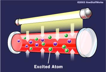
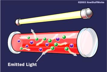
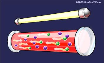
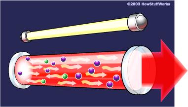
7 How Lasers Work
.")
8 Fluorescence Stokes shift Fluorescein Isothiocyanate Following absorption, a number of vibrational levels of the excited state are populated. Molecules in these higher vibrational levels then relax to the lowest vibrational level of the excited state (vibrational relaxation). From the lowest vibrational level, there can be an emission of photon of lower energy (fluorescence)
 photons can interact simultaneously with a molecule adding their energies")

9 One Photon vs. Two Photon Florescence? Two (or more) photons can interact simultaneously with a molecule adding their energies to produce an excitation equal to the sum of their individual energies. Lifetimes ~ns timescale i.e. 2 red photons can = 1 blue photon 1 photon excitation Fluorescence 2 photon excitation Increasing Wavelength Increasing Energy
10 S 1 S 1 Two/(Multi)-Photon-Excitation virtual state ca s Single photon excitation (488 nm) Two photon excitation (900 nm) hν hν/2 Fluorescence hν f S 0 Idea: Simultaneous (10-15 s) absorption of n photons of wavelength Major advantage: Inherent spatial sectioning by I n - dependency of excitation probability. Excitation only in vicinity of focal spot Pulsed excitation = (100 fs, 80 MHz)

 declines from z (red")

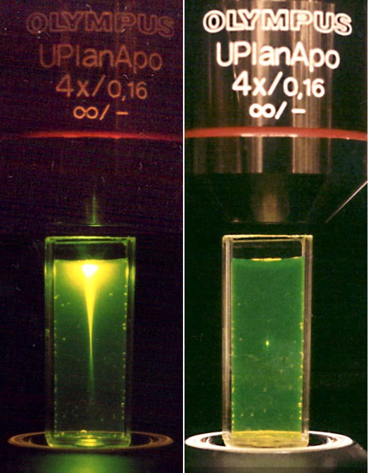
11 MPE is inherently localized to the focus of a high NA objective w 0 λ N. A μm The intensity (squared) declines from z (red arrows) as z -4 Calculated intensity of 740 nm light near focus of 1.2 NA objective
 I peak = = 10 fτ ( 5 I( t) 10-13 s Average Intensity")
12 Pulsed laser excitation enhances two-photon absorption 1/f 10-8 s τ I t) I peak = = 10 fτ ( 5 I( t) s Average Intensity I(t)


13 Two Photon Excitation (2PE) S 1 S 0 OPE A F S 1 S 0 A TPE F Advantages For intracellular work: 1. Small focal volume 2. Decreased photobleaching 3. Decreased phototoxicity 4. Increased viability 5. Increased focus depth For cross-correlation work: 6. Single laser line 7. No pinhole necessary 8. Good S/N ratio Disadvantages 1. Greater average Illumination intensities 2. Loss of resolution 3. High cost of pulse laser
14 Fluorescent Probes Uses of fluorescent molecules: 1. Labels - free dyes that may partition to a specific region of a cell or tissue, or fluorescent molecules that are bound to antibodies, receptor proteins or other biomolecules of interest. 2. Indicators dyes - the probes dynamically bind an ion (Ca ++, H +, Mg ++ ) and then change in either fluorescence intensity, emission or excitation spectrum. 3. Fluorescent proteins such as GFP, that are produced by the organism after the DNA for GFP, or more commonly a GFP fusion protein, is introduced into the cell.


15 Fluorescent Probes molecules EGFP quantum dot silica nanoparticle (rhodamine) N NC CN N 1 nm 3 nm Single fluorophores 6 nm 50 nm Multiple fluorophores


16 Two Photon Laser Scanning Microscopy Coupled to Spectroscopy Techniques

17 How do you generate an image? Scanning mirrors move the laser beam back and forth across the sample A detector collects the photons that come out of a single area and map them onto an X-Y Image as pixels.


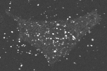
18 In vivo imaging - example: transgenic mouse models of Alzheimer's disease. β amyloid plaque stained with Thio-S, excitation at 760 nm

19 Two-photon Imaging in Live Animals Neurons imaged in the brain of a live animal 18 months apart Arrows-spines eliminated; Arrowheads, spines formed From Zuo et al., Neuron 2005
 3.")
20 Optical Methods Applied to Study Protein Dynamics 1. Two-Photon Fluorescence Photobleaching Recovery (TPFPR) 2. Two-Photon Fluorescence Correlation Spectroscopy (TPFCS) 3. Two-Photon Fluorescence Dual-Color Cross-Correlation (TPCCS) crosscorrelation
21
 of faster diffusing species Species Soma Neurite 10 kd dextran 29.2 29.0 Alexa-488-CaM 28.5 22.")
22 Diffusion Mapping of Alexa-488-Labeled Calmodulin in Neurons Using MPFPR Alexa-488-CaM in solution D(t) = 54 µm 2 /sec D(t) of faster diffusing species Species Soma Neurite 10 kd dextran Alexa-488-CaM

23 D = kt 6πηr

24 Intracellular Diffusion: Far from Simple 500 nm

25 Applications of Single Molecule Approach to Biochemistry and Cell Biology Fluorescence Correlation Spec./Fluorescence Cross Correlation Spec.
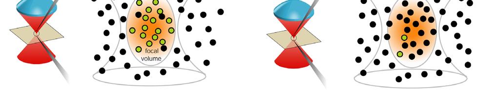
 Small volume elements achieved through confocal or multiphoton optics bursts time I em Primary measurement parameter is signal")
26 How to detect single molecules? Low concentrations of fluor (<10-9 M) Small volume elements achieved through confocal or multiphoton optics bursts time I em Primary measurement parameter is signal fluctuations induced by: Diffusion of molecules through the open measurement volume Intramolecular dynamics which affect the fluorescence emission
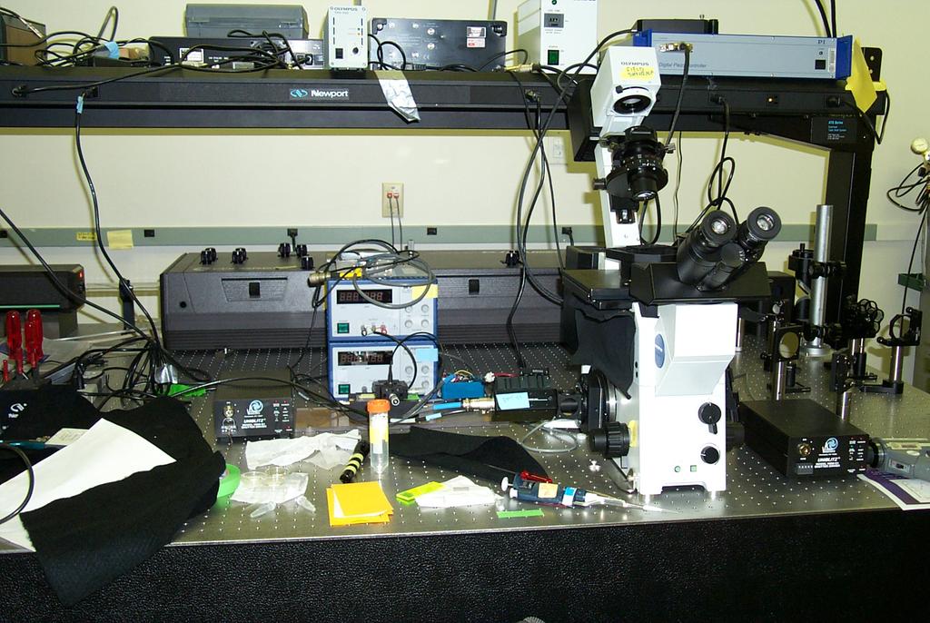
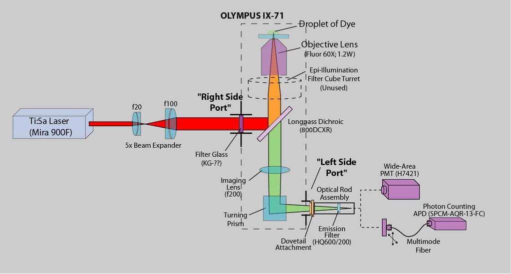
27 Experimental Apparatus

28 Analysis of Fluorescence Fluctuations G( τ ) = δf( t) δf( t + τ ) F( t) 2 Temporal analysis of spontaneous fluorescence fluctuations -δf- Signal fluctuations are induced by: Diffusion of molecules through the open measurement volume Intramolecular dynamics which affect the fluorescence emission
29 Parameters Provided by Fluorescence Correlation Spectroscopy (FCS)
 and its mutants: many of")

30 Examples for fast internal dynamics: flickering Molecules under study: GFP Green Fluorescent Protein) and its mutants: many of them show ph-dependent emission exc. 475nm pk= ph λ[nm]

31 FCS measurements of GFP-a ph sensor GFP blinks on a single molecule scale. Fast dynamics are strongly ph dependent reversible protonation dark fraction ph time constant τ [μs] k f R O + H + R OH k b Protonation Diffusion A λ abs,deprot = 488nm λ abs,prot = 400 nm pk-5.7 AH GFP can be employed as single molecule ph meter!
32 What to determine by diffusion analysis? D = kt 6πη R h Analysis of molecular structure: diffusion properties depend on hydrodynamic radius 1.0 Correlation G(τ) τ 1 τ Y(t) E Analysis of association/dissociation processes by change in molecular mass 0.0 t [s] k ass =10 4 M -1 s -1 to 10 6 M -1 s -1
 membrane (IgE receptor) D=3*10-10 cm 2 /s G(τ) 0.")

33 Assessing molecular mobility in different cellular compartments buffer cytosol membrane (lipid) membrane (IgE receptor) D=3*10-10 cm 2 /s G(τ) 0.4 Requirement: specific labeling of regions of interest Precision: 0.3 um in XY 1.0 um in Z fast 0.2 D=3*10-6 cm 2 /s 0.0 1E τ [ms] slow Determination of molecular speed
34 Detection of single molecules in membranes + 2 μm + 1 μm ±0 μm -1 μm -2 μm time [ms] time [s] Only labeled regions contribute to the measured signal
35 Dual-color cross-correlation analysis FCCS Advantage: mobility independent analysis of molecular interactions D = kt 6πη R h RECALL R h ~ (MW) 1/3 Correlation G(τ) E Principle: only doubly labeled species contributes to cross-correlation signal Red channel: Blue channel: + + cross-c. G ij ( τ ) = δf() t δf ( t+ τ ) i F() t F () t i j j Denominator Numerator
 G(τ) 0.03 0.02 0.01 Auto Red Auto Green Cross Fit 0.00 1E-3 0.01 0.")
36 Experimental setup for TPCCS Inherent overlap of excitation volumes Simplified alignment of detection volumes (no pinholes required) G(τ) Auto Red Auto Green Cross Fit E τ [ms]
 (4) (1) (2)")
37 Dual-color two-photon cross-correlation (TPCCS) Concept: Excitation of spectrally separable fluorophores with a single IR line Requirement: both dyes show similar emission on a single molecule scale emission η [khz/molecule] Ideal: 830 nm, < 30mW Rhodamine Green Texas Red λ [nm] % transmission/emission (3) (4) (1) (2) (5) Emission bei λ ex =830 nm λ [nm]
38 Analysis of DNA-DNA association G RB ( τ ) = RB BR [ V ( C + C )( C + C )] eff R C Diff RB B RB t 1 t 2 t 3 GCCGTCTCTGACTGCTGATGACTACTATCGTATAGTGCGG CGGCAGAGACTGACGACTACTGATGATAGCATATCACGCC Greater specificity for reaction product observation

39 Intracellular FCCS applications: The toxin system CTX-Cholera ST-Shigella Bacia et al., 2002, Biophy. J. System: Bacterial protein toxins entering the cell in a retrograde fashion Objective: to simultaneously study the endocytic trafficking of Cholera (red label) and Shiga (green label) Toxin
40 Comparing Endocytic Pathways for CTX and ST G(τ) G(τ) ST CTX Distinct autocorrelations, no cross-correlation: different pathways 0.0 1E τ[ms] 0.0 1E τ[ms] CTX-Cholera ST-Shigella G(τ) G(τ) CTX CTX Comparable autocorrelations, existing crosscorrelation: same pathway E τ / ms E τ / ms
 Cy-3 G(τ) (1) Cell membrane (2) τ[ms]")
![G(τ) (3) Golgi τ[ms] Cy-5 (3) Dissociation](/docs-images/77/75204369/images/41-1.jpg "of A and B 5 subunits required to induce")

41 FCCS reveals where the subunits dissociate (1) Cy-3 G(τ) (1) Cell membrane (2) τ[ms] G(τ) (3) Golgi τ[ms] Cy-5 (3) Dissociation of A and B 5 subunits required to induce toxicity of A (2) Endosomes Cross-correlation finally decays to zero in the Golgi G(τ) τ[ms]
42 Other Applications of MPE Uncaging of fluorescent compounds: inherent spatial localization provides excellent spatial selectivity for uncaging In vivo imaging over long time scales (months) deep in living tissue
Simultaneous multi-color, multiphoton fluorophore excitation using dual-color fiber lasers
 Multiphoton Microscopy / Fiber Laser Simultaneous multi-color, multiphoton fluorophore excitation using dual-color fiber lasers Matthias Handloser, Tim Paasch-Colberg, Bernhard Wolfring TOPTICA Photonics
Confocal Microscopy of Electronic Devices. James Saczuk. Consumer Optical Electronics EE594 02/22/2000
 Confocal Microscopy of Electronic Devices James Saczuk Consumer Optical Electronics EE594 02/22/2000 Introduction! Review of confocal principles! Why is CM used to examine electronics?! Several methods
Resolution of Microscopes Visible light is nm Dry lens(0.5na), green(530nm light)=0.65µm=650nm for oil lens (1.4NA) UV light (300nm) = 0.13µm f
, green(530nm light)=0.65µm=650nm for oil lens (1.4NA) UV light (300nm) = 0.13µm f") Microscopes and Microscopy MCB 380 Good information sources: Alberts-Molecular Biology of the Cell http://micro.magnet.fsu.edu/primer/ http://www.microscopyu.com/ Approaches to Problems in Cell Biology
Microscopes and Microscopy MCB 380 Good information sources: Alberts-Molecular Biology of the Cell http://micro.magnet.fsu.edu/primer/ http://www.microscopyu.com/ Approaches to Problems in Cell Biology
Introduction to Fluorescence Jablonski Diagram
 ntroduction to Fluorescence Jablonski Diagram Excited Singlet Manifold S1 internal conversion S2 k -isc k isc Excited riplet Manifold 1 S0 k nr k k' f nr fluorescence k p phosphorescence Singlet round
Genetically targeted all-optical electrophysiology with a transgenic Credependent
 Genetically targeted all-optical electrophysiology with a transgenic Credependent Optopatch mouse Short title: Transgenic Optopatch mouse Shan Lou 1, Yoav Adam 1, Eli N. Weinstein 1,4, Erika Williams 2,
Flow Cytometry - The Essentials
 Flow Cytometry - The Essentials Pocket Guide to Flow Cytometry: 1. Know your Cytometer 2. Understanding Fluorescence and Fluorophores 3. Gating Process 4. Controls 5. Optimization 6. Panel Building 7.
Practical light microscopy: an introduction
 Practical light microscopy: an introduction Dr. Mark Leake, Oxford University www.physics.ox.ac.uk/users/leake Aim of today s talk: Explanation of the very (very) basics of how a light microscope works
Monitoring and Optimizing the Lipopolysaccharides-plasmid DNA interaction by FLIM-FRET
 Transactions on Science and Technology Vol. 4, No. 3-3, 342-347, 2017 Monitoring and Optimizing the Lipopolysaccharides-plasmid DNA interaction by FLIM-FRET Nur Syahadatain Abdul Razak 1#, Clarence M.
Confocal Microscopy Analyzes Cells
 Choosing Filters for Fluorescence A Laurin Publication Photonic Solutions for Biotechnology and Medicine November 2002 Confocal Microscopy Analyzes Cells Reprinted from the November 2002 issue of Biophotonics
Challenges to measuring intracellular Ca 2+ Calmodulin: nature s Ca 2+ sensor
 Calcium Signals in Biological Systems Lecture 3 (2/9/0) Measuring intracellular Ca 2+ signals II: Genetically encoded Ca 2+ sensors Henry M. Colecraft, Ph.D. Challenges to measuring intracellular Ca 2+
Confocal Microscopes. Evolution of Imaging
 Confocal Microscopes and Evolution of Imaging Judi Reilly Hans Richter Massachusetts Institute of Technology Environment, Health & Safety Office Radiation Protection What is Confocal? Pinhole diaphragm
Time-resolved Measurements Using the Agilent Cary Eclipse Fluorescence Spectrophotometer A Versatile Instrument for Accurate Measurements
 Time-resolved Measurements Using the Agilent Cary Eclipse Fluorescence Spectrophotometer A Versatile Instrument for Accurate Measurements Technical Overview Authors Dr. Fabian Zieschang, Katherine MacNamara,
Nodes of regulation in cellular systems
 Nodes of regulation in cellular systems cell membrane signal transduction ligands receptors oligomerization transport signal transduction modified protein Golgi transcription factor transport ER transport
Multiphoton Microscopy: Seeing deeper and clearer
 Multiphoton Microscopy: Seeing deeper and clearer Since the invention of simple microscope by Leuwenhoek and Hooke in the 17th century, different types of light microscopy techniques (such as phase contrast,
Direct visualization, sizing and concentration measurement of fluorescently labeled nanoparticles using NTA
 Direct visualization, sizing and concentration measurement of fluorescently labeled nanoparticles using NTA NANOSIGHT RANGE Visualize and Measure Nanoparticle Size and Concentration PARTICLE SIZE PARTICLE
TARGETED IMAGING. Maureen Chan and Ruwani Mahathantila
 TARGETED IMAGING Maureen Chan and Ruwani Mahathantila Overview 2 Introduction to fluorescent imaging Fluorescent agents Quantum Dots Physical properties How QDs work In Vivo QD imaging Future Video What
Con-focal and Multi-photon Microscope Experiment Fundamental. Qian Hu, Lab of Laser Scanning Confocal & Two-Photon Microscopy, ION, CAS
 Con-focal and Multi-photon Microscope Experiment Fundamental Qian Hu, Lab of Laser Scanning Confocal & Two-Photon Microscopy, ION, CAS 1. Light is Electromagnetic Wave ν = c / λ 2. Image of a Point Source
Welcome! openmicberkeley.wordpress.com. Open Berkeley
 Welcome! openmicberkeley.wordpress.com Agenda Jen Lee: Introduction to FRET Marla Feller: Using FRET sensors to look at time resolved measurements Becky Lamason: Using FRET to determine if a bacterial
Partha Roy
 Fluorescence microscopy http://micro.magnet.fsu.edu/primer/index.html Partha Roy 1 Lecture Outline Definition of fluorescence Common fluorescent reagents Construction ti of a fluorescence microscope Optical
Performance of the Micro Photon Devices PDM 50CT SPAD detector with PicoQuant TCSPC systems
 Technical Note Performance of the Micro Photon Devices PDM 5CT SPAD detector with PicoQuant TCSPC systems Rolf Krahl, Andreas Bülter, Felix Koberling, PicoQuant GmbH These measurements were performed to
Nanophotonics: principle and application. Khai Q. Le Lecture 11 Optical biosensors
 Nanophotonics: principle and application Khai Q. Le Lecture 11 Optical biosensors Outline Biosensors: Introduction Optical Biosensors Label-Free Biosensor: Ringresonator Theory Measurements: Bulk sensing
Imagerie et spectroscopie de fluorescence par excitation non radiative
 Imagerie et spectroscopie de fluorescence par excitation non radiative comment s affranchir de la limite de diffraction Rodolphe Jaffiol, Cyrille Vézy, Marcelina Cardoso Dos Santos LNIO, UTT, Troyes NanoBioPhotonics
Measurement of surface concentration of fluorophores using. fluorescence fluctuation spectroscopy
 Measurement of surface concentration of fluorophores using fluorescence fluctuation spectroscopy A. Delon 1, J. Derouard 1, G. Delapierre and R. Jaffiol 1 (1) Laboratoire de Spectrométrie Physique (UMR
Tracking Cellular Protein Localization and Movement in Cells with a Flexible Fluorescent Labeling Technology. Chad Zimprich January 2015
 Tracking Cellular Protein Localization and Movement in Cells with a Flexible Fluorescent Labeling Technology Chad Zimprich January 2015 Presentation verview HaloTag Fusion Technology Design Functionality
SUPPLEMENTARY FIGURES
 SYNERGISTIC STRATEGY FOR MULTICOLOR TWO-PHOTON MICROSCOPY: APPLICATION TO THE ANALYSIS OF GERMINAL CENTER REACTIONS IN VIVO ASYLKHAN RAKHYMZHAN, RUTH LEBEN, HANNA ZIMMERMANN, ROBERT GÜNTHER, PEGGY MEX,
Fluorescence Measurements of Duplex DNA Oligomers under Conditions Conducive for Forming M-DNA (a Metal-DNA Complex)
") 10040 J. Phys. Chem. B 2007, 111, 10040-10052 Fluorescence Measurements of Duplex DNA Oligomers under Conditions Conducive for Forming M-DNA (a Metal-DNA Complex) Bryan Q. Spring and Robert M. Clegg*,,
10040 J. Phys. Chem. B 2007, 111, 10040-10052 Fluorescence Measurements of Duplex DNA Oligomers under Conditions Conducive for Forming M-DNA (a Metal-DNA Complex) Bryan Q. Spring and Robert M. Clegg*,,
Imaging & analysis with the LSM780 NLO Discover the secrets beyond the twilight zone
 Imaging & analysis with the LSM780 NLO Discover the secrets beyond the twilight zone Sven Terclavers LSM780 System overview The Scan Module - Core of the LSM 780 1 V/tunable PTC laser ports (405/440, cw/ps;
Towards Probing Skin Cancer using Endogenous Melanin Fluorescence
 8 Towards Probing Skin Cancer using Endogenous Melanin Fluorescence Andra Colbert, McNair Scholar, Virginia State University Faculty Research Advisor: Dr. Ahmed A. Heikal Associate Professor of Bioengineering
FLUORESCENT PEPTIDES. Outstanding Performance and Wide Application Range
 FLUORESCENT PEPTIDES Peptides and amino acids labeled with and Tide Quencher TM We offer peptides and amino acids tagged with fluorescent dyes. They meet highest demands in fluorescence intensity and photo-stability,
ADVANCED PRACTICAL COURSE IN BIOPHYSICS: FRET
 : FRET 1 INTRODUCTION Fluorescence spectroscopy and fluorescence microscopy are essential tools in biology. Biological molecules can be labeled with fluorescent molecules and thus, their localization and
Lighting research Toulouse team (France) Ludovic VANQUIN Ikbal MARGHAD Lydie AREXIS BOISSON
 Ludovic VANQUIN Ikbal MARGHAD Lydie AREXIS BOISSON") 1 Lighting research Toulouse team (France) Ludovic VANQUIN Ikbal MARGHAD Lydie AREXIS BOISSON Plan 2 INTRODUCTION I. Medical Imaging for the diagnosis of the Alzheimer s disease (Ludovic) II.Lighting display:
1 Lighting research Toulouse team (France) Ludovic VANQUIN Ikbal MARGHAD Lydie AREXIS BOISSON Plan 2 INTRODUCTION I. Medical Imaging for the diagnosis of the Alzheimer s disease (Ludovic) II.Lighting display:
Fs- Using Ultrafast Lasers to Add New Functionality to Glass
 An IMI Video Reproduction of Invited Lectures from the 17th University Glass Conference Fs- Using Ultrafast Lasers to Add New Functionality to Glass Denise M. Krol University of California, Davis 17th
Quantum Dot applications in Fluorescence Imaging for Calibration and Molecular Imaging
 Quantum Dot applications in Fluorescence Imaging for Calibration and Molecular Imaging Introduction In this application note, we will discuss the application of quantum dots in fluorescence imaging, both
Innovations To Meet Your Needs
 Innovations To Meet Your Needs Cooled CCD Camera 1340 x 1037 pixel resolution for greatest image quality 12-bit precision provides 3 orders of linear dynamic range Windows and Power Macintosh Software
CENTER FOR BRAIN EXPERIMENT
 CENTER FOR BRAIN EXPERIMENT Section of Brain Structure Associate Professor: ARII, Tatsuo, PhD 1967 Graduated from Tohoku University, Faculty of Science. Completed the doctoral course in Engineering, Nagoya
The analysis of fluorescence microscopy images for FRET detection
 The analysis of fluorescence microscopy images for FRET detection Ela Claridge, Dale J. Powner and Michael J.O. Wakelam School of Computer Science, The University of Birmingham B5 2TT Institute for Cancer
A simple introduction to multiphoton microscopy
 Journal of Microscopy, Vol. 243, Pt 3 2011, pp. 221 226 Received 29 April 2011; accepted 28 June 2011 doi: 10.1111/j.1365-2818.2011.03532.x A simple introduction to multiphoton microscopy A. USTIONE &
QImaging Camera Application Notes Multicolor Immunofluorescence Imaging
 QImaging Camera Application Notes Multicolor Immunofluorescence Imaging In order to image localization of intracellular proteins with high specificity, it is frequently necessary to multiplex antibody
SIL-based confocal fluorescence microscope for investigating individual nanostructures
 Cent. Eur. J. Phys. 9(2) 2011 293-299 DOI: 10.2478/s11534-010-0098-5 Central European Journal of Physics SIL-based confocal fluorescence microscope for investigating individual nanostructures Research
CBI Toolbox Tour 2015
 CBI Toolbox Tour 2015 Thermophoresis (NanoTemper) NT.115 & NT.LabelFree Images: NanoTemper Circular Dichroism Jasco J-1500 Spectrometer Six Position Turreted Peltier Temperature Control System Automated
Application of Quantum Mechanics to Biology
 Application of Quantum Mechanics to Biology How can we apply quantum mechanics to biology? Polymers of nucleotides and amino acids - millions of atoms bounded into a large molecule Visual System Must turn
Super-resolution Microscopy
 Semr oc kwhi t epaperser i es : 1. Introduction Super-resolution Microscopy Fluorescence microscopy has revolutionized the study of biological samples. Ever since the invention of fluorescence microscopy
Hard Coated Silica/Silica (Low OH) Radius
 Radius") DESCRIPTION When looking for a silica core and silica clad fiber with a hard polymer coating that allows a high core-to-clad ratio and a numerical aperture (N.A.) of 0.22 for efficient light coupling,
DESCRIPTION When looking for a silica core and silica clad fiber with a hard polymer coating that allows a high core-to-clad ratio and a numerical aperture (N.A.) of 0.22 for efficient light coupling,
Analysis of receptor oligomerization by FRAP microscopy
 TIGP CBMB Student Seminar Analysis of receptor oligomerization by FRAP microscopy Dorsch S, Klotz KN, Engelhardt S, Lohse MJ, Bünemann M Nat Methods. 2009 Mar;6(3):225 30. K. Vijayasarathy March 10 th
TWO-PHOTON EXCITATION FLUORESCENCE MICROSCOPY
 TWO-PHOTON EXCITATION FLUORESCENCE MICROSCOPY PeterT.C.So 1,ChenY.Dong 1, Barry R. Masters 2, and Keith M. Berland 3 1 Department of Mechanical Engineering, Massachusetts Institute of Technology, Cambridge,
Fluorescence Nanoscopy
 Fluorescence Nanoscopy Keith A. Lidke University of New Mexico panda3.phys.unm.edu/~klidke/index.html Optical Microscopy http://en.wikipedia.org/wiki/k%c3%b6hler_illumination 30 µm Fluorescent Probes Michalet
Flow Cytometry. Flow Cytometry Basics Guide
 Flow Cytometry Flow Cytometry Basics Guide Table of Contents Chapter 1 Chapter 2 Chapter 3 Chapter 4 Chapter 5 Principles of the Flow Cytometer Fluidics System.... 3 Optics and Detection.... 4 Signal and
Supporting Information for Matching nanoantenna field confinement to FRET distances enhances Förster energy transfer rates
 Supporting Information for Matching nanoantenna field confinement to FRET distances enhances Förster energy transfer rates Petru Ghenuche, Mathieu Mivelle, Juan de Torres, Satish Babu Moparthi, Hervé Rigneault,
CF Dyes Next Generation Fluorescent Dyes Secondary antibody
 CF Dyes Next Generation Fluorescent Dyes Secondary antibody OZYME 10 AVENUE AMPÈRE - CS 30268-78053 ST QUENTIN EN YVELINES CEDEX Tél. : 01 34 60 24 24 - Fax : 01 34 60 92 12 - www.ozyme.fr/info CF Dyes
Advantages of Photodiode Array
 Advantages of Photodiode Array Hun Choi SCINCO. Co., Ltd. 74-27 Nonhyun-dong, Kangnam-ku, Seoul, Korea, 135-010 Preface All products are continuously being improved for better performance. Spectrometers
SUPPLEMENTARY MATERIAL
 SUPPLEMENTARY MATERIAL Materials and Methods Circular dichroism (CD) spectroscopy. Far ultraviolet (UV) CD spectra of apo- and holo- CaM and the CaM mutants were recorded on a Jasco J-715 spectropolarimeter
Plasmonics: Application-oriented fabrication. Part 1. Introduction
 Plasmonics: Application-oriented fabrication Part 1. Introduction Victor Ovchinnikov Department of Aalto Nanofab Aalto University Espoo, Finland Alvar Aalto was a famous Finnish architect and designer
Masayoshi Honda, Jeehae Park, Robert A. Pugh, Taekjip Ha, and Maria Spies
 Molecular Cell, Volume 35 Supplemental Data Single-Molecule Analysis Reveals Differential Effect of ssdna-binding Proteins on DNA Translocation by XPD Helicase Masayoshi Honda, Jeehae Park, Robert A. Pugh,
Non-linear Optical Microscopy and Spectroscopy for Biomedical Studies
 Doctoral Thesis Non-linear Optical Microscopy and Spectroscopy for Biomedical Studies Stina Guldbrand Department of Physics University of Gothenburg SE-412 96 Göteborg, Sweden 2012 Non-linear Optical Microscopy
SAPIENZA Università di Roma Laurea magistrale in Ingegneria delle Nanotecnologie A.A Biophotonics Laboratory Course
 SAPIENZA Università di Roma Laurea magistrale in Ingegneria delle Nanotecnologie A.A. 2016-2017 Biophotonics Laboratory Course Prof. Francesco Michelotti SAPIENZA Università di Roma Facoltà di Ingegneria
Picosecond Transient Absorption Spectroscopy System. picotas
 Picosecond Transient Absorption Spectroscopy System Can Easily Measure Short-Lived Intermediates. In most of light induced phenomena, intermediates (transient species) play important roles to determine
The mechanism(s) of protein folding. What is meant by mechanism. Experimental approaches
 of protein folding. What is meant by mechanism. Experimental approaches") The mechanism(s) of protein folding What is meant by mechanism Computational approaches Experimental approaches Questions: What events occur and in what time sequence when a protein folds Is there a specified
The mechanism(s) of protein folding What is meant by mechanism Computational approaches Experimental approaches Questions: What events occur and in what time sequence when a protein folds Is there a specified
Lecture 13: Analysis of 2D gels
 Lecture 13: Analysis of 2D gels A complete proteomic analysis aims at collecting quantitative information about all protein in a sample. A normal 2 Dimensional gel electrophoresis results are analyzed
A Survey of Laser Types. Gas Lasers
 Mihail Pivtoraiko Andrei Rozhkov Applied Optics Winter 2003 A Survey of Laser Types Laser technology is available to us since 1960 s, and since then has been quite well developed. Currently, there is a
Chapter 10: Classification of Microorganisms
 Chapter 10: Classification of Microorganisms 1. The Taxonomic Hierarchy 2. Methods of Identification 1. The Taxonomic Hierarchy Phylogenetic Tree of the 3 Domains Taxonomic Hierarchy 8 successive taxa
Optical Fiber Sensors for Biomedical Applications
 Optical Fiber Sensors for Biomedical Applications Xingwei (Vivian) Wang, Ph.D. Assistant Professor Department of Electrical and Computer Engineering University of Massachusetts Lowell Phone: (978) 934-1981
Immunostaining Protocols
 Immunostaining Protocols Lula L. Hilenski, Ph.D. Director Microscopy in Medicine Core Emory University Variables in standard immunostaining protocol 2-step or indirect immunofluorescence 1. Substrate on
Post-expansion antibody delivery, after epitope-preserving homogenization.
 Supplementary Figure 1 Post-expansion antibody delivery, after epitope-preserving homogenization. (a, b) Wide-field fluorescence images of Thy1-YFP-expressing mouse brain hemisphere slice before expansion
Assembly of synapses by neuronal adhesion molecules: single molecule studies
 Assembly of synapses by neuronal adhesion molecules: single molecule studies Olivier Thoumine Interdisciplinary Institute of Neurosciences CNRS - University of Bordeaux Connectivity in the brain 300 nm
Introduction to N-STORM
 Introduction to N-STORM Dan Metcalf Advanced Imaging Manager Outline Introduction Principles of STORM Applications N-STORM overview Biological Scale Mitochondrion Microtubule Amino Acid 1Å Kinesin 1nm
Lab module 6b Receptor-mediated endocytosis
 Goal for the module Lab module 6b Receptor-mediated endocytosis To follow the movement of a degraded ligand, LDL, and a recycled ligand, transferrin, as they undergo endocytic processing. Pre-lab homework
Boundary-breaking acoustic focusing cytometry
 Boundary-breaking acoustic focusing cytometry Introducing the Attune NxT Acoustic Focusing Cytometer a high-performance system that s flexible enough for any lab One of the main projects in my laboratory
Applicability of Hyperspectral Fluorescence Imaging to Mineral Sorting
 Institute of Industrial Information Technology Applicability of Hyperspectral Fluorescence Imaging to Mineral Sorting Optical Characterization of Materials, March 19, 2015 Sebastian Bauer, M.Sc. (Head:
PHT1;2-CFP YFP-PHF + PHT1;2-CFP YFP-PHF
 YFP-PHF1 CFP-PHT1;2 PHT1;2-CFP YFP-PHF + PHT1;2-CFP YFP-PHF + CFP-PHT1;2 Negative control!-gfp Supplemental Figure 1: PHT1;2 accumulation is PHF1 dependent. Immunoblot analysis on total protein extract
Spectral Separation of Multifluorescence Labels with the LSM 510 META
 Microscopy from Carl Zeiss Spectral Separation of Multifluorescence Labels with the LSM 510 META Indians living in the South American rain forest can distinguish between almost 200 hues of green in their
Fundamentals and Applications of Biofilms Analysis, Structure and Physiology of Bacterial Biofilms Ching-Tsan Huang ( 黃慶璨 ) Office: Agronomy
 Office: Agronomy") 1 Fundamentals and Applications of Biofilms Analysis, Structure and Physiology of Bacterial Biofilms Ching-Tsan Huang ( 黃慶璨 ) Office: Agronomy Building, Room 111 Tel: (02) 33664454 E-mail: cthuang@ntu.edu.tw
1 Fundamentals and Applications of Biofilms Analysis, Structure and Physiology of Bacterial Biofilms Ching-Tsan Huang ( 黃慶璨 ) Office: Agronomy Building, Room 111 Tel: (02) 33664454 E-mail: cthuang@ntu.edu.tw
Next Level of Super Resolution Fluorescence Microscopy
 Work in your familiar GFP/YFP/RFP system from the first experiment to the nanoimage Bwcon business award winner: Inventor Prof Christoph Cremer Next Level of Super Resolution Fluorescence Microscopy Resolution:
Cell Imaging. Localization: Robust protein labeling of live or fixed cells
 F eat u r es & B e n efits ptions Cell Imaging with the alotag platform Localization: Robust protein labeling of live or fixed cells Trafficking & Turnover: Directly observe proteins with one or two colors
Exchange Kinetics and Activity analysis of GTP-Binding Proteins with Fluorescent GTP-Analogs
 Exchange Kinetics and Activity analysis of GTP-Binding Proteins with Fluorescent GTP-Analogs Chandandeep Kaur, Ahmed A. Heikal Department of Biomedical Engineering, University of California, Irvine CA92617;
AURORA AIRY BEAM LIGHT SHEET IMAGING SYSTEM THE CUSTOM DEVELOPMENT PROGRAMME
 AURORA AIRY BEAM LIGHT SHEET IMAGING SYSTEM THE CUSTOM DEVELOPMENT PROGRAMME The Custom Development Programme Collaboration breeds innovation Our aim at M Squared Life, a new Biophotonics division within
Transferrin Conjugates
 Transferrin Conjugates Table 1 Contents and storage Material Formulation Storage Stability Transferrin conjugates* lyophilized powder containing transferrin conjugate, lyophilized in phosphate-buffered
High-throughput three-dimensional (3D) lithographic microfabrication in biomedical applications
 lithographic microfabrication in biomedical applications") High-throughput three-dimensional (3D) lithographic microfabrication in biomedical applications The MIT Faculty has made this article openly available. Please share how this access benefits you. Your story
High-throughput three-dimensional (3D) lithographic microfabrication in biomedical applications The MIT Faculty has made this article openly available. Please share how this access benefits you. Your story
nanodsf 2bind: Your service provider for biophysical characterization of proteins Precisely revealing protein folding and stability
 nanodsf Precisely revealing protein folding and stability 2bind: Your service provider for biophysical characterization of proteins This booklet was written and designed by 2bind 08 2015 Any reproduction
Satoshi Kawata. Near-Field Optic s and Surface Plasmon Polaritons
 Satoshi Kawata Near-Field Optic s and Surface Plasmon Polaritons Near-Field Optics and the Surface Plasmon Polariton Dieter W. Pohl 1 1. Introduction 1 2. Back to the Roots 1 2.1. Rayleigh and Mie Scattering
Cationic Vector Intercalation into the Lipid Membrane Enables Intact Polyplex DNA Escape from Endosomes for Gene Delivery
 Cationic Vector Intercalation into the Lipid Membrane Enables Intact Polyplex DNA Escape from Endosomes for Gene Delivery Sriram Vaidyanathan, 1 Junjie Chen, 2 Bradford G. Orr, 3 Mark M. Banaszak Holl
How to perform-control immunostaining experiment - microscopist subjective point of view. Pawel Pasierbek
 How to perform-control immunostaining experiment - microscopist subjective point of view. Pawel Pasierbek Immunolabeling and fluorescent detection became such a standard procedure in the biomedical research
Two-photon excitation (TPE) microscopy 1 has evolved as. Two-Photon Microscopy of Cells and Tissue. Michael Rubart
 microscopy 1 has evolved as. Two-Photon Microscopy of Cells and Tissue. Michael Rubart") This Review is part of a thematic series on Imaging of Cardiovascular Cells and Tissues, which includes the following articles: Use of Chimeric Fluorescent Proteins and Fluorescence Resonance Energy Transfer
This Review is part of a thematic series on Imaging of Cardiovascular Cells and Tissues, which includes the following articles: Use of Chimeric Fluorescent Proteins and Fluorescence Resonance Energy Transfer
Bioinstrumentation Light Sources Lasers or LEDs?
 Bioinstrumentation Light Sources Lasers or LEDs? A comprehensive analysis of all the factors involved in designing and building life sciences instrumentation reveals that lasers provide superior performance
Double-pulse fluorescence lifetime measurements
 Journal of Microscopy, Vol. 186, Pt 3, June 1997, pp. 212 220. Received 15 August 1996; accepted 14 January 1997 Double-pulse fluorescence lifetime measurements A. H. BUIST,* M. MÜLLER,* E. J. GIJSBERS,*
Chapter 4 Fluorescence Resonance Energy Transfer (FRET) by Minor Groove-Associated Cyanine-Polyamide Conjugates
 by Minor Groove-Associated Cyanine-Polyamide Conjugates") Chapter 4 Fluorescence Resonance Energy Transfer (FRET) by Minor Groove-Associated Cyanine-Polyamide Conjugates The work described in this chapter was accomplished in collaboration with V. Rucker (Dervan
Chapter 4 Fluorescence Resonance Energy Transfer (FRET) by Minor Groove-Associated Cyanine-Polyamide Conjugates The work described in this chapter was accomplished in collaboration with V. Rucker (Dervan
Plastic Coated Silica/Silica (Low OH) FIBER CROSS SECTION Polyimide and Acrylate Coated. Nylon and Tefzel Coated
 FIBER CROSS SECTION Polyimide and Acrylate Coated. Nylon and Tefzel Coated") DESCRIPTION When looking for a high quality fiber with superior transmission and a numerical aperture (N.A.) of 0.22 for efficient light coupling, the is the fiber of choice. The Anhydroguide fiber is
DESCRIPTION When looking for a high quality fiber with superior transmission and a numerical aperture (N.A.) of 0.22 for efficient light coupling, the is the fiber of choice. The Anhydroguide fiber is
SI8000 Live Cell Imaging System
 SI8000 Live Cell Imaging System Sony Biotechnology Inc. SI8000 Cell Motion Imaging System The Sony SI8000 Live Cell Imaging System detects and quantifies cellular motion using proprietary video processing
Investigating Fluorescence Lifetime Spectroscopy & Imaging. David Birch Department of Physics University of Strathclyde Glasgow G4 0NG Scotland
 Investigating Fluorescence Lifetime Spectroscopy & Imaging David Birch Department of Physics University of Strathclyde Glasgow G4 0NG Scotland Content Fluorescence lifetime and its measurement Use in spectroscopy
EdU Flow Cytometry Kit. User Manual
 User Manual Ordering information: (for detailed kit content see Table 2) EdU Flow Cytometry Kits for 50 assays: Product number EdU Used fluorescent dye BCK-FC488-50 10 mg 6-FAM Azide BCK-FC555-50 10 mg
Identification of red and white blood cells from whole blood samples using the Agilent 2100 bioanalyzer. Application Note
 Identification of red and white blood cells from whole blood samples using the Agilent 2100 bioanalyzer Application Note Sylvie Veriac Valérie Perrone Madeleine Avon Abstract Agilent Equipment: 2100 bioanalyzer
Digitally Programmed Cells
 Digitally Programmed Cells Ron Weiss PI: Tom Knight MIT Artificial Intelligence Laboratory Goal Process-Control Cellular Computers -- Microbial Robotics Unique features: small, self-replicating, energy-efficient
LOW TEMPERATURE PHOTONIC SINTERING FOR PRINTED ELECTRONICS. Dr. Saad Ahmed XENON Corporation November 19, 2015
 LOW TEMPERATURE PHOTONIC SINTERING FOR PRINTED ELECTRONICS Dr. Saad Ahmed XENON Corporation November 19, 2015 Topics Introduction to Pulsed Light Photonic sintering for Printed Electronics R&D Tools for
LUPAS Luminescent Polymers for in vivo Imaging of Amyloid Signatures
 LUPAS Luminescent Polymers for in vivo Imaging of Amyloid Signatures A research project for innovative diagnostics for neurodegenerative disorders Funded by the European Union under the 7 th Framework
Experiment 2b X-Ray Diffraction* Optical Diffraction Experiments
 * Experiment 2b X-Ray Diffraction* Adapted from Teaching General Chemistry: A Materials Science Companion by A. B. Ellis et al.: ACS, Washington, DC (1993). Introduction Inorganic chemists, physicists,
AFM-Raman Characterization of Pharmaceutical Tablets
 AFM-Raman Characterization of Pharmaceutical Tablets Compound Distribution Studies in Pharmaceutical Tablets by Integrated AFM-Raman Instrument 1,2 1 Sergey Shashkov and Pavel Dorozhkin, 1 NT-MDT Co.,
Supplementary Information and Figures
 Supplementary Information and Figures Multicolor two-photon imaging of endogenous fluorophores in living tissues by wavelength mixing Chiara Stringari 1, Lamiae Abdeladim 1, Guy Malkinson 1, Pierre Mahou
A quantitative protocol for intensity-based live cell FRET imaging.
 A quantitative protocol for intensity-based live cell FRET imaging. Kaminski CF, Rees EJ, Schierle GS. Methods Mol Biol. 2014; 1076:445-454. Department of Chemical Engineering and Biotechnology, Pembroke
Measurement of Molecular Diffusion in Solution by Multiphoton Fluorescence Photobleaching Recovery
 Biophysical Journal Volume 77 November 999 2837 2849 2837 Measurement of Molecular Diffusion in Solution by Multiphoton Fluorescence Photobleaching Recovery Edward B. Brown,* En Shinn Wu, # Warren Zipfel,