Supplementary Fig. 5
|
|
|
- Brent Mark Johnston
- 6 years ago
- Views:
Transcription
1

2

3

4

5 Supplementary Fig. 5

6

7
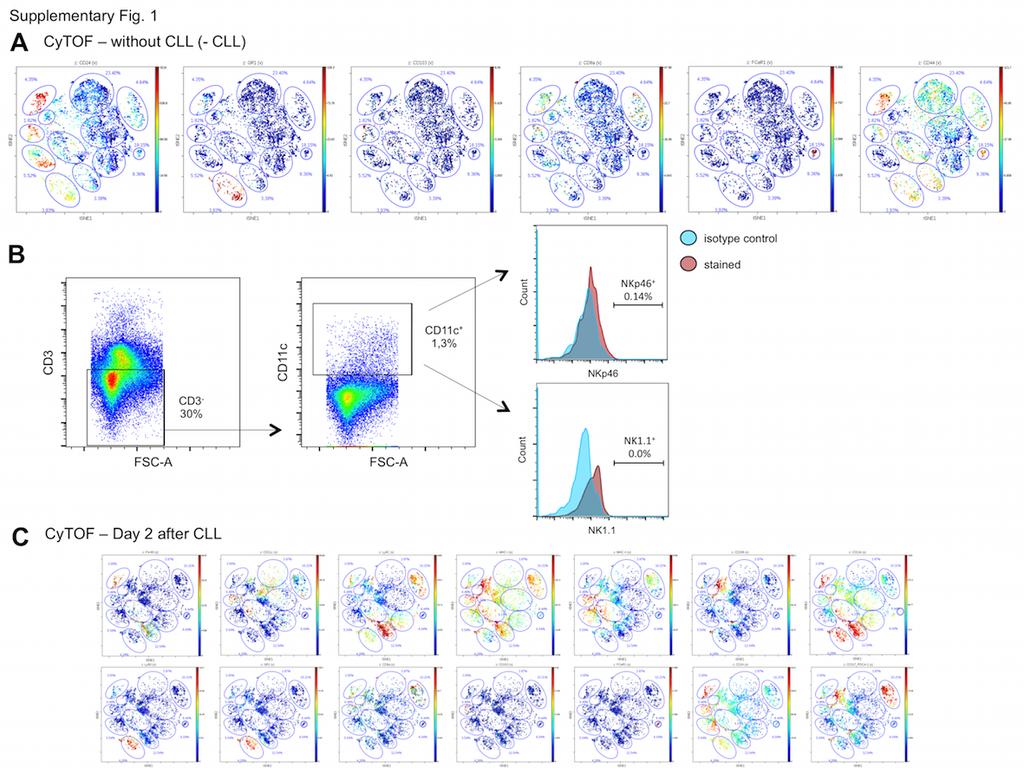
8 Supplemental Figures legends Supplementary Figure 1 (A) Additional dot plots from CyTOF analysis from untreated group. (B) Gating strategy for assessment of CD11c + NK cells frequency in liver non-parenchymal cells samples. CD11c + NK cells were defined as CD3 - CD11c + NKp46 + NK1.1 + events. (C) Additional dot plots from CyTOF analysis from mice 2 days after CLL treatment. Supplementary Figure 2 Identification of main hepatic vascular arrangements. Snapshots of chronological distribution of FITC-albumin within the liver (5mg/Kg). Portal spaces were identified as the first vascular region evidenced by fluorescence, and central vein were defined as the secondary draining vessels. Scale bars, 120 µm. Supplementary Figure 3 Expression of different cell surface markers in liver phagocytes. (A) Liver macrophage expression of lysozyme M (LysM-EGFP mice) and F4/80. (B) Same for A, but for CD11c (CD11c-EYFP mice). (C) Depletion of both intravascular CD11c + F4/80 + and extravascular CD11c + cells upon diphtheria toxin (DT) treatment in CD11c-DTR-EYFP mice. (D) Expression of EYFP and DTR under control of CX3CR1 in liver dendritic cells and selective depletion following diphtheria toxin administration. Note that only CX3CR1+ cells are depleted after DT treatment, with no effect on intravascular F4/80+ cells. Scale bars, 120 µm. Illustrative images from different experiments (N>5). Supplementary Figure 4 Liver dendritic cells are located in the subcapsular compartment and are CD11c + (A) Liver intravital microscopy of CX3CR1 gfp/wt mouse showing the distribution of CD11c + dendritic (anti-cd11) cells and
9 (B,C) confirmation of sub-mesothelial location of CX3CR1 + cells by intravital microscopy (B) immunohistochemistry (C). Illustrative images from different experiments (N>5). Supplementary Figure 5 Characterization of different cell surface markers and CCR2 role in phagocytes (A) Liver ex vivo transversal fragment showing that subcapsular neither CX3CR1 + nor CD11c + cells are positive for desmin (anti-desmin antibody), excluding this population as hepatic stellate cells. (B) Real-time PCR from isolated macrophages and dendritic cells showing that hepatic stellate cells (HSCs) did not contaminate samples. The following HSCs genes were measured: lecithin retinol acyltransferase (Lrat), desmin (Des) and alpha-actin-2 (Acta2). Data were normalized by a constitutive gene (Gapdh) and Itgax (CD11c) was used as positive control. (C) Liver intravital microscopy showing that neither CX3CR1 + cells nor F4/80 + have vitamin A granules (auto-fluorescence in 405nm laser). (D) Flow cytometry investigation of the absence of vitamin A in CX3CR1 + population (auto-fluorescence in 405nm laser). (E) CCR2 -/- mouse have normal KC density (anti-f4/80) and (F) location. Scale bars in A, 10 µm; B, 65µm and E, 120µm. Supplementary Figure 6 Additional dot plots from CyTOF analysis from mice 7 and 17 days after phagocyte depletion. Expression of cell surface markers in liver non-parenchymal cells clodronate depleted mice after 7 days (A) and after 17 days (B). CyTOF was performed and events were clustered as described in Methods and Results. Supplementary Figure 7 (A) Replenishment of liver phagocytes is driven by a bone marrow-derived precursor. Different focal planes imaged by intravital
10 confocal microscopy from GFP-expressing bone marrow chimeras showing that all Kupffer cells (F4/80 + GFP + cells in intravascular focus) and dendritic cells (GFP + cells in capsule focus) were derived from the bone marrow. (B) Acetaminophen (APAP) treatment caused a significant depletion of KCs (red; F4/80 + cells) and normal cell density and location were restored after days. In blue, DAPI stains for necrosis. Scale bars in KC focus, 35 µm, in DC focus, 20 µm and in APAP-treated group 120 µm. *P < 0.05 (unpaired t-test) in comparison to vehicle. Legends for Tables Table 1 Antibody panel for CyTOF and for genes for Real Time PCR. For PCR, cells were isolated and RNA was extracted with a mirneasy kit (Qiagen), then was reverse-transcribed with a high capacity cdna reverse transcription kit and analyzed by quantitative RT-PCR with a Vii 7 Real-time PCR system with Taqman primers. The following HSCs genes were measured: lecithin retinol acyltransferase (Lrat), desmin (Des) and alphaactin-2 (Acta2). Data were normalized by a constitutive gene (Gapdh) and Itgax (CD11c) was used as positive control. The comparative threshold cycle method and the internal control Gapdh was used for normalization of the target genes. Table 2 Absolute cell numbers (events) from time-of-flight flow cytometry (CyTOF) experiments. Liver samples from different groups were collected and processed for CyTOF. Events were normalized in all samples.
11 Table 3 Quantification of liver cytokine expression by multiplexed Luminex array. Liver samples from different groups were collected and processed for multiplexed cytokine array. Data are displayed as individual samples from different experiments. GEO accession number: GSE Gene expression assessed by Nanostring experiments. Intravascular CX3CR1 - F4/80 + and extravascular CX3CR1 + F4/80 - cells were isolated by sorting (FACS) and immune systemrelated genes expression was quantified using Nanostring. Statically relevant results consist in p-value < 0.05 and a fold change of at least 50% higher or lower. Pathways and functional classification were done by cross association using KEGG Pathways and KEGG Brite databases. Legends for Supplementary Movies Supplementary Movie 1 Identification of main hepatic vascular arrangements. Temporal distribution of FITC-albumin (green) within the liver (5mg/Kg). Portal spaces were identified as the first vascular region evidenced by fluorescence, and central vein were defined as the secondary draining vessels. Total experimenta time: 1 minute. Supplementary movie 2 Three-dimensional rendering of liver intravital imaging. Spatial distribution of Kupffer cells. Vessels are in blue (anti-pecam- 1 antibody) and KCs in red (anti-f4/80 antibody). Images were collect from a 3D section of µm of depth.
12 Supplementary movie 3 Three-dimensional rendering of liver intravital imaging. Liver dendritic cell (DCs) morphology and distribution in CD11c- EYFP mice. Vessels are in blue (anti-pecam-1 antibody) and DCs in yellow (EYFP). Images were collect from a 3D section of µm of depth. Supplementary movie 4 Three-dimensional rendering of liver intravital imaging. Liver dendritic cell morphology and distribution in CX3CR1-EGFP mice. Vessels are in blue (anti-pecam-1 antibody) and DCs in green (EGFP). Images were collect from a 3D section of µm of depth. Supplementary movie 5: KCs instantaneously trap circulating E. coli from the circulation. Merged video comparing E. coli arresting by a control KC (non-depleted mouse) versus an immature KC (7 days after CLL). Note that E. coli are arrested at the first passage through the liver, while immature KC are unable to proper catch and internalize bacteria. Kupffer cells (KCs) are in red and E. coli in green. Total video time: 10 minutes E.coli were injected in the beginning of the imaging procedure. Supplementary movie 6 Circulating E. coli capture by Kupffer cells. Merged video showing E. coli arresting by control group (non-depleted mouse) and after different timepoints of replenishment period (7, 17, 30 and 60 days). Kupffer cells (KCs) are in red and E. coli in green. Due to photobleaching issues, a still frame from KC channel was used as reference during the video. Total video time: 10 minutes each experimental group E.coli were injected in the beginning of the imaging procedure
13 Supplementary movie 7 E. coli are exclusively arrested by intravascular cells Three-dimensional rendering of liver confocal intravital microscopy showing GFP expressing E coli inside sinusoids. Sinusoids are in blue (anti-pecam-1 antibody) and E. coli gfp in green. Images were collect from a 3D section of µm of depth.
Nature Immunology: doi: /ni Supplementary Figure 1
 Supplementary Figure 1 BALB/c LYVE1-deficient mice exhibited reduced lymphatic trafficking of all DC subsets after oxazolone-induced sensitization. (a) Schematic overview of the mouse skin oxazolone contact
Nature Immunology: doi: /ni Supplementary Figure 1
 Supplementary Figure 1 PPAR-γ is dispensable for the development of tissue macrophages in the heart, kidneys, lamina propria and white adipose tissue. Plots show the expression of F4/80 and CD11b (a) or
Real-time PCR. Total RNA was isolated from purified splenic or LP macrophages using
 Supplementary Methods Real-time PCR. Total RNA was isolated from purified splenic or LP macrophages using the Qiagen RNeasy Mini Kit, according to the manufacturer s protocol with on-column DNase digestion
Mayumi Egawa, Kaori Mukai, Soichiro Yoshikawa, Misako Iki, Naofumi Mukaida, Yohei Kawano, Yoshiyuki Minegishi, and Hajime Karasuyama
 Immunity, Volume 38 Supplemental Information Inflammatory Monocytes Recruited to Allergic Skin Acquire an Anti-inflammatory M2 Phenotype via Basophil-Derived Interleukin-4 Mayumi Egawa, Kaori Mukai, Soichiro
Supporting Information
 Supporting Information Cieslewicz et al. 10.1073/pnas.1312197110 SI Results Human and mouse lesions of atherosclerosis contain both M1 and M2 macrophage phenotypes (1, 2). Previous work has suggested the
SUPPLEMENTARY FIGURES
 SYNERGISTIC STRATEGY FOR MULTICOLOR TWO-PHOTON MICROSCOPY: APPLICATION TO THE ANALYSIS OF GERMINAL CENTER REACTIONS IN VIVO ASYLKHAN RAKHYMZHAN, RUTH LEBEN, HANNA ZIMMERMANN, ROBERT GÜNTHER, PEGGY MEX,
Supplement Figure 1. Plin5 Plin2 Plin1. KDEL-DSRed. Plin-YFP. Merge
 Supplement Figure 1 Plin5 Plin2 Plin1 KDEL-DSRed Plin-YFP Merge Supplement Figure 2 A. Plin5-Ab MitoTracker Merge AML12 B. Plin5-YFP Cytochrome c-cfp merge Supplement Figure 3 Ad.GFP Ad.Plin5 Supplement
SUPPLEMENTARY INFORMATION
 a before amputation regeneration regenerated limb DERMIS SKELETON MUSCLE SCHWANN CELLS EPIDERMIS DERMIS SKELETON MUSCLE SCHWANN CELLS EPIDERMIS developmental origin: lateral plate mesoderm presomitic mesoderm
Supplementary Figure and Table Legends
 1 Supplementary Figure and Table Legends Figure S1: Whole-animal metabolic analysis. 12 week old WT and Dvl1 / were singly housed in CLAMS cages (Comprehensive Laboratory Animals Monitoring System) for
GFP CCD2 GFP IP:GFP
 D1 D2 1 75 95 148 178 492 GFP CCD1 CCD2 CCD2 GFP D1 D2 GFP D1 D2 Beclin 1 IB:GFP IP:GFP Supplementary Figure 1: Mapping domains required for binding to HEK293T cells are transfected with EGFP-tagged mutant
Cell culture and drug treatment. Lineage - Sca-1+ CD31+ EPCs were cultured on
 Supplemental Material Detailed Methods Cell culture and drug treatment. Lineage - Sca-1+ CD31+ EPCs were cultured on 5µg/mL human fibronectin coated plates in DMEM supplemented with 10% FBS and penicillin/streptomycin
Supplementary Figure 1
 Supplementary Figure 1 Supplementary Figure 1: Vector maps of TRMPV and TRMPVIR variants. Many derivatives of TRMPV have been generated and tested. Unless otherwise noted, experiments in this paper use
Supplemental Information Inventory
 Cell Stem Cell, Volume 6 Supplemental Information Distinct Hematopoietic Stem Cell Subtypes Are Differentially Regulated by TGF-β1 Grant A. Challen, Nathan C. Boles, Stuart M. Chambers, and Margaret A.
Supplementary Materials and Methods:
 Supplementary Materials and Methods: Preclinical chemoprevention experimental design Mice were weighed and each mammary tumor was manually palpated and measured with digital calipers once a week from 4
Supplemental Materials and Methods
 Supplemental Materials and Methods 125 I-CXCL12 binding assay KG1 cells (2 10 6 ) were preincubated on ice with cold CXCL12 (1.6µg/mL corresponding to 200nM), CXCL11 (1.66µg/mL corresponding to 200nM),
Supplementary Information
 Supplementary Information Live imaging reveals the dynamics and regulation of mitochondrial nucleoids during the cell cycle in Fucci2-HeLa cells Taeko Sasaki 1, Yoshikatsu Sato 2, Tetsuya Higashiyama 1,2,
Nature Methods: doi: /nmeth Supplementary Figure 1
 Supplementary Figure 1 Workflow for multimodal analysis using sc-gem on a programmable microfluidic device (Fluidigm). 1) Cells are captured and lysed, 2) RNA from lysed single cell is reverse-transcribed
Nature Medicine: doi: /nm.4464
 Supplementary Fig. 1. Amino acid transporters and substrates used for selectivity screening. (A) Common transporters and amino acid substrates shown. Amino acids designated by one-letter codes. Transporters
SUPPLEMENTARY INFORMATION
 SUPPLEMENTARY INFORMATION Legends for Supplementary Tables. Supplementary Table 1. An excel file containing primary screen data. Worksheet 1, Normalized quantification data from a duplicated screen: valid
TF-1a lymphoblastic leukemia cell line: marking with GFP, phenotyping and sorting
 Supplemental Material Supplemental Methods TF-1a lymphoblastic leukemia cell line: marking with GFP, phenotyping and sorting In order to determine if the multi-parameter FACS approach would be successful
Reagents and cell culture Mcl-1 gene expression: real-time quantitative RT-PCR In vitro PP2A phosphatase assay Detection of Mcl-1 in vivo
 Reagents and cell culture Antibodies specific for caspase 3, PARP and GAPDH were purchased from Cell Signaling Technology Inc. (Beverly, MA). Caspase inhibitor z-vad-fmk and ROS scavenger N-acetyl-Lcysteine
Nature Methods: doi: /nmeth Supplementary Figure 1. Retention of RNA with LabelX.
 Supplementary Figure 1 Retention of RNA with LabelX. (a) Epi-fluorescence image of single molecule FISH (smfish) against GAPDH on HeLa cells expanded without LabelX treatment. (b) Epi-fluorescence image
DEPArray Technology. Sorting and Recovery of Rare Cells
 DEPArray Technology Sorting and Recovery of Rare Cells Delivering pure, single, viable cells The DEPArray system from Silicon Biosystems is the only automated instrument that can identify, quantify, and
Phagocytosis Assay Kit (IgG PE)
") Phagocytosis Assay Kit (IgG PE) Item No. 600540 www.caymanchem.com Customer Service 800.364.9897 Technical Support 888.526.5351 1180 E. Ellsworth Rd Ann Arbor, MI USA TABLE OF CONTENTS GENERAL INFORMATION
Phagocytosis Assay Kit (IgG PE) Item No. 600540 www.caymanchem.com Customer Service 800.364.9897 Technical Support 888.526.5351 1180 E. Ellsworth Rd Ann Arbor, MI USA TABLE OF CONTENTS GENERAL INFORMATION
Supplemental Information
 Supplemental Information Itemized List Materials and Methods, Related to Supplemental Figures S5A-C and S6. Supplemental Figure S1, Related to Figures 1 and 2. Supplemental Figure S2, Related to Figure
Nature Methods doi: /nmeth Supplementary Figure 1. Screening for cancer stem cell marker(s)
") Supplementary Figure 1 Screening for cancer stem cell marker(s) (a) Flow cytometry analysis of CD133 at increasing times to analysis. Cells were trypsinized, stained, transferred to RPMI medium and then
Supplementary Figure 1 Screening for cancer stem cell marker(s) (a) Flow cytometry analysis of CD133 at increasing times to analysis. Cells were trypsinized, stained, transferred to RPMI medium and then
Supplemental Information. Inflammatory Ly6C high Monocytes Protect. against Candidiasis through IL-15-Driven. NK Cell/Neutrophil Activation
 Immunity, Volume 46 Supplemental Information Inflammatory Ly6C high Monocytes Protect against Candidiasis through IL-15-Driven NK Cell/Neutrophil Activation Jorge Domínguez-Andrés, Lidia Feo-Lucas, María
SUPPLEMENTARY INFORMATION
 A diphtheria toxin resistance marker for in vitro and in vivo selection of stably transduced human cells Gabriele Picco, Consalvo Petti, Livio Trusolino, Andrea Bertotti and Enzo Medico SUPPLEMENTARY INFORMATION
Supplementary Figure 1. Bone density was decreased in osteoclast-lineage cell specific Gna13 deficient mice. (a-c) PCR genotyping of mice by mouse
 PCR genotyping of mice by mouse") Supplementary Figure 1. Bone density was decreased in osteoclast-lineage cell specific Gna13 deficient mice. (a-c) PCR genotyping of mice by mouse tail DNA. Primers were designed to detect Gna13-WT/f (~400bp/470bp)
Supplementary Figure 1. Bone density was decreased in osteoclast-lineage cell specific Gna13 deficient mice. (a-c) PCR genotyping of mice by mouse tail DNA. Primers were designed to detect Gna13-WT/f (~400bp/470bp)
Description: Nuclear morphology and dynamics in nontargeting sirna transfected cells. HeLa Kyoto
 Title of file for HTML: Supplementary Information Description: Supplementary Figures and Supplementary Tables Title of file for HTML: Supplementary Movie 1 Description: Nuclear morphology and dynamics
Supplementary Figure 1
 Supplementary Figure 1 Ex2 promotor region Cre IRES cherry pa Ex4 Ex5 Ex1 untranslated Ex3 Ex5 untranslated EYFP pa Rosa26 STOP loxp loxp Cre recombinase EYFP pa Rosa26 loxp 1 kb Interleukin-9 fate reporter
PHT1;2-CFP YFP-PHF + PHT1;2-CFP YFP-PHF
 YFP-PHF1 CFP-PHT1;2 PHT1;2-CFP YFP-PHF + PHT1;2-CFP YFP-PHF + CFP-PHT1;2 Negative control!-gfp Supplemental Figure 1: PHT1;2 accumulation is PHF1 dependent. Immunoblot analysis on total protein extract
B Vehicle 1V270 (35 μg) 1V270 (100 μg) Days post tumor implantation. Vehicle 100μg 1V270 biweekly 100μg 1V270 daily
 1V270 (100 μg) Days post tumor implantation. Vehicle 100μg 1V270 biweekly 100μg 1V270 daily") Supplemental Figure 1 A 1 1V7 (8 μg) 1 1V7 (16 μg) 8 6 4 B 1 1 8 6 4 1V7 (35 μg) 1V7 (1 μg) 5 1 15 5 1 15 C Biweekly 8 11 14 17 Days Implant SCC7 cells Daily 8 9 1 11 1 Implant SCC7 cells 1V7 i.t. treatment
Supplemental Figure 1 A 1 1V7 (8 μg) 1 1V7 (16 μg) 8 6 4 B 1 1 8 6 4 1V7 (35 μg) 1V7 (1 μg) 5 1 15 5 1 15 C Biweekly 8 11 14 17 Days Implant SCC7 cells Daily 8 9 1 11 1 Implant SCC7 cells 1V7 i.t. treatment
Parthanatos mediates AIMP2-activated age-dependent dopaminergic neuronal loss
 SUPPLEMENTARY INFORMATION Parthanatos mediates AIMP2-activated age-dependent dopaminergic neuronal loss Yunjong Lee, Senthilkumar S. Karuppagounder, Joo-Ho Shin, Yun-Il Lee, Han Seok Ko, Debbie Swing,
Technical Review. Real time PCR
 Technical Review Real time PCR Normal PCR: Analyze with agarose gel Normal PCR vs Real time PCR Real-time PCR, also known as quantitative PCR (qpcr) or kinetic PCR Key feature: Used to amplify and simultaneously
qpcr Quantitative PCR or Real-time PCR Gives a measurement of PCR product at end of each cycle real time
 qpcr qpcr Quantitative PCR or Real-time PCR Gives a measurement of PCR product at end of each cycle real time Differs from endpoint PCR gel on last cycle Used to determines relative amount of template
Supplemental Information. The TRAIL-Induced Cancer Secretome. Promotes a Tumor-Supportive Immune. Microenvironment via CCR2
 Molecular Cell, Volume 65 Supplemental Information The TRAIL-Induced Cancer Secretome Promotes a Tumor-Supportive Immune Microenvironment via CCR2 Torsten Hartwig, Antonella Montinaro, Silvia von Karstedt,
Real-Time PCR Workshop Gene Expression. Applications Absolute and Relative Quantitation
 Real-Time PCR Workshop Gene Expression Applications Absolute and Relative Quantitation Absolute Quantitation Easy to understand the data, difficult to develop/qualify the standards Relative Quantitation
Figure S1. Figure S2. Figure S3 HB Anti-FSP27 (COOH-terminal peptide) Ab. Anti-GST-FSP27(45-127) Ab.
 Ab. Anti-GST-FSP27(45-127) Ab.") / 36B4 mrna ratio Figure S1 * 2. 1.6 1.2.8 *.4 control TNFα BRL49653 Figure S2 Su bw AT p iw Anti- (COOH-terminal peptide) Ab Blot : Anti-GST-(45-127) Ab β-actin Figure S3 HB2 HW AT BA T Figure S4 A TAG
/ 36B4 mrna ratio Figure S1 * 2. 1.6 1.2.8 *.4 control TNFα BRL49653 Figure S2 Su bw AT p iw Anti- (COOH-terminal peptide) Ab Blot : Anti-GST-(45-127) Ab β-actin Figure S3 HB2 HW AT BA T Figure S4 A TAG
Nature Immunology: doi: /ni Supplementary Figure 1
 Supplementary Figure 1 Validation of the monoclonal antibody to mouse ACKR1 and expression of ACKR1 by BM hematopoietic cells. (a to d) Comparison of immunostaining of BM cells by anti-mouse ACKR1 antibodies:
Confocal immunofluorescence microscopy
 Confocal immunofluorescence microscopy HL-6 and cells were cultured and cytospun onto glass slides. The cells were double immunofluorescence stained for Mt NPM1 and fibrillarin (nucleolar marker). Briefly,
SUPPLEMENTARY INFORMATION
 doi:.38/nature899 Supplementary Figure Suzuki et al. a c p7 -/- / WT ratio (+)/(-) p7 -/- / WT ratio Log X 3. Fold change by treatment ( (+)/(-)) Log X.5 3-3. -. b Fold change by treatment ( (+)/(-)) 8
Table S1. Primer sequences
 Table S1. Primer sequences Primers for quantitative PCR Tgf 1 Forward Tgf 1 Reverse Tgf 2Forward Tgf 2Reverse Tgf 3 Forward Tgf 3 Reverse Tgf r1 Forward Tgf r1 Reverse Tgf r2 Forward Tgf r2 Reverse Thbs1
Supplementary Figure S1. Immunodetection of full-length XA21 and the XA21 C-terminal cleavage product.
 Supplementary Information Supplementary Figure S1. Immunodetection of full-length XA21 and the XA21 C-terminal cleavage product. Total protein extracted from Kitaake wild type and rice plants carrying
Strategies for Assessment of Immunotoxicology in Preclinical Drug Development
 Strategies for Assessment of Immunotoxicology in Preclinical Drug Development Rebecca Brunette, PhD Scientist, Analytical Biology SNBL USA Preclinical Immunotoxicology The study of evaluating adverse effects
Genetically targeted all-optical electrophysiology with a transgenic Credependent
 Genetically targeted all-optical electrophysiology with a transgenic Credependent Optopatch mouse Short title: Transgenic Optopatch mouse Shan Lou 1, Yoav Adam 1, Eli N. Weinstein 1,4, Erika Williams 2,
Rorα is essential for nuocyte development
 Rorα is essential for nuocyte development See Heng Wong,,, Jennifer A. Walker,, Helen E. Jolin,, Lesley F. Drynan, Emily Hams 3, Ana Camelo, Jillian L. Barlow, Daniel R. Neill,6, Veera Panova, Ute Koch,
HeLa cells stably transfected with an empty pcdna3 vector (HeLa Neo) or with a plasmid
 or with a plasmid") SUPPLEMENTAL MATERIALS AND METHODS Cell culture, transfection and treatments. HeLa cells stably transfected with an empty pcdna3 vector (HeLa Neo) or with a plasmid encoding vmia (HeLa vmia) 1 were cultured
SUPPLEMENTAL MATERIALS AND METHODS Cell culture, transfection and treatments. HeLa cells stably transfected with an empty pcdna3 vector (HeLa Neo) or with a plasmid encoding vmia (HeLa vmia) 1 were cultured
Supporting Information for. Bongseo Choi, 1, Hyojin Moon, 1, Sung Joon Hong, 1 Changsik Shin, 1 Yoonkyung Do, 1 Seongho Ryu, 2,* Sebyung Kang 1,*
 Supporting Information for Effective Delivery of Antigen-Encapsulin Nanoparticle Fusions to Dendritic Cells Leads to Antigen-Specific Cytotoxic T Cell Activation and Tumor Rejection Bongseo Choi, 1, Hyojin
Kwon et al., Supplemental Figure 1
 Kwon et al., Supplemental Figure 1 Mock, 14 dpi AvrRpm1, 14 dpi RabG3bDN RabG3bRNAi Supplemental Figure S1. Cell death phenotypes of leaves inoculated with Pst DC3000 (AvrRpm1) in 5-week-old young plants.
Percent survival. Supplementary fig. S3 A.
 Supplementary fig. S3 A. B. 100 Percent survival 80 60 40 20 Ml 0 0 100 C. Fig. S3 Comparison of leukaemia incidence rate in the triple targeted chimaeric mice and germline-transmission translocator mice
E. coli Phagocytosis Assay Kit
 E. coli Phagocytosis Assay Kit Item No. 601370 www.caymanchem.com Customer Service 800.364.9897 Technical Support 888.526.5351 1180 E. Ellsworth Rd Ann Arbor, MI USA TABLE OF CONTENTS GENERAL INFORMATION
Supporting Information
 Supporting Information Narni-Mancinelli et al. 10.1073/pnas.1112064108 SI Materials and Methods Cell Preparation. Mice were anesthetized and immediately perfused with PBS before organs were collected.
Supplemental Information. Macrophages Mediate the Repair of Brain. Vascular Rupture through Direct Physical Adhesion. and Mechanical Traction
 Immunity, Volume 44 Supplemental Information Macrophages Mediate the Repair of Brain Vascular Rupture through Direct Physical Adhesion and Mechanical Traction Chi Liu, Chuan Wu, Qifen Yang, Jing Gao, Li
Cytomics in Action: Cytokine Network Cytometry
 Cytomics in Action: Cytokine Network Cytometry Jonni S. Moore, Ph.D. Director, Clinical and Research Flow Cytometry and PathBioResource Associate Professor of Pathology & Laboratory Medicine University
Nature Neuroscience: doi: /nn Supplementary Figure 1
 Supplementary Figure 1 PCR-genotyping of the three mouse models used in this study and controls for behavioral experiments after semi-chronic Pten inhibition. a-c. DNA from App/Psen1 (a), Pten tg (b) and
Supplementary Figure 1. Isolation of GFPHigh cells.
 Supplementary Figure 1. Isolation of GFP High cells. (A) Schematic diagram of cell isolation based on Wnt signaling activity. Colorectal cancer (CRC) cell lines were stably transduced with lentivirus encoding
SUPPLEMENTARY INFORMATION. Integrin alpha 11 in regulation of myofibroblasts phenotype: Implication for fibrotic diseases
 SUPPLEMENTARY INFORMATION Integrin alpha 11 in regulation of myofibroblasts phenotype: Implication for fibrotic diseases Ruchi Bansal 1, Shigeki Nakagawa 2, Saleh Yazdani 1, Joop van Baarlen 3, Anu Venkatesh
Supporting information. Single-cell and subcellular pharmacokinetic imaging allows insight into drug action in vivo
 Supporting information Single-cell and subcellular pharmacokinetic imaging allows insight into drug action in vivo Greg Thurber 1, Katy Yang 1, Thomas Reiner 1, Rainer Kohler 1, Peter Sorger 2, Tim Mitchison
SUPPLEMENTARY INFORMATION
 DOI: 10.1038/ncb2774 Figure S1 TRF2 dosage modulates the tumorigenicity of mouse and human tumor cells. (a) Left: immunoblotting with antibodies directed against the Myc tag of the transduced TRF2 forms
ViewRNA ISH Cell Assays. Visualize RNA with single-molecule sensitivity and single-cell resolution
 ViewRNA ISH Cell Assays Visualize RNA with single-molecule sensitivity and single-cell resolution ViewRNA ISH Cell Assays Analyze sample heterogeneity Study noncoding RNAs, including mirna and lncrna,
Gene Sequence Fragment size ΔEGFR F 5' GGGCTCTGGAGGAAAAGAAAG GT 3' 116 bp R 5' CTTCTTACACTTGCGGACGC 3'
 Supplementary Table 1: Real-time PCR primer sequences for ΔEGFR, wtegfr, IL-6, LIF and GAPDH. Gene Sequence Fragment size ΔEGFR F 5' GGGCTCTGGAGGAAAAGAAAG GT 3' 116 bp R 5' CTTCTTACACTTGCGGACGC 3' wtegfr
sirna Transfection Into Primary Neurons Using Fuse-It-siRNA
 sirna Transfection Into Primary Neurons Using Fuse-It-siRNA This Application Note describes a protocol for sirna transfection into sensitive, primary cortical neurons using Fuse-It-siRNA. This innovative
Figure S2. Response of mouse ES cells to GSK3 inhibition. Mentioned in discussion
 Stem Cell Reports, Volume 1 Supplemental Information Robust Self-Renewal of Rat Embryonic Stem Cells Requires Fine-Tuning of Glycogen Synthase Kinase-3 Inhibition Yaoyao Chen, Kathryn Blair, and Austin
Quantitative Real Time PCR USING SYBR GREEN
 Quantitative Real Time PCR USING SYBR GREEN SYBR Green SYBR Green is a cyanine dye that binds to double stranded DNA. When it is bound to D.S. DNA it has a much greater fluorescence than when bound to
Supplementary Fig. 1 related to Fig. 1 Clinical relevance of lncrna candidate
 Supplementary Figure Legends Supplementary Fig. 1 related to Fig. 1 Clinical relevance of lncrna candidate BC041951 in gastric cancer. (A) The flow chart for selected candidate lncrnas in 660 up-regulated
Page Bioanalyzer Assays and Applications
 Page 18 2100 Bioanalyzer Assays and Applications RNA Applications RNA QA/QC Microarrays Gene Expression RNA QA/QC qpcr RNA QA/QC mpcr smallrna QA/QC Page 19 RNA QC Workflow Analysis of Total RNA Integrity
Supplementary Materials
 Supplementary Materials Construction of Synthetic Nucleoli in Human Cells Reveals How a Major Functional Nuclear Domain is Formed and Propagated Through Cell Divisision Authors: Alice Grob, Christine Colleran
Supplementary Information
 Supplementary Information MLL histone methylases regulate expression of HDLR- in presence of estrogen and control plasma cholesterol in vivo Khairul I. Ansari 1, Sahba Kasiri 1, Imran Hussain 1, Samara
Intestinal Epithelial Cell-Specific Deletion of PLD2 Alleviates DSS-Induced Colitis by. Regulating Occludin
 Intestinal Epithelial Cell-Specific Deletion of PLD2 Alleviates DSS-Induced Colitis by Regulating Occludin Chaithanya Chelakkot 1,ǂ, Jaewang Ghim 2,3,ǂ, Nirmal Rajasekaran 4, Jong-Sun Choi 5, Jung-Hwan
Supplementary Appendix
 Supplementary Appendix This appendix has been provided by the authors to give readers additional information about their work. Supplement to: Svegliati Baroni S, Santillo M, Bevilacqua F, et al. Stimulatory
Supplementary Figure 1: Derivation and characterization of RN ips cell lines. (a) RN ips cells maintain expression of pluripotency markers OCT4 and
 RN ips cells maintain expression of pluripotency markers OCT4 and") Supplementary Figure 1: Derivation and characterization of RN ips cell lines. (a) RN ips cells maintain expression of pluripotency markers OCT4 and SSEA4 after 10 passages in mtesr 1 medium. (b) Schematic
Supplementary Figure 1: Derivation and characterization of RN ips cell lines. (a) RN ips cells maintain expression of pluripotency markers OCT4 and SSEA4 after 10 passages in mtesr 1 medium. (b) Schematic
Supplemental Materials. Matrix Proteases Contribute to Progression of Pelvic Organ Prolapse in Mice and Humans
 Supplemental Materials Matrix Proteases Contribute to Progression of Pelvic Organ Prolapse in Mice and Humans Madhusudhan Budatha, Shayzreen Roshanravan, Qian Zheng, Cecilia Weislander, Shelby L. Chapman,
Targeted modification of gene function exploiting homology directed repair of TALENmediated double strand breaks in barley
 Targeted modification of gene function exploiting homology directed repair of TALENmediated double strand breaks in barley Nagaveni Budhagatapalli a, Twan Rutten b, Maia Gurushidze a, Jochen Kumlehn a,
Cycles of vascular plexus formation within the nephrogenic zone of the developing mouse kidney
 1 Supplementary text and data for: 2 3 4 5 Cycles of vascular plexus formation within the nephrogenic zone of the developing mouse kidney Authors: David A. D. Munro 1*, Peter Hohenstein 2, and Jamie A.
Supplementary information to accompany: A novel role for the DNA repair gene Rad51 in Netrin-1 signalling
 Supplementary information to accompany: A novel role for the DNA repair gene Rad51 in Netrin-1 signalling Glendining KA 1, Markie D 2, Gardner RJM 4, Franz EA 3, Robertson SP 4, Jasoni CL 1 Supplementary
Modulation of the anti-tumor immune response by complement. Maciej M. Markiewski, Robert A. DeAngelis, Fabian Benencia, Salome K.
 Modulation of the anti-tumor immune response by complement Maciej M. Markiewski, Robert A. DeAngelis, Fabian Benencia, Salome K. Ricklin- Lichtsteiner, Anna Koutoulaki, Craig Gerard, George Coukos & John
Supplementary material
 Supplementary material Tracking mesenchymal stem cell contributions to regeneration in an immunocompetent cartilage regeneration model Daniela Zwolanek, María Satué, Verena Proell, Josè R. Godoy, Kathrin
(A) Schematic illustration of sciatic nerve ligation. P, proximal; D, distal to the ligation site.
 Schematic illustration of sciatic nerve ligation. P, proximal; D, distal to the ligation site.") SUPPLEMENTRY INFORMTION SUPPLEMENTL FIGURES Figure S1. () Schematic illustration of sciatic nerve ligation. P, proximal; D, distal to the ligation site. () Western blot of ligated and unligated sciatic
SUPPLEMENTRY INFORMTION SUPPLEMENTL FIGURES Figure S1. () Schematic illustration of sciatic nerve ligation. P, proximal; D, distal to the ligation site. () Western blot of ligated and unligated sciatic
SUPPLEMENTARY MATERIAL AND METHODS
 SUPPLEMENTARY MATERIAL AND METHODS Amplification of HEV ORF1, ORF2 and ORF3 genome regions Total RNA was extracted from 200 µl EDTA plasma using Cobas AmpliPrep total nucleic acid isolation kit (Roche,
Yoshida et al. Extra-hepatic PDGFB, Delivered by Platelets, Promotes Activation of Hepatic Stellate Cells and Biliary Fibrosis in Mice
 Yoshida et al. Extra-hepatic PDGFB, Delivered by Platelets, Promotes Activation of Hepatic Stellate Cells and Biliary Fibrosis in Mice SUPPLEMENTARY MATERIAL PDGF receptor β is upregulated across experimental
Supplementary Figures Montero et al._supplementary Figure 1
 Montero et al_suppl. Info 1 Supplementary Figures Montero et al._supplementary Figure 1 Montero et al_suppl. Info 2 Supplementary Figure 1. Transcripts arising from the structurally conserved subtelomeres
Supplementary Materials for
 www.sciencesignaling.org/cgi/content/full/9/426/ra46/dc1 Supplementary Materials for Transforming growth factor and Notch ligands act as opposing environmental cues in regulating the plasticity of type
ingenio electroporation kits & solution
 ingenio electroporation kits & solution Electroporation x DEFINITION and OPTIMIZATION What is ELECTROPORATION? Electroporation is a physical method of nucleic acid transfer wherein the cells and nucleic
ab Hypoxic Response Human Flow Cytometry Kit
 ab126585 Hypoxic Response Human Flow Cytometry Kit Instructions for Use For measuring protein levels by flow cytometry: hypoxia-inducible factor 1-alpha (HIF1A) and BCL2/adenovirus E1B 19 kda proteininteracting
Initial genotyping of all new litters was performed by Transnetyx (Memphis,
 SUPPLEMENTAL INFORMATION MURILLO ET AL. SUPPLEMENTAL EXPERIMENTAL PROCEDURES Mouse Genotyping Initial genotyping of all new litters was performed by Transnetyx (Memphis, USA). Assessment of LoxPpα recombination
Supplementary Information Design of small molecule-responsive micrornas based on structural requirements for Drosha processing
 Supplementary Information Design of small molecule-responsive micrornas based on structural requirements for Drosha processing Chase L. Beisel, Yvonne Y. Chen, Stephanie J. Culler, Kevin G. Hoff, & Christina
GENESDEV/2007/ Supplementary Figure 1 Elkabetz et al.,
 GENESDEV/2007/089581 Supplementary Figure 1 Elkabetz et al., GENESDEV/2007/089581 Supplementary Figure 2 Elkabetz et al., GENESDEV/2007/089581 Supplementary Figure 3 Elkabetz et al., GENESDEV/2007/089581
efluor Organic Dyes 450/50 BP Fluorescence Intensity Wavelength (nm)
") efluor Organic Dyes efluor Organic Dyes Catalog No. Description Clone Application Anti-Mouse efluor 450 Products 48-0042 Anti-mouse CD4 RM4-5 Flow Cytometry 48-0081 Anti-mouse CD8 53-6.7 Flow Cytometry
efluor Organic Dyes efluor Organic Dyes Catalog No. Description Clone Application Anti-Mouse efluor 450 Products 48-0042 Anti-mouse CD4 RM4-5 Flow Cytometry 48-0081 Anti-mouse CD8 53-6.7 Flow Cytometry
Isolation, culture, and transfection of primary mammary epithelial organoids
 Supplementary Experimental Procedures Isolation, culture, and transfection of primary mammary epithelial organoids Primary mammary epithelial organoids were prepared from 8-week-old CD1 mice (Charles River)
SUPPLEMENTARY INFORMATION
 In the format provided by the authors and unedited. SUPPLEMENTARY INFORMATION ARTICLE NUMBER: 16206 DOI: 10.1038/NMICROBIOL.2016.206 Single cell RNA seq ties macrophage polarization to growth rate of intracellular
Electron microscopy technology of reticulocytes after sorting with
 Electron microscopy technology of reticulocytes after sorting with magnetic beads The Cell Analysis Center Scientific Bulletin Part 2 For efficient analysis of cells, sorting of the target cells is crucial.
Premix Ex Taq (Probe qpcr)
") For Research Use Premix Ex Taq (Probe qpcr) Product Manual Table of Contents I. Description... 3 II. Principle... 4 III. Components... 5 IV. Materials Required but not Provided... 5 V. Storage... 5 VI.
For Research Use Premix Ex Taq (Probe qpcr) Product Manual Table of Contents I. Description... 3 II. Principle... 4 III. Components... 5 IV. Materials Required but not Provided... 5 V. Storage... 5 VI.
Supplemental figures Supplemental Figure 1: Fluorescence recovery for FRAP experiments depicted in Figure 1.
 Supplemental figures Supplemental Figure 1: Fluorescence recovery for FRAP experiments depicted in Figure 1. Percent of original fluorescence was plotted as a function of time following photobleaching
CyTOF 2. Mass Cytometry System
 CyTOF 2 Mass Cytometry System Discover more. Imagine more. CyTOF Applications Mass Cytometry. The Future of Cytometry Today. Mass cytometry resolves multiple metal probes per cell with minimal signal overlap,
Table S1. Primers used in RT-PCR studies (all in 5 to 3 direction)
") Table S1. Primers used in RT-PCR studies (all in 5 to 3 direction) Epo Fw CTGTATCATGGACCACCTCGG Epo Rw TGAAGCACAGAAGCTCTTCGG Jak2 Fw ATCTGACCTTTCCATCTGGGG Jak2 Rw TGGTTGGGTGGATACCAGATC Stat5A Fw TTACTGAAGATCAAGCTGGGG
Table S1. Primers used in RT-PCR studies (all in 5 to 3 direction) Epo Fw CTGTATCATGGACCACCTCGG Epo Rw TGAAGCACAGAAGCTCTTCGG Jak2 Fw ATCTGACCTTTCCATCTGGGG Jak2 Rw TGGTTGGGTGGATACCAGATC Stat5A Fw TTACTGAAGATCAAGCTGGGG
Boundary-breaking acoustic focusing cytometry
 Boundary-breaking acoustic focusing cytometry Introducing the Attune NxT Acoustic Focusing Cytometer a high-performance system that s flexible enough for any lab One of the main projects in my laboratory
Title: Production and characterisation of monoclonal antibodies against RAI3 and its expression in human breast cancer
 Author's response to reviews Title: Production and characterisation of monoclonal antibodies against RAI3 and its expression in human breast cancer Authors: Hannah Jörißen (hannah.joerissen@molbiotech.rwth-aachen.de)
Single-cell analysis reveals the continuum of human lympho-myeloid progenitor cells
 SUPPLEMENTARY INFORMATION Articles https://doi.org/10.1038/s41590-017-0001-2 In the format provided by the authors and unedited. Single-cell analysis reveals the continuum of human lympho-myeloid progenitor