Rictor Forms a Complex with Cullin-1 to Promote SGK1 Ubiquitination and Destruction
|
|
|
- Sybil Hamilton
- 6 years ago
- Views:
Transcription
1 Molecular Cell, Volume 39 Supplemental Information Rictor Forms a Complex with Cullin-1 to Promote SGK1 Ubiquitination and Destruction Daming Gao, Lixin Wan, Hiroyuki Inuzuka, Anders H. Berg, Alan Tseng, Bo Zhai, Shavali Shaik, Eric Bennett, Adriana E. Tron, Jessica A. Gasser, Alan Lau, Steven Gygi, J. Wade Harper, James A. DeCaprio, Alex Toker, and Wenyi Wei

2

3

4

5

6
7

8
9
10

11
12 Supplemental Figure Legends Figure S1. Rictor regulates SGK1 protein expression (related to Figure 1). A. Immunoblot analysis to examine the relative SGK1 expression levels in rictor +/+ and -/- MEFs. Results are shown as means + s.d. for three sets of experiments. A representative anti-sgk1 immunoblot was shown for rictor +/+ and -/- MEFs cultured in 10% FBS-DMEM medium. B. Real-time RT-PCR analysis to examine the relative sgk1 mrna expression levels in Rictor +/+ and -/- MEFs. Results are shown as means + s.d. for three sets of experiments. C. Real-time RT-PCR analysis to examine the relative sgk1 mrna expression levels in HeLa cells infected with the lentiviral shrictor construct (with shgfp as a negative control) and selected with 1 µg /ml puromycin to eliminate the non-infected cells. Results are shown as means + s.d. for three sets of experiments. D. HeLa cells were infected with the indicated lentiviral shrna constructs (with shgfp as a negative control) and selected with 1 µg /ml puromycin to eliminate the non-infected cells. Whole cell lystates were collected for immunoblot analysis. *: Please note that shsin1-a could not efficiently deplete endogenous Sin1 protein, which is included as a negative control. E. Immunoblot (IB) analysis of whole cell lysates (WCL) derived from HeLa cells treated with 20 M LY for the indicated durations of time. Where indicated, 10 M MG132 was added together with LY F. Immunoblot (IB) analysis of whole cell lysates (WCL) derived from HeLa cells treated with 200 nm Wortmannin for the indicated durations of time. Where indicated, 10 M MG132 was added together with Wortmannin. G. Immunoblot (IB) analysis of whole cell lysates (WCL) derived from HeLa cells treated with 100nM Rapamycin for the indicated durations of time. Where indicated, 10 M MG132 was added together with Rapamycin.
13 H. HeLa cells were infected with shrictor lentiviral construct (with shgfp as a negative control) and selected with 1 µg /ml puromycin to eliminate the non-infected cells. The resulting two cell lines were treated with 20 g/ml cycloheximide. At the indicated time points, whole-cell lysates were prepared and immunoblots were probed with indicated antibodies. The SGK1 band intensity was normalized to tubulin, then normalized to the t=0 controls. I-J. Wild type or Rictor-/- MEFs were serum starved for 24 hours. After addition of 100 nm Insulin (I) or 100 ng/ml IGF-1 (J), whole cell lysates were collected at the indicated time points for immunoblot analysis with the indicated antibodies. Figure S2. Rictor interacts with Cullin-1 to promote SGK1 ubiquitination (related to Figure 2). A. Immunoblot (IB) analysis of whole cell lysates (WCL) and anti-ha immunoprecipitates of HCT116 cells transfected with the indicated plasmids. Twenty hours post-transfection, cells were treated with the proteasome inhibitor MG132 overnight before harvesting. B. Immunoblot (IB) analysis of whole cell lysates (WCL) and anti-ha immunoprecipitates of 293T cells transfected with the indicated plasmids. Twenty hours post-transfection, cells were treated with the proteasome inhibitor MG132 overnight before harvesting. C. Immunoblot (IB) analysis of whole cell lysates (WCL) and anti-myc immunoprecipitates derived from 293T cells transfected with HA-Rictor and the indicated Myc-Cullin plasmids. Twenty hours posttransfection, cells were treated with the proteasome inhibitor MG132 overnight before harvesting. D. Immunoblot (IB) analysis of whole cell lysates (WCL) and anti-myc immunoprecipitates derived from 293T cells transfected with the indicated Myc-Cullin plasmids. Twenty hours post-transfection, cells were treated with the proteasome inhibitor MG132 overnight before harvesting.
14 Figure S3. Rictor forms a novel complex with Cullin-1 and Rbx1 to control SGK1 turnover (related to Figure 3). A. Immunoblot (IB) analysis of 293T cell whole cell lysates (WCL) and anti-rictor and anti-mtor immunoprecipitates (IP). Rabbit IgG was used as a negative control for the immunoprecipitation procedure. WCL were collected with CHAPS buffer and IPs were washed with CHAPS buffer. B. Immunoblot (IB) analysis of whole cell lysates (WCL) and anti-ha immunoprecipitates derived from 293T cells transfected with HA-Cullin-1 and the indicated Myc-mTOR or Myc-Rictor plasmids. WCL were collected with CHAPS buffer and IPs were washed with CHAPS buffer. C. Immunoblot (IB) analysis of whole cell lysates (WCL) and anti-myc immunoprecipitates derived from 293T cells transfected with Myc-Cullin or Myc-mTOR plasmid. Twenty hours post-transfection, cells were treated with the proteasome inhibitor MG132 overnight before harvesting. IPs were performed with either NP40-containing EBC buffer or CHAPS buffer. D. Immunoblot (IB) analysis of whole cell lysates (WCL) and anti-ha immunoprecipitates derived from 293T cells transfected with HA-Cullin or HA-Sin1 plasmid. Twenty hours post-transfection, cells were treated with the proteasome inhibitor MG132 overnight before harvesting. IPs were performed with either NP40-containing EBC buffer or CHAPS buffer. E. Immunoblot (IB) analysis of whole cell lysates (WCL) and anti-myc immunoprecipitates derived from 293T cells transfected with HA-Rictor and the indicated Myc-Cullin-1 plasmids. Twenty hours posttransfection, cells were treated with the proteasome inhibitor MG132 overnight before harvesting. F. Immunoblot analysis of HeLa cells transfected with the indicated sirna oligos, after synchronization with nocodazole and release. G. Gel filtration experiment to illustrate that there might be two different pools of Rictor containing complexes and that Rictor comigrates with Cullin-1 and Rbx1. Immunoblot analysis of the indicated fractionations derived from the gel filtration experiment with HeLa whole cell lysates harvested in CHAPS buffer. Prior to running cell lysates, the molecular weight resolution of the column was first estimated by running native molecular weight markers (Urease ~550KD, mouse IgG ~180KD and
15 human serum albumin ~68KD) and determining their retention times on coomassie-stained SDS- PAGE protein gels. H. Immunoblot (IB) analysis of whole cell lysates (WCL) and anti-ha immunoprecipitates of 293T cells transfected with the HA-Rbx1 and Myc-Rictor plasmids (or empty vector as a negative control). Twenty hours post-transfection, cells were treated with the proteasome inhibitor MG132 overnight before harvesting. I. Immunoblot (IB) analysis of whole cell lysates (WCL) and anti-ha immunoprecipitates derived from 293T cells transfected with the indicated HA-Rbx1 plasmids. Twenty hours post-transfection, cells were treated with the proteasome inhibitor MG132 overnight before harvesting. J. Immunoblot (IB) analysis of whole cell lysates (WCL) and anti-myc immunoprecipitates derived from 293T cells transfected with Myc-Rictor plasmid (with empty vector plasmid as a negative control). Twenty hours post-transfection, cells were treated with the proteasome inhibitor MG132 overnight before harvesting. The immunopurified Cullin-1/Rictor complexes were incubated with purified recombinant SGK proteins (from Genway), purified E1, E2 and ubiquitin in 30 C for 45 minutes. The ubiquitination reaction products were resolved by SDS-PAGE and probed with anti-sgk1 antibody. *: non-specific band. K. Immunoblot (IB) analysis of whole cell lysates (WCL) and immunoprecipitates (IP) derived from 293T cells transfected with HA-Rictor or HA-Fbw7 constructs together with the indicated Myc- Cullin-1 or Myc-Skp1 constructs. Thirty hours post-transfection, cells were pretreated with 10 µm MG132 for 10 hours to block the proteasome pathway before harvesting. L. U2-OS cells were infected with lentiviral shrbx1 construct (with shgfp as a negative control) and selected with 1 µg /ml puromycin to eliminate the non-infected cells. Whole cell lystates were collected for immunoblot analysis. M. Immunoblot (IB) analysis of whole cell lysates (WCL) and immunoprecipitates (IP) derived from 293T cells transfected with the indicated Myc-Rictor and HA-Cullin-1 constructs together with the indicated sirna oligos.
16 N. Immunoblot (IB) analysis of whole cell lysates (WCL) and immunoprecipitates (IP) derived from 293T cells transfected with the indicated Myc-Rictor, Flag-Ub and HA- 60-SGK1 constructs together with the indicated sirna oligos. Thirty hours post-transfection, cells were pretreated with 10 µm MG132 for 10 hours to block the proteasome pathway before harvesting. O. Schematic representation of the various Myc-Rictor constructs used in B-D. P. Immunoblot (IB) analysis of whole cell lysates (WCL) and Myc-immunoprecipitates derived from 293T cells transfected with the indicated Myc-Rictor plasmids. Q. Immunoblot (IB) analysis of whole cell lysates (WCL) and anti-myc immunoprecipitates of 293T cells transfected with HA-Cullin-1 and the indicated Myc-Rictor plasmids. Twenty hours post-transfection, cells were treated with the proteasome inhibitor MG132 overnight before harvesting. R. Immunoblot (IB) analysis of whole cell lysates (WCL) and immunoprecipitates (IP) derived from 293T cells transfected with HA- 60-SGK1 together with His-Ub and various Myc-Rictor constructs. Twenty hours post-transfection, cells were treated with the proteasome inhibitor MG132 overnight before harvesting. The whole cell lysates were collected in EDTA-free lysis buffer and the His-pull down was carried out in the presence of 8 M Urea to disrupt possible protein interactions. Figure S4. Rictor is phosphorylated in vivo at T1135 (related to Figure 4). A. Rictor, but not Raptor can be phosphorylated by multiple AGC family of kinases in vivo. Immunoblot (IB) analysis of whole cell lysates (WCL) and immunoprecipitates (IP) derived from 293T cells transfected with Myc-Rictor or HA-Raptor together with the indicated HA-tagged AGC family of kinases. B. Detection of in vivo Rictor T1135 phosphorylation by mass spectrometry analysis. C. Immunoblot analysis (IB) of whole cell lysates (WCL) and immunoprecipitates (IP) derived from 293T cells infected with HA-tagged wild type or T1135A Rictor constructs.
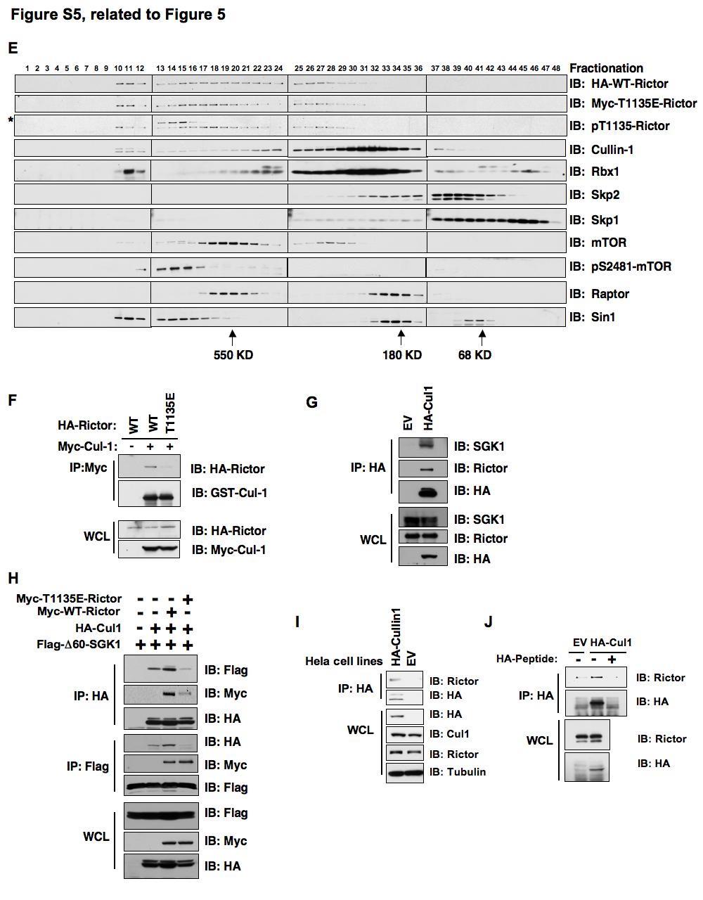
17 D. Immunoblot (IB) analysis of whole cell lysates (WCL) derived from HeLa cells with or without - phosphatase treatment. E. HeLa cells were serum starved for 24 hours followed by addition of 100 nm Insulin. Various pharmacological kinase inhibitors (100 nm Rapamycin, 20 M LY294002, 200 nm Wortmannin and 10 M Akt inhibitor, with DMSO as a negative control) were added together with Insulin. Forty minutes later, whole cell lysates (WCL) were collected for immunoblot analysis. Figure S5. Phosphorylation of Rictor at T1135 disrupts the interaction between Cullin-1 and Rictor (related to Figure 5). A. Immunoblot (IB) analysis of whole cell lysates (WCL) and immunoprecipitates (IP) derived from 293T cells transfected with the indicated Myc-Rictor constructs in the presence or absence of HA- 60-SGK1. B. Immunoblot (IB) analysis of whole cell lysates (WCL) and anti-myc immunoprecipitates derived from 293T cells transfected with the indicated Myc-Rictor plasmids in the presence of Flag Twenty hours post-transfection, cells were either serum starved for 36 hours or 10% FBS was added to the serum starved cells for 1.5 hours before harvesting. Where indicated, 20 M LY was added to further suppress Rictor T1135 phosphorylation. C. 293T cells were transiently tranfected with Myc-tagged WT, T1135A and T1135E Rictor constructs. Thirty hours post-transfection, whole cell lystates were collected for immunoblots with the indicated antibodies, and also for GST pull-down assays to examine their ability to interact with GST protein (with GST protein as a negative control). D. Immunoblot (IB) analysis of whole cell lysates (WCL) and immunoprecipitates (IP) derived from 293T cells transfected with the indicated Myc-Rictor constructs in the presence or absence of HA- 60-SGK1. Where indicated, high-affinity interacting R18 peptides were added to the whole cell lysates 30 minutes prior to the Myc-IP.
18 E. HeLa cells were transiently transfected with HA-WT-Rictor and Myc-T1135E-Rictor constructs. Thirty hours post-transfection, whole cell lysates were collected in CHAPS buffer and subjected to gel filtration chromography. Tandem size exclusion columns (Superose 6 and Superdex 200 columns in series) were used to enhance the separation efficiency. Immunonblot analysis were performed with various antibodies for the indicated fractionations. Prior to running the cell lysates, the molecular weight resolution of the column was first estimated by running native molecular weight markers (Urease ~550KD, mouse IgG ~180KD and human serum albumin ~68KD) and determining their retention times on coomassie-stained SDS-PAGE protein gels. *: non-specific band. F. Immunoblot (IB) analysis of whole cell lysates (WCL) and immunoprecipitates (IP) derived from 293T cells transfected with the indicated HA-Rictor and Myc-Cullin-1 constructs. G. Immunoblot (IB) analysis of whole cell lysates (WCL) and immunoprecipitates (IP) derived from HeLa cells transfected with the indicated HA-Cullin-1 construct (with Empty Vector plasmid as a negative control). Thirty hours post-transfection, cells were pretreated with 10 µm MG132 for 10 hours to block the proteasome pathway before harvesting. H. Immunoblot (IB) analysis of whole cell lysates (WCL) and immunoprecipitates (IP) derived from 293T cells transfected with the indicated Flag- 60-SGK1, HA-Cullin-1 and Myc-Rictor (WT or T1135E) constructs. Thirty hours post-transfection, cells were pretreated with 10 µm MG132 for 10 hours to block the proteasome pathway before harvesting. I. HeLa cells were transfected with the pcdna3-ha-cullin-1 constuct (with empty pcdna3 vector as a negative control). Forty hours post-transfection, cells were selected with 800 g/ml G418 for 2 weeks to generate the stable cell lines expressing HA-Cullin-1. Whole cell lysates were collected for HAimmunoprecipitations and immunoblots with the indicated antibodies. J. Immunoblot (IB) analysis of whole cell lysates (WCL) and HA-immunoprecipitates (IP) derived from stable HA-Cullin-1 HeLa cell line. Where indicated, HA-peptides were used as a negative control to
19 indicate that the interaction between Rictor and HA-Cullin-1 depends on successful HA immunoprecipitation. Figure S6. Phosphorylation of Rictor at T1135 impairs its ability to promote SGK1 ubiquitination and destruction (related to Figure 6). A. Immunoblot (IB) analysis of whole cell lysates (WCL) and immunoprecipitates (IP) derived from 293T cells transfected with HA- 60-SGK1, HA-Akt1 or HA-S6K together with Flag-Ub and the indicated Myc-Rictor constructs. Twenty hours post-transfection, cells were treated with the proteasome inhibitor MG132 overnight before harvesting. B. Immunoblot (IB) analysis of whole cell lysates (WCL) and anti-ha immunoprecipitates derived from 293T cells transfected with the HA- 60-SGK1 construct together with the indicated Myc-Rictor constructs. Twenty hours post-transfection, cells were treated with the proteasome inhibitor MG132 overnight before harvesting. C. Wild type or rictor-/- MEFs were transfected with the indicated Rictor plasmids (with empty vector as a negative control) together with the pbabe-puro retroviral empty vector. Twenty-four hours posttransfection, the cells were treated with 1 g/ml puromycin for hours to kill the non-transfected cells prior to collecting the whole cell lysates for immunoblots. D. Immunoblot analysis (IB) of HeLa cells transfected with the indicated sirna oligos. The pt1135- Rictor and SGK1 band intensities were normalized against tubulin, then normalized against the luciferase sirna treated sample. The quantification of the band intensities for pt1135-rictor and SGK1 in each lane is shown under the respective immunoblot image as indicated in the figure panel. E. Immunoblot analysis (IB) of HeLa cells transfected with the indicated sirna oligos, after synchronization with nocodazole and release. F. Immunoblot (IB) analysis of whole cell lysates (WCL) derived from a panel of breast cancer cell lines to demonstrate the positive correlation between Rictor T1135 phoshorylation and SGK1 abundance.
20 Supplemental Experimental Procedures Plasmids: Plasmids to express myc-rictor, myc-raptor, HA-Raptor, myc-mtor and myc-mtor kinase dead were obtained from Addgene. The HA-Rictor vector was a kind gift from Dr. Do-Hyung Kim. The myc-rictor truncations and were gifts from Dr. Brendan Manning. Plasmids to express HA-myr- Akt were described previously (Gao et al., 2009); the HA-S6K1-CA and HA-S6K1-KD constructs were obtained from Dr. John Blenis; the HA-SGK1 and HA- 60-SGK1 constructs were a kind gift from Dr. Suzanne Conzen. pcdna3-ha-cullin1 was generated by inserting the Cullin-1 coding sequence into the pcdna3-ha vector. HA-tagged WT, N14, N23 and N41-Rbx1 constructs were kind gifts from Dr. Yue Xiong (Furukawa et al., 2002). The HA-Sin1 plasmid was a kind gift from Dr. Bing Su. Flag and pgex were kind gifts from Dr. Lewis Cantley and Dr. Hui-Kuan Lin, respectively. The Myc- K720R-Cullin-1 mutant, Rictor mutants and truncations were generated using the QuikChange XL Site- Directed Mutagenesis Kit (Stratagene) according to the manufacturer s instructions. Antibodies and Reagents: Anti c-myc antibody (SC-40), polyclonal anti-ha antibody (SC-805), anti-cyclin A antibody (SC-751), anti-skp1 antibody (sc-7163), anti-cullin-1 antibody (sc-70895), anti-rictor antibody (sc-81538), anti- Raptor antibody (sc-81537), anti-cyclin B antibody (SC-245), anti-phospho-ser422-sgk antibody (SC R) and anti antibody (SC-629) were purchased from Santa Cruz. Anti-mTOR antibody (2972), anti-pser2481-mtor antibody (2974), anti-pthr346-ndrg1 antibody (3217), anti-raptor antibody (2280), anti-s6k antibody (9202), anti-phospho-ser473-akt antibody (4051), anti-phospho- Thr389-S6K antibody (9205), anti-phospho-akt substrate antibody (9614), anti-phospho binding motif antibody (9608) were purchased from Cell Signaling. Anti-Rictor antibodies (A A, A A), anti-mtor antibodies (A A, A A), anti-raptor antibody (A A) and anti- Sin1 antibody (A A) were purchased from Bethyl. Anti-SGK1 antibody (KAP-PK015) was from
21 Stressgen. Anti-phospho-Thr1135- Rictor antibody was a kind gift from Cell Signaling. Anti-tubulin antibody (T-5168), polyclonal anti-flag antibody (F2425), monoclonal anti-flag antibody (F-3165), anti-vinculin antibody (V4505), peroxidase-conjugated anti-mouse secondary antibody (A4416) and peroxidase-conjugated anti-rabbit secondary antibody (A4914) were purchased from Sigma. Monoclonal anti-ha antibody (MMS-101P) was purchased from Covance. Monoclonal anti-cullin-1 antibody ( ) and anti-skp2 antibody ( ) were purchased from Invitrogen. Anti-Brdu antibody (555627) was purchased from BD Biosciences. Anti-Rbx1 anti-body (RB-069P1) was purchased from Neomarker. sirna and shrna treatment: shrna vectors to knock down Rictor, Raptor, mtor and Sin1 were purchased from Addgene. shrbx1 lentiviral vector was purchased from Open Biosystems. plko lentiviral expression vectors to knock down S6K and SGK were kind gifts from Dr. William Hahn. plko lentiviral vector to knock down Akt1 was described previously (Gao et al., 2009), and virus generated in 293T cells for infection, as described (Boehm et al., 2005). A luciferase GL2 sirna oligo was purchased from Dharmacon, and the sirnas to knock down PTEN were described previously (Gao et al., 2009). sirna oligos to deplete endogenous Rbx1 (AACUGUGCCAUCUGCAGGAACAA) (Jia et al., 2009), Cullin1 (GGUCGCUUCAUAAAC AACAUU) (Benmaamar and Pagano, 2005), and Rictor (AAACUUGUGAAGAAUCGUAUCUU (Sarbassov et al., 2005), AAAGACUACAGCAACAAAGAAUU (Joshi et al., 2008)) were synthesized by Dharmacon. Cocktailed sirnas targeting Skp1 were purchased from Invitrogen ( ). sirna oligos were transfected into subconfluent cells using Oligofectamine or Lipofectamine 2000 (Invitrogen) according to the manufacturer s instructions (Wei et al., 2004; Wei et al., 2005). Immunoblots and Immunoprecipitation: Cells were lysed in EBC (50 mm Tris ph 7.5, 120 mm NaCl, 0.5% NP-40) buffer or CHAPS buffer (25 mm HEPES ph7.4, 150 mm NaCl, 1 mm EDTA, 0.3% CHAPS) supplemented with protease inhibitors (Complete Mini, Roche) and phosphatase inhibitors (phosphatase inhibitor cocktail set I and II,
22 Calbiochem). 2 mm 1,10-Phenanthroline was used for endogenous Cullin-1 immunoprecipitation to prevent the de-neddylation of Cullin-1. The protein concentrations of the lysates were measured using the Bio-Rad protein assay reagent on a Beckman Coulter DU-800 spectrophotometer. The lysates were then resolved by SDS-PAGE and immunoblotted with indicated antibodies. For immunoprecipitation, 800 g lysates were incubated with the appropriate antibody (1-2 g) for 3-4 hours at 4 C followed by one hour incubation with Protein A sepharose beads (GE Healthcare). Immuno-complexes were washed five times with NETN buffer (20 mm Tris, ph 8.0, 100 mm NaCl, 1 mm EDTA and 0.5% NP-40) before being resolved by SDS-PAGE and immunoblotted with indicated antibodies. MEF Transfection: rictor -/- or control MEFs were transfected with Lipofectamine LTX following the manufacturer s protocol (Invitrogen). Twenty four hours after transfection, cells were split into puromycin containing media and harvested 40 hours later to eliminate non-transfected cells. R18 Peptide Competition Assay R18 peptide was purchased from BIOMOL (P-214) and dissolved in water. Cell lysate was incubated with 25 M R18 peptide for 30min before the indicated IP was performed. Bromodeoxyuridine Labeling Bromodeoxyuridine (BrdU) labeling assay was performed as described previously (Wei et al., 2001). Experiments were repeated three times to generate the error bars.
23 Supplemental References Benmaamar, R., and Pagano, M. (2005). Involvement of the SCF complex in the control of Cdh1 degradation in S-phase. Cell Cycle 4, Furukawa, M., Ohta, T., and Xiong, Y. (2002). Activation of UBC5 ubiquitin-conjugating enzyme by the RING finger of ROC1 and assembly of active ubiquitin ligases by all cullins. J Biol Chem 277, Jia, L., Soengas, M. S., and Sun, Y. (2009). ROC1/RBX1 E3 ubiquitin ligase silencing suppresses tumor cell growth via sequential induction of G2-M arrest, apoptosis, and senescence. Cancer Res 69, Joshi, J., Fernandez-Marcos, P. J., Galvez, A., Amanchy, R., Linares, J. F., Duran, A., Pathrose, P., Leitges, M., Canamero, M., Collado, M., et al. (2008). Par-4 inhibits Akt and suppresses Ras-induced lung tumorigenesis. Embo J 27, Wei, W., Ayad, N. G., Wan, Y., Zhang, G. J., Kirschner, M. W., and Kaelin, W. G., Jr. (2004). Degradation of the SCF component Skp2 in cell-cycle phase G1 by the anaphase-promoting complex. Nature 428, Wei, W., Hemmer, R. M., and Sedivy, J. M. (2001). Role of p14(arf) in replicative and induced senescence of human fibroblasts. Mol Cell Biol 21, Wei, W., Jin, J., Schlisio, S., Harper, J. W., and Kaelin, W. G., Jr. (2005). The v-jun point mutation allows c-jun to escape GSK3-dependent recognition and destruction by the Fbw7 ubiquitin ligase. Cancer Cell 8,
SUPPLEMENTARY INFORMATION
 doi:10.1038/nature09732 Supplementary Figure 1: Depletion of Fbw7 results in elevated Mcl-1 abundance. a, Total thymocytes from 8-wk-old Lck-Cre/Fbw7 +/fl (Control) or Lck-Cre/Fbw7 fl/fl (Fbw7 KO) mice
SUPPLEMENTARY INFORMATION
 The Supplementary Information (SI) Methods Cell culture and transfections H1299, U2OS, 293, HeLa cells were maintained in DMEM medium supplemented with 10% fetal bovine serum. H1299 and 293 cells were
Supplementary data. sienigma. F-Enigma F-EnigmaSM. a-p53
 Supplementary data Supplemental Figure 1 A sienigma #2 sienigma sicontrol a-enigma - + ++ - - - - - - + ++ - - - - - - ++ B sienigma F-Enigma F-EnigmaSM a-flag HLK3 cells - - - + ++ + ++ - + - + + - -
Supplemental Information. Phosphorylation by Casein Kinase I Promotes. the Turnover of the Mdm2 Oncoprotein. via the SCF β-trcp Ubiquitin Ligase
 Cancer Cell, Volume 18 Supplemental Information Phosphorylation by Casein Kinase I Promotes the Turnover of the Mdm2 Oncoprotein via the SCF β-trcp Ubiquitin Ligase Hiroyuki Inuzuka, Alan Tseng, Daming
Supplemental Information
 Supplemental Information Intrinsic protein-protein interaction mediated and chaperonin assisted sequential assembly of a stable Bardet Biedl syndome protein complex, the BBSome * Qihong Zhang 1#, Dahai
T H E J O U R N A L O F C E L L B I O L O G Y
 T H E J O U R N A L O F C E L L B I O L O G Y Supplemental material Han et al., http://www.jcb.org/cgi/content/full/jcb.201311007/dc1 Figure S1. SIVA1 interacts with PCNA. (A) HEK293T cells were transiently
Supplementary information
 Supplementary information The E3 ligase RNF8 regulates KU80 removal and NHEJ repair Lin Feng 1, Junjie Chen 1 1 Department of Experimental Radiation Oncology, The University of Texas M. D. Anderson Cancer
Supporting Information
 Supporting Information Su et al. 10.1073/pnas.1211604110 SI Materials and Methods Cell Culture and Plasmids. Tera-1 and Tera-2 cells (ATCC: HTB- 105/106) were maintained in McCoy s 5A medium with 15% FBS
Supplementary Material
 Supplementary Material Supplementary Methods Cell synchronization. For synchronized cell growth, thymidine was added to 30% confluent U2OS cells to a final concentration of 2.5mM. Cells were incubated
Figure 1: TDP-43 is subject to lysine acetylation within the RNA-binding domain a) QBI-293 cells were transfected with TDP-43 in the presence or
 QBI-293 cells were transfected with TDP-43 in the presence or") Figure 1: TDP-43 is subject to lysine acetylation within the RNA-binding domain a) QBI-293 cells were transfected with TDP-43 in the presence or absence of the acetyltransferase CBP and acetylated TDP-43
Figure 1: TDP-43 is subject to lysine acetylation within the RNA-binding domain a) QBI-293 cells were transfected with TDP-43 in the presence or absence of the acetyltransferase CBP and acetylated TDP-43
Supplementary Materials
 Supplementary Materials Supplementary Figure 1. PKM2 interacts with MLC2 in cytokinesis. a, U87, U87/EGFRvIII, and HeLa cells in cytokinesis were immunostained with DAPI and an anti-pkm2 antibody. Thirty
Supplementary Materials and Methods
 Supplementary Materials and Methods sirna sequences used in this study The sequences of Stealth Select RNAi for ALK and FLOT-1 were as follows: ALK sense no.1 (ALK): 5 -AAUACUGACAGCCACAGGCAAUGUC-3 ; ALK
Supplementary Information: Materials and Methods. Immunoblot and immunoprecipitation. Cells were washed in phosphate buffered
 Supplementary Information: Materials and Methods Immunoblot and immunoprecipitation. Cells were washed in phosphate buffered saline (PBS) and lysed in TNN lysis buffer (50mM Tris at ph 8.0, 120mM NaCl
Supplementary Information: Materials and Methods. GST and GST-p53 were purified according to standard protocol after
 Supplementary Information: Materials and Methods Recombinant protein expression and in vitro kinase assay. GST and GST-p53 were purified according to standard protocol after induction with.5mm IPTG for
Supplementary methods Shoc2 In Vitro Ubiquitination Assay
 Supplementary methods Shoc2 In Vitro Ubiquitination Assay 35 S-labelled Shoc2 was prepared using a TNT quick Coupled transcription/ translation System (Promega) as recommended by manufacturer. For the
HPV E6 oncoprotein targets histone methyltransferases for modulating specific. Chih-Hung Hsu, Kai-Lin Peng, Hua-Ci Jhang, Chia-Hui Lin, Shwu-Yuan Wu,
 1 HPV E oncoprotein targets histone methyltransferases for modulating specific gene transcription 3 5 Chih-Hung Hsu, Kai-Lin Peng, Hua-Ci Jhang, Chia-Hui Lin, Shwu-Yuan Wu, Cheng-Ming Chiang, Sheng-Chung
supplementary information
 DOI: 10.1038/ncb2116 Figure S1 CDK phosphorylation of EZH2 in cells. (a) Comparison of candidate CDK phosphorylation sites on EZH2 with known CDK substrates by multiple sequence alignments. (b) CDK1 and
Supplementary Figure 1. GST pull-down analysis of the interaction of GST-cIAP1 (A, B), GSTcIAP1
, GSTcIAP1") Legends Supplementary Figure 1. GST pull-down analysis of the interaction of GST- (A, B), GST mutants (B) or GST- (C) with indicated proteins. A, B, Cell lysate from untransfected HeLa cells were loaded
Legends Supplementary Figure 1. GST pull-down analysis of the interaction of GST- (A, B), GST mutants (B) or GST- (C) with indicated proteins. A, B, Cell lysate from untransfected HeLa cells were loaded
Supplemental Information. Akt-Mediated Phosphorylation of XLF Impairs. Non-Homologous End-Joining DNA Repair. Molecular Cell, Volume 57
 Molecular Cell, Volume 57 Supplemental Information Akt-Mediated Phosphorylation of XLF Impairs Non-Homologous End-Joining DNA Repair Pengda Liu, Wenjian Gan, Chunguang Guo, Anyong Xie, Daming Gao, Jianping
Nature Structural & Molecular Biology: doi: /nsmb.1583
 Acetylation by GCN5 regulates CDC6 phosphorylation in the S-phase of the cell cycle Roberta Paolinelli 1,2, Ramiro Mendoza-Maldonado 2, Anna Cereseto 1 and Mauro Giacca 2 1 Molecular Biology Laboratory,
Supplemental Information: Phosphorylation of CLIP-170 by Both Plk1 and CK2 Is Involved in the Timely Formation of Kinetochore-microtubule Attachments
 Supplemental Information: Phosphorylation of CLIP-170 by Both Plk1 and CK2 Is Involved in the Timely Formation of Kinetochore-microtubule Attachments Hongchang Li, X. Shawn Liu, Xiaoming Yang, Yingmin
ASPP1 Fw GGTTGGGAATCCACGTGTTG ASPP1 Rv GCCATATCTTGGAGCTCTGAGAG
 Supplemental Materials and Methods Plasmids: the following plasmids were used in the supplementary data: pwzl-myc- Lats2 (Aylon et al, 2006), pretrosuper-vector and pretrosuper-shp53 (generous gift of
Anna A. Sablina, Wen Chen, Jason D. Arroyo, Laura Corral, Melissa Hector, Sara E. Bulmer, James A. DeCaprio, and William C. Hahn
 Cell, Volume 129 Supplemental Data The Tumor Suppressor PP2A Aβ Regulates the RalA GTPase Anna A. Sablina, Wen Chen, Jason D. Arroyo, Laura Corral, Melissa Hector, Sara E. Bulmer, James A. DeCaprio, and
Coleman et al., Supplementary Figure 1
 Coleman et al., Supplementary Figure 1 BrdU Merge G1 Early S Mid S Supplementary Figure 1. Sequential destruction of CRL4 Cdt2 targets during the G1/S transition. HCT116 cells were synchronized by sequential
Attenuation of synaptic toxicity and MARK4/PAR1-mediated Tau phosphorylation by
 Supplementary Methods and Figures Attenuation of synaptic toxicity and MARK4/PAR1-mediated Tau phosphorylation by methylene blue for Alzheimer s disease treatment Wenchao Sun 1, Seongsoo Lee 1,2, Xiaoran
from Dr. David Livingston. Rabbit anti-myc, V5, and BACH1 were raised by immunizing rabbits with peptides EQKLISEEDI, GKPIPNPLLGLDST, and
 upporting Material Experimental Procedures: Cell culture and antibodies All cell lines were maintained in RPMI 164 medium with 1% fetal calf serum at 37 C in 5% CO 2 (v/v). For HCC 1937-BRCA1 cells and
SUPPLEMENTAL MATERIAL. Supplemental Methods. Cumate solution was from System Biosciences. Human complement C1q and complement
 SUPPLEMENTAL MATERIAL Supplemental Methods Reagents Cumate solution was from System Biosciences. Human complement Cq and complement C-esterase inhibitor (C-INH) were from Calbiochem. C-INH (Berinert) for
Supplementary Figure S1 Purification of deubiquitinases HEK293 cells were transfected with the indicated DUB-expressing plasmids.
 Supplementary Figure S1 Purification of deubiquitinases HEK293 cells were transfected with the indicated DUB-expressing plasmids. The cells were harvested 72 h after transfection. FLAG-tagged deubiquitinases
Supplementary methods
 Supplementary methods Cell culture, infection, transfection, and RNA interference HEK293 cells and its derivatives were grown in DMEM supplemented with 10% FBS. Various constructs were introduced into
Supplementary Table 1. The Q-PCR primer sequence is summarized in the following table.
 Supplementary Table 1. The Q-PCR primer sequence is summarized in the following table. Name Sequence (5-3 ) Application Flag-u ggactacaaggacgacgatgac Shared upstream primer for all the amplifications of
Antibodies against PCNA were previously described [1]. To deplete PCNA from Xenopus egg
![Antibodies against PCNA were previously described [1]. To deplete PCNA from Xenopus egg](/thumbs/84/90687567.jpg "Antibodies against PCNA were previously described [1]. To deplete PCNA from Xenopus egg") Supplementary information Supplementary methods PCNA antibody and immunodepletion Antibodies against PCNA were previously described [1]. To deplete PCNA from Xenopus egg extracts, one volume of protein
Supplementary information Supplementary methods PCNA antibody and immunodepletion Antibodies against PCNA were previously described [1]. To deplete PCNA from Xenopus egg extracts, one volume of protein
USP19 modulates autophagy and antiviral immune responses by. deubiquitinating Beclin-1
 USP19 modulates autophagy and antiviral immune responses by deubiquitinating Beclin-1 Shouheng Jin 1,2,, Shuo Tian 1,, Yamei Chen 1,, Chuanxia Zhang 1,2, Weihong Xie, 1 Xiaojun Xia 3,4, Jun Cui 1,3* &
supplementary information
 DOI: 1.138/ncb1839 a b Control 1 2 3 Control 1 2 3 Fbw7 Smad3 1 2 3 4 1 2 3 4 c d IGF-1 IGF-1Rβ IGF-1Rβ-P Control / 1 2 3 4 Real-time RT-PCR Relative quantity (IGF-1/ mrna) 2 1 IGF-1 1 2 3 4 Control /
T H E J O U R N A L O F C E L L B I O L O G Y
 T H E J O U R N A L O F C E L L B I O L O G Y Supplemental material Nakajima and Tanoue, http://www.jcb.org/cgi/content/full/jcb.201104118/dc1 Figure S1. DLD-1 cells exhibit the characteristic morphology
Supplemental Figure 1
 Supplemental Fig. 1. Kinetics of,,, AKT and ERK activation in BMMCs following SCF stimulation. Starved BMMCs were stimulated with 250ng/mL of SCF for the indicated time. Soluble Cell Lysates (SCLs) were
Transcriptional regulation of BRCA1 expression by a metabolic switch: Di, Fernandez, De Siervi, Longo, and Gardner. H3K4Me3
 ChIP H3K4Me3 enrichment.25.2.15.1.5 H3K4Me3 H3K4Me3 ctrl H3K4Me3 + E2 NS + E2 1. kb kb +82 kb Figure S1. Estrogen promotes entry of MCF-7 into the cell cycle but does not significantly change activation-associated
Recombinant adenoviruses. A TRB3-expressing adenovirus was generated through
 Materials and Methods Recombinant adenoviruses. A -expressing adenovirus was generated through homologous recombination between a linearized transfer vector pad-track and the adenoviral backbone vector
hours after food deprivation hours after food deprivation
 Figure S.6 protein (fasted / control).2.8.4 p47 p97 Rpt Ufd 3 24 48 hours after food deprivation mrn (fasted / control) C mrn (denervated / control) 2.5 2.5.5 3.5 3 2.5 2.5.5 p97 ** Npl4 Ufd p47 2 3 4
Flag-Rac Vector V12 V12 N17 C40. Vector C40 pakt (T308) Akt1. Myc-DN-PAK1 (N-SP)
 Akt1. Myc-DN-PAK1 (N-SP)") a b FlagRac FlagRac V2 V2 N7 C4 V2 V2 N7 C4 p (T38) p (S99, S24) p Flag (Rac) NIH 3T3 COS c +Serum p (T38) MycDN (NSP) Mycp27 3 6 2 3 6 2 3 6 2 min p Myc ( or p27) Figure S (a) Effects of Rac mutants on
a b FlagRac FlagRac V2 V2 N7 C4 V2 V2 N7 C4 p (T38) p (S99, S24) p Flag (Rac) NIH 3T3 COS c +Serum p (T38) MycDN (NSP) Mycp27 3 6 2 3 6 2 3 6 2 min p Myc ( or p27) Figure S (a) Effects of Rac mutants on
Supplemental Data Supplementary Figure Legends and Scheme Figure S1.
 Supplemental Data Supplementary Figure Legends and Scheme Figure S1. UTK1 inhibits the second EGF-induced wave of lamellipodia formation in TT cells. A and B, EGF-induced lamellipodia formation in TT cells,
Supplemental Data. LMO4 Controls the Balance between Excitatory. and Inhibitory Spinal V2 Interneurons
 Neuron, Volume 61 Supplemental Data LMO4 Controls the Balance between Excitatory and Inhibitory Spinal V2 Interneurons Kaumudi Joshi, Seunghee Lee, Bora Lee, Jae W. Lee, and Soo-Kyung Lee Supplemental
b alternative classical none
 Supplementary Figure. 1: Related to Figure.1 a d e b alternative classical none NIK P-IkBa Total IkBa Tubulin P52 (Lighter) P52 (Darker) RelB (Lighter) RelB (Darker) HDAC1 Control-Sh RelB-Sh NF-kB2-Sh
SUPPLEMENTARY INFORMATION. Small molecule activation of the TRAIL receptor DR5 in human cancer cells
 SUPPLEMENTARY INFORMATION Small molecule activation of the TRAIL receptor DR5 in human cancer cells Gelin Wang 1*, Xiaoming Wang 2, Hong Yu 1, Shuguang Wei 1, Noelle Williams 1, Daniel L. Holmes 1, Randal
SUPPLEMENTAL MATERIALS SIRTUIN 1 PROMOTES HYPEROXIA-INDUCED LUNG EPITHELIAL DEATH INDEPENDENT OF NRF2 ACTIVATION
 SUPPLEMENTAL MATERIALS SIRTUIN PROMOTES HYPEROXIA-INDUCED LUNG EPITHELIAL DEATH INDEPENDENT OF NRF ACTIVATION Haranatha R. Potteti*, Subbiah Rajasekaran*, Senthilkumar B. Rajamohan*, Chandramohan R. Tamatam,
SUPPLEMENTARY INFORMATION
 SUPPLEMENTARY INFORMATION SUPPLEMENTARY MATERIALS AND METHODS Antibodies and sirna The antibodies against BAF170, BAF155, BAF60a, BAF57 and BAF53 were purchased from Santa Cruz (TX), and the antibodies
SANTA CRUZ BIOTECHNOLOGY, INC.
 TECHNICAL SERVICE GUIDE: Western Blotting 2. What size bands were expected and what size bands were detected? 3. Was the blot blank or was a dark background or non-specific bands seen? 4. Did this same
used at a final concentration of 5 ng/ml. Rabbit anti-bim and mouse anti-mkp2 antibodies were
 1 Supplemental Methods Reagents and chemicals: TGFβ was a generous gift from Genzyme Inc. (Cambridge, MA) and was used at a final concentration of 5 ng/ml. Rabbit anti-bim and mouse anti-mkp2 antibodies
SUPPLEMENTAL EXPERIMENTAL PROCEDURES
 SUPPLEMENTAL EXPERIMENTAL PROCEDURES Luciferase Assays Cells were seeded on 24well plates and grown to 7% confluency. Cells were then transfected with ng of reporter constructs and 1 ng of the renilla
SUPPLEMENTARY INFORMATION
 SUPPLEMENTARY INFORMATION Dynamic Phosphorylation of HP1 Regulates Mitotic Progression in Human Cells Supplementary Figures Supplementary Figure 1. NDR1 interacts with HP1. (a) Immunoprecipitation using
Table S1. Alteration of ZNF322A and FBXW7 protein expression levels in relation to clinicopathological parameters in 135 lung cancer patients.
 SUPPLEMENTARY TABLES Table S1. Alteration of ZNF322A and FBXW7 protein expression levels in relation to clinicopathological parameters in 135 lung cancer patients. ZNF322A Normal Overexpression expression
Supplementary Figure 1 - Characterization of rbag3 binding on macrophages cell surface.
 Supplementary Figure 1 - Characterization of rbag3 binding on macrophages cell surface. (a) Human PDAC cell lines were treated as indicated in Figure 1 panel F. Cells were analyzed for FITC-rBAG3 binding
SUPPLEMENTARY INFORMATION
 Figure S1 The effect of T198A mutation on p27 stability. a, Hoechst 33342 staining for nuclei (see Fig 1d). Scale bar, 100 μm. b, Densitometric analysis of wild type and mutant p27 protein levels represented
EXPERIMENTAL PROCEDURES
 EXPERIMENTAL PROCEDURES Cell culture and antibodies-human colorectal cancer HCT-116 cells, human embryonic kidney 293 cells and human normal breast epithelial cells MCF-10A cells were cultured as recommended
Sarker et al. Supplementary Material. Subcellular Fractionation
 Supplementary Material Subcellular Fractionation Transfected 293T cells were harvested with phosphate buffered saline (PBS) and centrifuged at 2000 rpm (500g) for 3 min. The pellet was washed, re-centrifuged
Fig. S1 TGF RI inhibitor SB effectively blocks phosphorylation of Smad2 induced by TGF. FET cells were treated with TGF in the presence of
 Fig. S1 TGF RI inhibitor SB525334 effectively blocks phosphorylation of Smad2 induced by TGF. FET cells were treated with TGF in the presence of different concentrations of SB525334. Cells were lysed and
Anti-Pim-1 (Cat#3247), anti-met (Cat#3127), anti-ron (Cat#2654), Anti-EGFR
, anti-met (Cat#3127), anti-ron (Cat#2654), Anti-EGFR") Supplementary Methods Antibodies Anti-Pim-1 (Cat#3247), anti-met (Cat#3127), anti-ron (Cat#2654), Anti-EGFR (Cat#2646), anti-igf1r (Cat#3018), anti-insr (Cat#3020), anti-akt (pan, Cat#4691), anti-phospho-akt
Supplementary Methods Antibodies Anti-Pim-1 (Cat#3247), anti-met (Cat#3127), anti-ron (Cat#2654), Anti-EGFR (Cat#2646), anti-igf1r (Cat#3018), anti-insr (Cat#3020), anti-akt (pan, Cat#4691), anti-phospho-akt
Supporting Online Material for
 Supporting Online Material for Spatiotemporal dynamics of Aurora B-PLK1-MCAK signaling axis orchestrates kinetochore bi-orientation and faithful chromosome segregation Hengyi Shao, Yuejia Huang, Liangyu
Supplementary Figure 1 Phosphorylated tau accumulates in Nrf2 (-/-) mice. Hippocampal tissues obtained from Nrf2 (-/-) (10 months old, 4 male; 2
 mice. Hippocampal tissues obtained from Nrf2 (-/-) (10 months old, 4 male; 2") Supplementary Figure 1 Phosphorylated tau accumulates in Nrf2 (-/-) mice. Hippocampal tissues obtained from Nrf2 (-/-) (10 months old, 4 male; 2 female) or wild-type (5 months old, 1 male; 11 months old,
Supplementary Figure 1 Phosphorylated tau accumulates in Nrf2 (-/-) mice. Hippocampal tissues obtained from Nrf2 (-/-) (10 months old, 4 male; 2 female) or wild-type (5 months old, 1 male; 11 months old,
Supplementary Materials for
 www.sciencesignaling.org/cgi/content/full/8/404/ra120/dc1 Supplementary Materials for The subcellular localization and activity of cortactin is regulated by acetylation and interaction with Keap1 Akihiro
supplementary information
 DOI: 10.1038/ncb2172 Figure S1 p53 regulates cellular NADPH and lipid levels via inhibition of G6PD. (a) U2OS cells stably expressing p53 shrna or a control shrna were transfected with control sirna or
seminal vesicle thymus kidney lung liver
 a 1 HEK293 1 B 3 3 T GFPSMAD TGFβ (T)/ BMP (B) IB: SMAD1/3TP IB: GFP b wild type mouse tissue kidney thymus seminal vesicle liver lung brain adipose tissue muscle pancreas heart uterus spleen testis IB:
Supporting Information
 Supporting Information Krieg et al. 10.1073/pnas.0907131106 SI Text Reagents. Recombinant human TNF- was from Peprotech. Monoclonal rat anti-rip2 was purchased from Alexis, whereas monoclonal mouse anti-xiap
Angeles Duran, Juan F. Linares, Anita S. Galvez, Kathryn Wikenheiser, Juana M. Flores, Maria T. Diaz-Meco, and Jorge Moscat
 Cancer Cell, Volume 13 Supplemental Data The Signaling Adaptor p62 Is an Important NF-κB Mediator in Tumorigenesis Angeles Duran, Juan F. Linares, Anita S. Galvez, Kathryn Wikenheiser, Juana M. Flores,
Supplementary Fig. 1 Identification of Nedd4 as an IRS-2-associated protein in camp-treated FRTL-5 cells.
 Supplementary Fig. 1 Supplementary Fig. 1 Identification of Nedd4 as an IRS-2-associated protein in camp-treated FRTL-5 cells. (a) FRTL-5 cells were treated with 1 mm dibutyryl camp for 24 h, and the lysates
Supplemental Methods Cell lines and culture
 Supplemental Methods Cell lines and culture AGS, CL5, BT549, and SKBR were propagated in RPMI 64 medium (Mediatech Inc., Manassas, VA) supplemented with % fetal bovine serum (FBS, Atlanta Biologicals,
Cell culture and drug treatment. HEK 293 cells were cultured in DMEM (Gibco-BRL)
") Supplementary materials Detailed methods Cell culture and drug treatment. HEK 293 cells were cultured in DMEM (Gibco-BRL) supplemented with 10% fetal bovine serum. To inhibit glucosidase Ι and ΙΙ, castanospermine
Supplementary materials Detailed methods Cell culture and drug treatment. HEK 293 cells were cultured in DMEM (Gibco-BRL) supplemented with 10% fetal bovine serum. To inhibit glucosidase Ι and ΙΙ, castanospermine
Supplementary information
 Supplementary information Table of Content: Supplementary Results... 2 Supplementary Figure S1: Experimental validation of AP-MS results by coimmunprecipitation Western blot analysis.... 3 Supplementary
supplementary information
 DOI: 10.1038/ncb1864 Figure S1 Apak specifically inhibits p53 transcriptional activity. Transcription activity of p53 was measured in U2OS (p53 wild-type) and H1299 (p53 deficient) cells which were transfected
Supporting Online Material for
 www.sciencemag.org/cgi/content/full/1154040/dc1 Supporting Online Material for Selective Blockade of MicroRNA Processing by Lin-28 Srinivas R. Viswanathan, George Q. Daley,* Richard I. Gregory* *To whom
Regulation of nucleolar signalling to p53 through NEDDylation of L11. Anders Sundqvist, Geng Liu, Antonis Mirsaliotis and Dimitris P.
 Regulation of nucleolar signalling to p53 through NEDDylation of L11 Anders Sundqvist, Geng Liu, Antonis Mirsaliotis and Dimitris P. Xirodimas Supplementary Information Methods Plasmids All plasmids used
transcription and the promoter occupancy of Smad proteins. (A) HepG2 cells were co-transfected with the wwp-luc reporter, and FLAG-tagged FHL1,
 HepG2 cells were co-transfected with the wwp-luc reporter, and FLAG-tagged FHL1,") Supplementary Data Supplementary Figure Legends Supplementary Figure 1 FHL-mediated TGFβ-responsive reporter transcription and the promoter occupancy of Smad proteins. (A) HepG2 cells were co-transfected
Supplementary Data Supplementary Figure Legends Supplementary Figure 1 FHL-mediated TGFβ-responsive reporter transcription and the promoter occupancy of Smad proteins. (A) HepG2 cells were co-transfected
Hossain_Supplemental Figure 1
 Hossain_Supplemental Figure 1 GFP-PACT GFP-PACT Motif I GFP-PACT Motif II A. MG132 (1µM) GFP Tubulin GFP-PACT Pericentrin GFP-PACT GFP-PACT Pericentrin Fig. S1. Expression and localization of Orc1 PACT
SUPPLEMENTAL FIGURES AND TABLES
 SUPPLEMENTAL FIGURES AND TABLES A B Flag-ALDH1A1 IP: α-ac HEK293T WT 91R 128R 252Q 367R 41/ 419R 435R 495R 412R C Flag-ALDH1A1 NAM IP: HEK293T + + - + D NAM - + + E Relative ALDH1A1 activity 1..8.6.4.2
T H E J O U R N A L O F C E L L B I O L O G Y
 Supplemental material Thompson et al., http://www.jcb.org/cgi/content/full/jcb.200909067/dc1 T H E J O U R N A L O F C E L L B I O L O G Y Figure S1. Modification-specific antibodies do not detect unmodified
Supplementary material for: Materials and Methods:
 Supplementary material for: Iron-responsive degradation of iron regulatory protein 1 does not require the Fe-S cluster: S.L. Clarke, et al. Materials and Methods: Fe-S Cluster Reconstitution: Cells treated
Supplementary Figure 1. TSA (10 nmol/l), non-class-selective HDAC inhibitor, potentiates
, non-class-selective HDAC inhibitor, potentiates") Supplementary Figure 1. TSA (10 nmol/l), non-class-selective HDAC inhibitor, potentiates vascular calcification (VC). (a) Von Kossa staining shows that TSA potentiated the Pi-induced VC. Scale bar, 100
Supplementary Figure 1. TSA (10 nmol/l), non-class-selective HDAC inhibitor, potentiates vascular calcification (VC). (a) Von Kossa staining shows that TSA potentiated the Pi-induced VC. Scale bar, 100
Mechanism of Induction and Suppression of Antiviral Immunity Directed by Virus-Derived Small RNAs in Drosophila
 Cell Host & Microbe, Volume 4 Supplemental Data Mechanism of Induction and Suppression of Antiviral Immunity Directed by Virus-Derived Small RNAs in Drosophila Roghiyh Aliyari, Qingfa Wu, Hong-Wei Li,
At E17.5, the embryos were rinsed in phosphate-buffered saline (PBS) and immersed in
 and immersed in") Supplementary Materials and Methods Barrier function assays At E17.5, the embryos were rinsed in phosphate-buffered saline (PBS) and immersed in acidic X-gal mix (100 mm phosphate buffer at ph4.3, 3 mm
Supplementary Materials and Methods Barrier function assays At E17.5, the embryos were rinsed in phosphate-buffered saline (PBS) and immersed in acidic X-gal mix (100 mm phosphate buffer at ph4.3, 3 mm
Supplementary Information for. Regulation of Rev1 by the Fanconi Anemia Core Complex
 Supplementary Information for Regulation of Rev1 by the Fanconi Anemia Core Complex Hyungjin Kim, Kailin Yang, Donniphat Dejsuphong, Alan D. D Andrea* *Corresponding Author: Alan D. D Andrea, M.D. Alan_dandrea@dfci.harvard.edu
Supplemental Information
 Molecular Cell, Volume 54 Supplemental Information BRD7, a Tumor Suppressor, Interacts with p85 and Regulates PI3K Activity Yu-Hsin Chiu, Jennifer Y. Lee, and Lewis C. Cantley Supplemental Materials Supplemental
Supplementary Figure 1, related to Figure 1. GAS5 is highly expressed in the cytoplasm of hescs, and positively correlates with pluripotency.
 Supplementary Figure 1, related to Figure 1. GAS5 is highly expressed in the cytoplasm of hescs, and positively correlates with pluripotency. (a) Transfection of different concentration of GAS5-overexpressing
SUPPLEMENTARY INFORMATION
 Figure S1 UVRAG amino acid sequence. Bold letters indicate the peptides identified from mass spectrometry analysis. Symbols indicate the prolines in the N-terminal proline-rich sequence (*), the C2 domain
Supplemental Materials and Methods
 Supplemental Materials and Methods In situ hybridization In situ hybridization analysis of HFE2 and genin mrna in rat liver tissues was performed as previously described (1). Briefly, the digoxigenin-labeled
Supplemental Information
 FOOT-AND-MOUTH DISEASE VIRUS PROTEIN 2C IS A HEXAMERIC AAA+ PROTEIN WITH A COORDINATED ATP HYDROLYSIS MECHANISM. Trevor R. Sweeney 1, Valentina Cisnetto 1, Daniel Bose 2, Matthew Bailey 3, Jon R. Wilson
Post-translational modification
 Protein expression Western blotting, is a widely used and accepted technique to detect levels of protein expression in a cell or tissue extract. This technique measures protein levels in a biological sample
Supplementary Figure 1
 Supplementary Figure 1 PTEN promotes virus-induced expression of IFNB1 and its downstream genes. (a) Quantitative RT-PCR analysis of IFNB1 mrna (left) and ELISA of IFN-β (right) in HEK 293 cells (2 10
IgG TrueBlot Protocol for Mouse, Rabbit or Goatderived Antibodies - For Research Use Only
 IgG TrueBlot Protocol for Mouse, Rabbit or Goatderived Antibodies - For Research Use Only Introduction The IgG TrueBlot for mouse, rabbit, or goat-derived antibodies represents unique series of respective
Cell extracts and western blotting RNA isolation and real-time PCR Chromatin immunoprecipitation (ChIP)
") Cell extracts and western blotting Cells were washed with ice-cold phosphate-buffered saline (PBS) and lysed with lysis buffer. 1 Total cell extracts were separated by SDS-PAGE and transferred to nitrocellulose
Cell extracts and western blotting Cells were washed with ice-cold phosphate-buffered saline (PBS) and lysed with lysis buffer. 1 Total cell extracts were separated by SDS-PAGE and transferred to nitrocellulose
Supplemental material
 Supplemental material THE JOURNAL OF CELL BIOLOGY Gillespie et al., http://www.jcb.org/cgi/content/full/jcb.200907037/dc1 repressor complex induced by p38- Gillespie et al. Figure S1. Reduced fiber size
Online Supplementary Information
 Online Supplementary Information NLRP4 negatively regulates type I interferon signaling by targeting TBK1 for degradation via E3 ubiquitin ligase DTX4 Jun Cui 1,4,6,7, Yinyin Li 1,5,6,7, Liang Zhu 1, Dan
For Research Use Only. Not for use in diagnostic procedures.
 Printed December 13, 2011 Version 1.0 For Research Use Only. Not for use in diagnostic procedures. DDDDK-tagged Protein PURIFICATION GEL with Elution Peptide (MoAb. clone FLA-1) CODE No. 3326 / 3327 PURIFICATION
Supplementary Fig. 1 Proteomic analysis of ATR-interacting proteins. ATR, ARID1A and
 Supplementary Figure Legend: Supplementary Fig. 1 Proteomic analysis of ATR-interacting proteins. ATR, ARID1A and ATRIP protein peptides identified from our mass spectrum analysis were shown. Supplementary
Xiaoqing Zhang, Guo Zhang, Hai Zhang, Michael Karin, Hua Bai, and Dongsheng Cai
 Cell, Volume 135 Supplemental Data Hypothalamic IKKβ/NF-κB and ER Stress Link Overnutrition to Energy Imbalance and Obesity Xiaoqing Zhang, Guo Zhang, Hai Zhang, Michael Karin, Hua Bai, and Dongsheng Cai
Supplementary Figure 1. TRIM9 does not affect AP-1, NF-AT or ISRE activity. (a,b) At 24h post-transfection with TRIM9 or vector and indicated
 At 24h post-transfection with TRIM9 or vector and indicated") Supplementary Figure 1. TRIM9 does not affect AP-1, NF-AT or ISRE activity. (a,b) At 24h post-transfection with TRIM9 or vector and indicated reporter luciferase constructs, HEK293T cells were stimulated
Supplementary Figure 1. TRIM9 does not affect AP-1, NF-AT or ISRE activity. (a,b) At 24h post-transfection with TRIM9 or vector and indicated reporter luciferase constructs, HEK293T cells were stimulated
Fig. S1. Effect of p120-catenin overexpression on the interaction of SCUBE2 with E-cadherin. The expression plasmid encoding FLAG.
 Fig. S1. Effect of p120-catenin overexpression on the interaction of SCUBE2 with E-cadherin. The expression plasmid encoding FLAG.SCUBE2, E-cadherin.Myc, or HA.p120-catenin was transfected in a combination
RNA was isolated using NucleoSpin RNA II (Macherey-Nagel, Bethlehem, PA) according to the
 according to the") Supplementary Methods RT-PCR and real-time PCR analysis RNA was isolated using NucleoSpin RNA II (Macherey-Nagel, Bethlehem, PA) according to the manufacturer s protocol and quantified by measuring the
Supplementary Methods RT-PCR and real-time PCR analysis RNA was isolated using NucleoSpin RNA II (Macherey-Nagel, Bethlehem, PA) according to the manufacturer s protocol and quantified by measuring the
Protocol for induction of expression and cell lysate production
 Protocol for induction of expression and cell lysate production AV-04 Doxycyclin induction and cell lysate 1.0 Introduction / Description This method is intended for the treatment of the previously transfected
SUPPLEMENTARY INFORMATION
 DOI: 10.1038/ncb3209 Supplementary Figure 1 IR induces the association of FH with chromatin. a, U2OS cells synchronized by thymidine double block (2 mm) underwent no release (G1 phase) or release for 2
Supplementary Figure 1. α-synuclein is truncated in PD and LBD brains. Nature Structural & Molecular Biology: doi: /nsmb.
 Supplementary Figure 1 α-synuclein is truncated in PD and LBD brains. (a) Specificity of anti-n103 antibody. Anti-N103 antibody was coated on an ELISA plate and different concentrations of full-length
Supplemental Figure Legends:
 Supplemental Figure Legends: Fig S1. GFP-ABRO1 localization. U2OS cells were infected with retrovirus expressing GFP- ABRO1. The cells were fixed with 3.6% formaldehyde and stained with antibodies against